 2019, Vol. 30


Noble metal nanoparticles (NPs) have unique localized surface plasmon resonance (LSPR) due to the collective oscillation of free electrons in the NPs under light irradiation, which lead to a high electromagnetic field (EF) in the vicinity of the NPs surface [1-3]. The resonance properties of metal NPs depend on their size, shape, composition as well as environment [4, 5]. In particular, when noble metal NPs are assembled in closely packed cluster, a giant EF enhancement can be generated due to the near-field plasmon coupling of individual NPs at the interparticle junctions, known as the "hotspots", which makes the NP cluster very attractive for SERS sensors [6-10]. It has been theoretically and experimentally demonstrated that the EF enhancement is concentrated mainly in the nanometer gap junction of adjacent particles and can be orders of magnitude stronger than on the individual particle surface [11, 12]. In this regard, due to the lack of efficient control on packing structure the random NP clusters as SERS substrate generally show limited uniformity and reproducibility in signal acquisition [13, 14]. By contrast, regular NP clusters with well-defined geometries exhibit reliable SERS sensing and detection [15].
Recently, the NP clusters were used as building blocks to construct periodic nanoparticle cluster arrays (NCAs) with the expectation of developing high performance SERS substrates [15-17]. For examples, Reinhard's group described a series of periodic Au NCAs with variable nanoparticle diameter and intercluster separation assembled on flat glass substrate. It was found that the multiscale structural feature could lead to a multiplicative EF enhancement effect in NCAs. The combination of the near-field EF enhancement given by nanoscale feature (individual clusters) with a periodical microscale structure capable of far-field electromagnetic coupling could generate an additional enhancement in hotspot region, thus leading to a significant improvement of SERS efficiency. Besides the size and shape of the NPs, the parameters of the array such as array size and intercluster distance could be further used as efficient means for modulating SERS substrate performance [15, 16]. Additionally, the periodic structure endows the substrate with the ability of producing a highly uniform and reproducible SERS signals. All of the results indicate that the array strategy is a promising approach to achieve high performance SERS substrates. Over past years, various methods, including top-down, bottom-up approach as well as the combined strategies, are employed for producing periodic array SERS systems with well-controlled features [15-24]. Although great progress has been made in recent years, the creation of new array SERS substrate or architecture with further improved performance is still a research hotspot.
Creating hotspots with significantly enhanced EF, efficiently placing target molecules in hotspot sites, and reliably achieving uniform and reproducible Raman signals are three critical issues for developing high-performance SERS substrates [15, 16, 24]. In this work, we report a new SERS array architecture, namely 3D Au nanoparticle cluster nanopillar array with a gold mirror (Au-NCPAGM) (Fig. 1d), which can address these key issues. Compared with the reported two-dimensional (2D) NCA on a planar substrate, this 3D periodic hierarchical architecture can generate more coupling modes, including near-field plasmon coupling of neighbouring Au nanoparticles, far-field coupling between clusters as well as farfield coupling between clusters and gold mirror. By employing the nanosphere lithography (NSL) and self-assembly approach [25], it is possible to precisely control and modulate the parameters of the created periodic array structure, which ensure the uniformity and reproducibility of the SERS signals. Attractively, in our case we used a photoresponsive polymer for the formation of pillar array structure. Its unique photoinduced deformation makes it possible to reversibly open and close the gaps of the closely packed Au NP arrays, thus enabling efficient placement or entrapment of probe molecules into hotspot sites between adjacent nanoparticles. Our results clearly indicate that this new active Au-NCPA-GM structure described here could be used as a new type of high-performance SERS substrates.

|
Download:
|
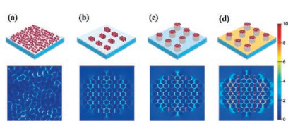
| Fig. 1. Comparison of the simulated EF intensity distributions of four different gold cluster structures. | |
Fig. S1 (Supporting information) outlined the fabrication procedure of the Au-NCPA-GM structure with controlled parameters. First the photoresponsive polymer film was coated onto a flat glass substrate through photo initiated polymerization of the acrylate monomers and cross-linker (Fig. S1), and utilized to fabricate the desired photoactive nanopillar arrays using NSL method. By employing the NSL, it is possible to precisely control the geometrical parameters of the resulting pillar structure by varying the experiment conditions. Then a gold film with a thickness of about 30 nm was further deposited on the etched substrate using an electron beam evaporation deposition system. The subsequent removal of all templates affords the polymer nanopillar array with a gold mirror at bottom. Finally, the gold cluster monolayer was assembled on the top of the prepared nanopillar array by peel-off transferring of the Au NPs monolayer deposited on PDMS substrate. The TEM images and UV-vis spectra of the prepared Au NPs with diameter of 20 nm, 35 nm, and 50 nm were given in Fig. S2 in Supporting information. Fig. S3 (Supporting information) shows the SEM images of the close packed uniform monolayers of the Au NPs with different sizes on a PDMA substrate. To evaluate the influence of structural parameters on the electromagnetic field enhancement in the prepared substrates, both theoretical simulations and experiments were carried out in our work.
To reveal the different near-field and far-field coupling interactions of Au-NCPA-GM compared to 2D random unpatterned gold NP cluster (Au-NC), 2D gold NP cluster array (Au-NCA) as well as gold NP nanopillar array without gold mirror (AuNCPA), FDTD simulations were carried out to study the EF intensity distribution of the four different structures. In this work, the EF is set as |E|2, and the incident light has |E|2 = 1 in all simulations. The colour scale bar in Fig. 1 represents the EF in liner scale. Thus, the EF value of each point can be obtained by referring to the scale bar. In the case of Au-NC (Fig. 1a), only the near-field surface plasmon coupling exists between adjacent Au NPs. It is obvious that the EF intensity in hotspot sites is relative weak in this structure. In the case of NCAs (Fig. 1b), however, it is found that the periodic array structure has an additional EF enhancement effect mainly due to the combine the electromagnetic near-field coupling with far-field coupling of clusters. It can be seen that the field intensity is stronger than that of the unpatterned random Au NPs cluster (Au-NC). Clearly, the far-field coupling interaction plays an important role in the NCA structure. The Au-NCPA (Fig. 1c) is a structure derived from expansion of the NCAs from two-dimension to three-dimension. We found that due to the longer propagation pathway of light in the third dimension the Au NPs close-packed on the top of pillars have a higher EF enhancement effect as compared with 2D planar counterparts. In particular, when gold film with a thickness of 30 nm was deposited on the bottom of pillars (Fig. 1d), further farfield coupling between the gold film and Au NPs was introduced. As the gold film acts as a mirror and reflects light, the strong coupling interaction between the gold film and Au NPs occurs in Au-NCPA-GM structure. As a result, the EF intensity between adjacent NPs in Au-NCPA-GM structure was significantly enhanced (Fig. 1d). These results indicate that the rational design of the multiscale periodic structure could make it possible to realize more abundant coupling interactions, thus leading to higher EF enhancement.
Certainly, the structural parameters of the Au-NCPA-GM architecture, including the Au NP size (d), the pillar array size (D) as well as the height of pillar (H), have a big influence on the EF intensity distribution in gold NP clusters. The FDTD simulations were carried out to study the influence of the different structural parameters on the electromagnetic field intensity. The results were shown in Fig. S4 in Supporting information.
Three surface structures prepared in our work, namely Au-NC, Au-NCPA and Au-NCPA-GM, were used as SERS substrates to confirm the FDTD simulation results. The SERS probe molecule, 4-mercaptophenol, was used in our case to assess the SERS performance of these substrates with the exciting light at 633 nm wavelength. Fig. 2d shows the comparison of SERS spectra collected on three different substrates under identical measurement conditions. Obviously, the periodic hierarchical Au-NCPA-GM substrate exhibits excellent SERS performance. Its SERS enhancement factor is one order of magnitude stronger as compared with the Au-CN. If we consider the two structures have the same number of the nanoparticles, the 3D Au-NCPA-GM structure will exhibit even more higher SERS enhancement than the 2D random structure. Notably, the introduction of gold mirror at the bottom of pillar array could further realize more coupling interactions, generating five times stronger SERS enhancement than Au-NCPA. These results are in consistent with the FDTD simulation, indicating that the multiple coupling strategy is an efficient approach to achieve the SERS substrate with improved SERS performance. In order to show the universality, another two probe molecules (4-mercaptobenzoic acid and 4-mercaptopyridine) were analyzed using our SERS substrates. As shown in Fig. S5 (Supporting information), our substrates is still suitable for analysis of more different compounds with high SERS enhancement.

|
Download:
|
| Fig. 2. SEM images of the three different substrates: (a) unpatterned Au-NCs; (b) Au-NCPAs; (c) Au-NCPAs-GM; (d) comparison of the SERS spectra performed on the three different substrates. Scale bars are 500 nm. | |
According to the simulation the NPs size has great influence on the SERS performance (Fig. S4a). Therefore, the Au NPs with size of 20, 35, and 50 nm were separately used to prepare the substrates (Fig. S6 in Supporting information). SEM images show that the uniform and close-packed Au NPs monolayer was assembled on the top of each nanopillar. Fig. S5d show the comparison of SERS spectra on the substrates consisting of different sizes of gold NPs. The experimental results show the SERS enhancement effect increases when large NPs were used, which is well consistent with the observation in the reference [26].
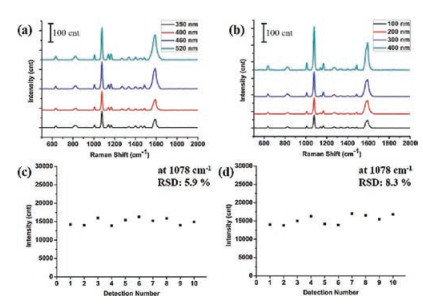
The size (diameter) of pillar is another parameter that can be broadly modulated by simply changing the etching time during the preparation and has an effect on the SERS efficacy. By changing the etching time of PS nanospheres, we prepared the nanopillars have diameter of 350, 400, 460, and 520 nm respectively, while the pillar heights were kept constant at 100 nm. Fig. S7 (Supporting information) shows the SEM images of the fabricated four substrates having different pillar sizes. In this case, gold NPs with a diameter of 50 nm were assembled on the top of these pillars to form different Au cluster sizes. Using these substrates the SERS performances were compared, as shown in Fig. 3a. In agreement with the simulation results the SERS signal intensity becomes stronger with the increase of the pillar size.

|
Download:
|
| Fig. 3. SERS spectra performed on the substrates with (a) different nanopillar size and (b) nanopillar height; (c) SERS signals at 1078 cm-1 collected at ten different area on the same substrate; (d) SERS signals at 1078 cm-1 collected on ten different substrates prepared under identical preparation conditions. | |
In our Au-NCPA-GM substrates the height of nanopillars determines the distance between the gold mirror and the Au clusters assembled on the top of pillars. Therefore, it is found that the height of pillars exerts an important influence on the EF coupling, thus the SERS perfermance. In this work, four different nanopillar arrays with the height of 100, 200, 300, and 400 nm, respectively, were fabricated (Fig. S8 in Supporting information), while the pillar size was kept constant at 460 nm. The crosssectional SEM views of these Au-NCPA-GM structures indicate that each of the substrates is highly uniform (Fig. S9 in Supporting information). Indeed, in the case of lower pillar array a stronger and efficient coupling between gold NP clusters and mirror occurs, thus facilitating improvement of SERS performance (Fig. 3b).
A high uniformity and reproducibility of SERS signals are of great importance and prerequisite for the practical application of SERS substrates. Fig. 3c shows the comparison of the SERS signals at 1078 cm-1 collected randomly at ten different spots on the same substrate. To check the reproducibility, ten substrates were prepared under identical preparation conditions and used for the SERS testing (Fig. 3d). We used the relative standard deviation (RSD) of SERS signal intensity at 1078 cm-1 to assess the uniformity and reproducibility of the fabricated substrates. According to ten measurements, the corresponding RSD values of SERS signal at 1078 cm-1 were calculated to be about 5.9% and 8.3%, respectively. Moreover, the SERS substrate was measured at intervals of 12 h to demonstrate the stability. Fig. S10 (Supporting information) shown the SERS signal intensity at 1078 cm-1 of ten measurements, and the corresponding RSD values was calculated to be about 5%, which indicated that our substrates with excellent stability. Similar to the observation in literature [23], the 3D ordered structure intrinsically endows our substrates with excellent signal uniformity and reproducibility.
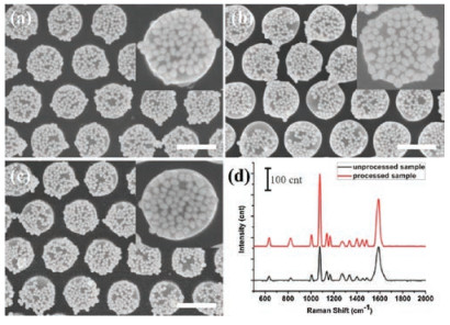
Besides creating highly enhanced EF and achieving uniform and reproducible signals, how to effectively place or entrap target molecules in hotspot region is another crucial issue for the development of high-performance SERS substrates. In case of the normal periodic cluster arrays, the nanoparticles were closely packed and the most of probe molecules were absorbed on the surface rather than inside the hotspots. In our work, a cross-linked polymer containing special azobenzene units was used for the formation of nanopillar arrays. The coupling of the photo-induced mass immigration effect and polymer network makes it possible for this polymer to expand the diameter of the created pillars by using circularly polarized light even far below glass temperature and to return its initial state by simple thermal treatment (Fig. S11 in Supporting information) [27]. This unique deformation property of the photoresponsive polymer implies that the nanogaps of the closely packed Au nanoparticles can be reversibly opened and closed through expanding and contracting the polymer pillars, and then the target molecule could be efficiently entrapped inside nanogaps (hotspots). Indeed, our experiments confirmed our concept by using a 532 nm wavelength laser beam with intensity of 10 mW/cm2. Fig. S10a shows the as-prepared Au-NCPA-GM structure, in which the gold nanoparticles are closely packed. It is found that a short illumination on the substrate induced the opening of the gaps of the adjacent nanoparticles (Fig. 4b). Interestingly, when the treated substrate was immersed into the solution of probe molecule (1 × 10⁻⁶ mol/L) for a short time and followed by a simple thermal treatment (Fig. 4c), more probe molecules were entrapped in the hotspots. As a result, with this active pillar array a higher SERS signal enhancement efficiency was achieved (Fig. 4d).

|
Download:
|
| Fig. 4. SEM images of (a) as-prepared Au-NCPAs-GM structure, (b) after light irradiation, (c) followed by thermal treatment; (d) comparison of the SERS spectra performed on as-prepared Au-NCPAs-GM structure and Au-NCPAs-GM structure after light illumination followed by thermal treatment. Scale bars are 500 nm. | |
In summary, by fabrication of 3D periodic and hierarchical gold nanoparticle cluster pillar arrays, we present a new strategy to address three critical issues for developing high-performance SERS substrates, namely creating hotspots with significantly enhanced EF, efficiently placing target molecules in hotspot region, and achieving uniform and reproducible Raman signals. Compared with 2D nanoparticle cluster array systems, it is theoretically and experimentally found that the rational design of 3D periodic and hierarchical array architecture could generate new opportunity to realize more abundant multiscale coupling interactions, thus affording the substrate with further improved SERS enhancement effect. Combined use of the top-down and bottom-up approach provide not only the precise control over the creation of 3D hierarchical array structure for ensuring the uniformity and reproducibility of the SERS signals, but also more parameters for broadly modulating array SERS performance. Moreover, the use of the novel photoresponsive polymer to produce nanopillar array endow our SERS substrate with active property. In our case, the unique photoinduced deformation of used polymer was employed to reversibly open and close the gaps of the closely packed Au NP arrays, thus enabling efficient placement or entrapment of probe molecules into hotspot sites between adjacent nanoparticles for further enhancing SERS efficiency. With these attractive features, we believe this new active Au-NCPA-GM array structure described here could be used as a new type of high-performance SERS substrates and hold great potential for practical SERS detecting applications.
AcknowledgmentsThe authors gratefully acknowledge the financial support from the Ministry of Science and Technology of the People's Republic of China (MOST, Nos. 2017YFA0204501, 2013CB834502), the National Natural Science Foundation of China (NSFC, Nos. 21473098, 21121004, and 21421064), and the Deutsche Forschungsgemeinschaft DFG (No. TRR61).
Appendix A. Supplementary dataSupplementary material related to this article can befound, in the online version, at doi:https://doi.org/10.1016/j.cclet.2018.06.013.
| [1] |
S.Y. Ding, E.M. You, Z.Q. Tian, M. Moskovits, Chem. Soc. Rev. 46 (2017) 4042-4076. DOI:10.1039/C7CS00238F |
| [2] |
C. Hamon, L.M. Liz-Marzán, J. Colloid Interface Sci. 512 (2018) 834-843. DOI:10.1016/j.jcis.2017.10.117 |
| [3] |
Y.Z. Chu, M.G. Banaee, K.B. Crozier, ACS Nano 4 (2010) 2804-2810. DOI:10.1021/nn901826q |
| [4] |
K.L. Kelly, E. Coronado, L.L. Zhao, G.C. Schatz, J. Phys. Chem. B 107 (2003) 668-677. |
| [5] |
E. Ringe, J.M. McMahon, K. Sohn, et al., J. Phys. Chem. C 114 (2010) 12511-12516. DOI:10.1021/jp104366r |
| [6] |
S.M. Nie, S.R. Emery, Science 275 (1997) 1102-1106. DOI:10.1126/science.275.5303.1102 |
| [7] |
S. Schlücker, Angew. Chem. Int. Ed. 53 (2014) 4756-4795. DOI:10.1002/anie.201205748 |
| [8] |
B. Yan, S.V. Boriskina, B.M. Reinhard, J. Phys. Chem. C 115 (2011) 4578-4583. DOI:10.1021/jp112146d |
| [9] |
J.M. Nam, J.W. Oh, H. Lee, Y.D. Suh, Acc. Chem. Res. 49 (2016) 2746-2755. DOI:10.1021/acs.accounts.6b00409 |
| [10] |
C. Wang, L. Tian, W. Zhu, et al., Chem. Sci. 9 (2018) 889-895. DOI:10.1039/C7SC03536E |
| [11] |
X.L. Zhang, X.H. Xiao, Z.G. Dai, et al., Nanoscale 9 (2017) 3114-3120. DOI:10.1039/C6NR09592E |
| [12] |
H.Y. Chen, M.H. Lin, C.Y. Wang, Y.M. Chang, S. Gwo, J. Am. Chem. Soc. 137 (2015) 13698-13705. DOI:10.1021/jacs.5b09111 |
| [13] |
E.C. Le Ru, P.G. Etchegoin, M. Meyer, J. Chem. Phys. 125 (2006) 204701. DOI:10.1063/1.2390694 |
| [14] |
D.D. Lin, Z.L. Wu, S.J. Li, et al., ACS Nano 11 (2017) 1478-1487. DOI:10.1021/acsnano.6b06778 |
| [15] |
B. Yan, A. Thubagere, W.R. Premasiri, et al., ACS Nano 3 (2009) 1190-1202. DOI:10.1021/nn800836f |
| [16] |
L.L. Yang, B. Yan, W.R. Premasiri, et al., Adv. Funct. Mater. 20 (2010) 2619-2628. DOI:10.1002/adfm.201000630 |
| [17] |
J. Wang, L.L. Yang, S. Boriskina, B. Yan, B.M. Reinhard, Anal. Chem. 83 (2011) 2243-2249. DOI:10.1021/ac103123r |
| [18] |
F.L. Yap, P. Thoniyot, S. Krishnan, S. Krishnamoorthy, ACS Nano 6 (2012) 2056-2070. DOI:10.1021/nn203661n |
| [19] |
C. Leordean, M. Potara, S. Boca-Farcau, et al., J. Raman Spectrosc. 45 (2014) 627-635. DOI:10.1002/jrs.v45.8 |
| [20] |
W.D. Liu, B. Yang, Chin. Chem. Lett. 28 (2017) 675-690. DOI:10.1016/j.cclet.2016.09.004 |
| [21] |
K.N. Kanipe, P.P. Chidester, G.D. Stucky, M. Moskovits, ACS Nano 10 (2016) 7566-7571. DOI:10.1021/acsnano.6b02564 |
| [22] |
W.D. Li, F. Ding, J. Hu, S.Y. Chou, Opt. Express 19 (2011) 3925-3936. DOI:10.1364/OE.19.003925 |
| [23] |
T. Lee, J.S. Wi, A. Oh, et al., Nanoscale 10 (2018) 3680-3687. DOI:10.1039/C7NR08066B |
| [24] |
M. Chirumamilla, A. Chirumamilla, A.S. Roberts, et al., Adv. Opt. Mater. 5 (2017) 1600836. DOI:10.1002/adom.v5.4 |
| [25] |
C.L. Haynes, R.P. Van Duyne, J. Phys. Chem. B 105 (2001) 5599-5611. DOI:10.1021/jp010657m |
| [26] |
P.N. Njoki, I.I.S. Lim, D. Mott, et al., J. Phys. Chem. C 111 (2007) 14664-14669. DOI:10.1021/jp074902z |
| [27] |
P. Rochon, E. Batalla, A. Natansohn, Appl. Phys. Lett. 66 (1995) 136-138. DOI:10.1063/1.113541 |