 2019, Vol. 30


b Department of Pharmaceutics, School of Pharmacy, East China University of Science and Technology, Shanghai 200237, China;
c Shanghai Key Laboratory of New Drug Design, East China University of Science and Technology, Shanghai 200237, China
Recently, morphology-controlled self-assembly of block and graft copolymers have attracted much interest, due to their potential applications in many fields such as in biomaterials and drug delivery [1-3]. Block or graft copolymers can be self- assembled to form micelles in their selective solvents. Compared with linear block copolymers, graft copolymer can add considerable functionalities on the branched chains with great advantage of convenient chemical modifications in advance of the self-assembly process [4, 5]. However, in most cases, amphiphilic graft copolymers tend to form compound spherical micelles [6, 7], whereas amphiphilic linear block copolymers can organize into abundant morphologies such as spheres, rods, vesicles, etc. [8-10]. Among many self-assembled morphologies, hollow spheres have drawn great attentions recently due to their unique large encapsulation capabilities with potential applications in the controlled release and targeted drug delivery [11-13]. Polymer-based hollow spheres of various dimensions can be fabricated by different methods, such as emulsion polymerization [14], layer by layer method [15], self-assembly of polyelectrolytes or rigid-coil copolymer [16, 17]. Among various methods, self-assembly of rigid-coil copolymer presents the most popular one due to its simple preparation procedure [18], as hollow spheres could be obtained directly in their selective solvent through self-assembly [19].
Chitosan (CS), an amino-polysaccharide from deacetylation of chitin, has been investigated for biomedical applications due to its low-toxicity, good biocompatibility and biodegradability [20-23]. Self-assembly behavior of CS-based biomaterials and their applications for drug delivery have been the focus of our research [24-34]. One major drawback of chitosan is its low solubility in physiological condition (pH 7.4), which greatly limits its utilities as it is only soluble in a few dilute acidic solutions such as in acetic acid and in dilute hydrochloric acid [20]. To improve chitosan solubility, methoxy polyethylene glycol (mPEG), a well-adopted hydrophilic, biocompatible, and biodegradable polymer for stabil- ityenhancement of nanoparticles, sustained release of protein, and reduction of severe side effects such as cytotoxicity in physico-chemical environment [35-38], could be grafted onto chitosan polysaccharide backbone. Moreover, the rigidity of the PEG chain could be further adjusted by incorporation of α-cyclodextrin (a-CD). First reported by Harada [39], PEG and α-CD forms rigid rod-like structure by threading α-CD molecules onto PEG chain. However, the further self-assembly of the formed α-CD/PEG rigid rod-like structure to more complex morphologies were seldom studied in literature [35, 41]. Recently, Zhang and coworkers [40] managed to use chitosan as the coil section and α-CD/PEG complex as the rigid section to afford hollow spheres loaded with magnetofluid in aqueous solution. We envisioned that the amount of α-CD may be crucial for the formation ofhollow spheres, but has not yet been fully explored.
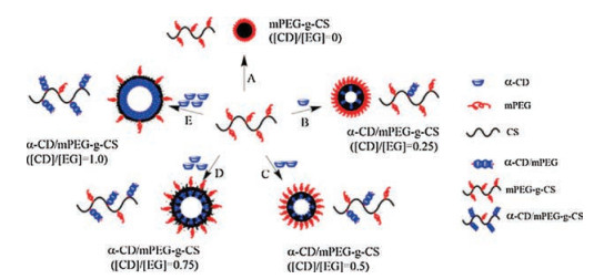
Herein, we fully investigated the effect of α-CD inclusion to the self-assemblybehavior of mPEG grafted chitosan. The α-CD/mPEG- g-CS system forms different morphology of nanoparticle and PEG density on the surface of nanoparticle at different ratio of[CD]/[EG] (Scheme 1). As far as we know, the current work presents the first report on the change of aggregation behavior of mPEG-g-CS graft copolymer induced by the different concentration of α-CD.

|
Download:
|
| Scheme 1. Schematic illustration of the formation of α-CD/mPEG-g-CS nanoparticles. | |
In the present studies, mPEG-g-CS was synthesized and characterized (Fig. S1 in Supporting information). The 1H NMR spectra of chitosan and mPEG-g-CS were shown in Fig. S2 (Supporting information). The proton assignment of chitosan (Fig. S2A) was as follows (ppm): 2.93 (CH, carbon b ofglucosamine ring), 3.4-3.8 (CH, carbon c, d, e and f of glucosamine ring). Compared with chitosan, the characteristic proton signals of mPEG-g-CS (Fig. S2B) appeared in the range of 3.1-3.5ppm. The peak at around 2.97 ppm was the proton signal of methoxyl of mPEG-g-CS. Therefore, degree of substitute (DS) of mPEG moiety could be calculated by comparing the ratio of methoxyl of mPEG protons (2.97 ppm) to sugar protons (2.72 ppm, CH, carbon 2 of glucosamine ring). The DS could be calculated by follows:

|
The calculalted DS of mPEG moiety on mPEG-g-CS was 21.5%.
Fig. S3 (Supporting information) showed the Fourier transform infrared spectroscopy (FT-IR)spectra of chitosan (a) and mPEG-g-CS (b). The chitosan spectrum (Fig. S3A) showed characteristic bands of amide Ⅰ (1683 cm-1) and amide Ⅱ (1587 cm-1). The peaks at 3420 cm-1 in chitosan scaffolds corresponded to O—H stretch overlapped with N-H stretch. For mPEG-g-CS (Fig. S3B), bands of amide Ⅰ (1683 cm-1) and amide Ⅱ (1587 cm-1) observably decreased. Furthermore, the increased intensity of the peaks at around 2920, 1739 and 1100cm-1 indicated the CH2, C=O and C—O—C stretch of mPEG. These results showed that the amino groups of chitosan were successfully substituted by mPEG groups.
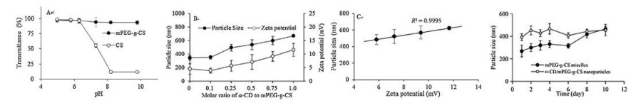
pH-dependent solubility study of mPEG-g-CS (Fig. 1A) revealed that solubility of the prepared polymer mPEG-g-CS at various pH from 5 to 11 in aqueous solution was studied and compared with chitosan. At pH 5, both chitosan and mPEG-g-CS showed an optical transmittance over 98%, indicating good solubility for both polymers. When pH > 6.1, chitosan started to form a milky solution due to reduced solubility and formation of large aggregates. In contrast, mPEG-g-CS remained a clear solution with an optical transmittance more than 90%, indicating a good solubility of mPEG-g-CS at the pH range tested from 5 to 11. Clearly, the introduction of mPEG to the main chain of chitosan greatly enhances the polymer's solubility at pH over 6.1 including physiological pH at 7.4.
Then, hollow spheres were prepared by adding α-CD solution into mPEG-g-CS, the threading of α-CD onto the flanking mPEG chains of the mPEG-g-CS copolymer, and inclusion formation between the α-CD and mPEG-g-CS were further confirmed with X-ray powder diffraction studies. As shown in Fig. S4 (Supporting information), the pattern of α-CD/mPEG-g-CS particles were similar to complex of α-CD/mPEG inclusion (2θ = 20.0°) [39], which has been reported to have a rod channel structure, while the mPEG-g-CS crystalline peaks (2θ = 19.1°and 23.2°)and α-CD crystalline peak (2θ = 21.7°) were absent. This implied that α-CD rings are stacked along the graft PEG chain axis to form a channel- type crystalline structure. As a result, the α-CD/mPEG-g-CS has both rod block (α-CD/mPEG complex) and coil block (protonated chitosan chain). It was reported that polymer with rod-coil structure prefers parallel packing to achieve the most efficient space-filling and resulted in the formation ofhollow spheres [19].
To explore the effect of molar ratio of α-CD to mPEG-g-CS to properties of α-CD/mPEG-g-CS nanoparticles, a series of α-CD/mPEG-g-CS nanoparticles with different molar (0, 0.1, 0.25, 0.5, 0.75, 1.0) ratio of [CD] to [EG], [CD] indicated molar concentration of α-CD and [EG] indicated molar concentration of ethylene glycol unit, were prepared in aqueous solution (pH 6.0). As shown in Fig. 1B, particle size of α-CD/mPEG-g-CS nanoparticles increased from 257.6nm to 699.7nm as the molar ratio of [CD]/[EG] increased from 0 to 1.0. To explain this phenomenon, a possible mechanism was proposed as shown in Scheme 1. mPEG-g-CS was able to form micelle composed of an inner core mainly consisting of chitosan with entangled intramolecular hydrogen bonding network within and an outside corona consisting of mainly mPEG chains for hydrophilic interaction with solvent molecules when no α-CD was added [30]. Upon addition of α-CD, the complexation of α-CD molecules with mPEG chains through sliding into the PEG chains forms rigid rod-like complexes [39], which tend to stick to each other and exclude solvent molecules, resulting in transloca tion of mPEG chains for better space-filling, reduced shielding of positive charges of chitosan, and the growth of a cavity within each nanospheres [19].

|
Download:
|
| Fig. 1. (A) The influence of pH on water solubility of chitosan and mPEG-g-CS. (B) The relationship of particle size and zeta potential of α-CD/mPEG-g-CS nanoparticle with different molar ratio of α-CD to mPEG-g-CS in pH 6.0 aqueous solution. (C) Correlation between zeta potential and particle size of α-CD/mPEG-g-CS nanoparticle with molar ratio of α-CD to mPEG-g-CS from 0 to 1.0 in pH 6.0 aqueous solution. (D) The size change of mPEG-g-CS micelles and α-CD/mPEG-g-CS ([CD]/[EG] = 0.25) nanoparticles in pH 7.4 aqueous solution at 4 ℃. Data were expressed as mean±SD (n = 3). | |
As shown in Fig. 1B, no significant physicochemical property change occurred until the molar ratio of [CD]/[EG] reached 0.25. And then starting from [CD]/[EG] = 0.25, α-CD/mPEG-g-CS nanoparticles ([CD]/[EG] = 0.25, 0.5, 0.75, 1.0) exhibited increased zeta potential from +4.5 mV to +11.6 mV along with the increased size from 495.2 nm to 669.7 nm. The increased zeta potential showed reduced shielding of the positive charges of chitosan, suggesting the reduced density of uncomplexed mPEG chain on the surface of nanoparticles. Meanwhile, the increased particle size indicated the growth of hollow sphere in the nanoparticles due to increased α-CD/mPEG complexation. Fig. 1C showed the correlation between zeta potential and particle size of α-CD/mPEG-g-CS nanoparticles, indicating varied PEG density on surface of nanoparticles and mPEG chain can transferred from outside to inside, leading to exposed positive charge of chitosan and increased particle size by efficient space-filling packing of α-CD/mPEG complexes.
To investigate the effect of pH on the self-asssembly behavior of α-CD/mPEG-g-CS nanoparticles, nanoparticles were prepared under various pH (4.0, 5.0, 6.0, 6.8, 7.4, 8.0). As shown in Table 1, α-CD/mPEG-g-CS ([CD]/[EG] =0.25) nanoparticles, when pH value increased from 4.0 to 8.0, the particle size and the zeta potential gradually decreased from 666.1 nm to 308.5 nm and from +28.3 mV to +2.0 mV, respectively. This phenomenon could be rationalzied as the following. α-CD/mPEG-g-CS ([CD]/[EG] = 0.25) nanoparticles contain a highly compact protonated inner chitosan core surrounded by a relative hydrophilic mPEG corona. At lower pH, particle size of α-CD/mPEG-g-CS ([CD]/[EG] = 0.25) nanoparticles were increased likely due to the increased electronic repulsion of protonated amino groups within the chitosan core. As for α-CD/mPEG-g-CS ([CD]/[EG] = 1.0) nanoparticles, the particle size increased gradually from around 504.8 nm to 768.2 nm, while the zeta potential decreased gradually from +31.1 mV to -3.4 mV when the pH value increased from 4.0 to 8.0. α-CD/mPEG-g-CS ([CD]/[EG] = 1.0) nanoparticles contained a hollow cavity inside due to efficient space-filling packing of rod-shaped α-CD/mPEG-g-CS, causing few free PEG chains and chitosan chains stretched outside as a stablizing corona. The chitosan (pKa = 6.5) chains were poor soluble because of the deprotonation of amino groups. Therefore, at high pH region (pH > 6.5), the α-CD/mPEG-g-CS nanoparticles were not stable and macrocosmic precipitation took place. As pH value decreased, the amino group was ionized and then the α-CD/mPEG-g-CS ([CD]/[EG] = 1.0) nanoparticles became more and more stable.
|
|
Table 1 Physicochemical properties of α-CD/mPEG-g-CS nanoparticles. Data were expressed as mean±SD (n = 3). |

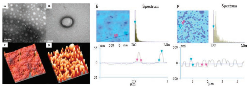
The morphology of mPEG-g-CS micelles were studied by transmission electron microscopy (TEM) and atomic force microscopy (AFM). The results were shown in Figs. 2A, C, and E. Micelles had spherical morphology and maintained in non-aggregated state. It should be noticed that the size of mPEG-g- CS estimated from TEM is about 50-100nm, which was much smaller than that from dynamic light scattering (DLS). The discrepancy of nanoparticle size measured between DLS and TEM was likely due to the shrinkage of nanoparticles under vacuum condition since TEM was performed under high vacuum. However, AFM images showed the diameters of mPEG-g-CS nanoparticles are around 300-350nm, which was even larger than that from DLS (257.6 nm). The seemingly discrepancy might be caused by the different sample preparation and measurement condition since DLS was performed in aqueous solution, while the AFM image were taken in dried sample in which nanoparticles tended to be flatten [42]. Upon addition of α-CD into mPEG-g-CS solution, hollow spheres could be obtained as shown in the TEM image (Fig. 2B). The contrast between central cavity and outer layer clearly indicated the presence of hollow sphere structure of the nanoparticles [17]. From AFM studies (Figs. 2D and F), the height/particle size ratio of α-CD/mPEG-g-CS (around 1:2) nanoparticle ([CD]/[EG] = 0.25) was higher than that of mPEG-g-CS (around 1:8), reflecting increased rigidity due to threading of α-CD into mPEG chains of mPEG-g-CS nanoparticles.

|
Download:
|
| Fig. 2. The TEM and AFM image ofmPEG-g-CS micelles (A, C, E), α-CD/mPEG-g-CS ([CD]/[EG] = 0.25) nanoparticles (B, D, F). | |
Stability study (Fig. 1D) showed that mPEG-g-CS nanoparticles showed good stability in the first seven days without much increase of particle size, while the particle size significantly increased from the day 7 to the day 10. In contrast, α-CD/mPEG-g-CS ([CD]/[EG] = 0.25) nanoparticles showed good stability up to ten days. The increased stability could be rationalized by the enhanced rigidity of nanoparticles upon addition of α-CD. Apparently, the conformational flexibility of mPEG-g-CS makes it easier to dissociate than a more rigid α-CD/mPEG-g-CS.
In conclusion, we presented an interesting nanoparticle-based drug-delivery system, α-CD/mPEG-g-CS, with morphology transition behavior depending on the different amount of α-CD added. The effect of α-CD inclusion on the self-assembly behavior of mPEG grafted chitosan was studied. The results showed that the mPEG-g-CS forms self-assembled nanoparticles with either micelle or hollow sphere morphology depending on the ratio of α-CD to EG, as characterized by DLS, AFM, TEM, and X-ray diffraction (XRD). The increase of nanoparticle size and zeta potential upon addition of α-CD correlated well with the decreasing amount of PEG density on the surface of nanoparticles. Due to its unique pH dependent properties and large internal cavity, the α-CD/mPEG-g-CS hollow spheres showed promising as a novel nanocarrier system for delivery of protein drugs. The method of controlling the PEG density on surface of nanoparticle by α-CD paved a path to investigate the in vivo circulation time of nanoparticle in the future.
AcknowledgmentsThis work was supported by Science and Technology Commission of Shanghai Municipality (Nos. 17ZR1406600, 10DZ2220500, 11DZ2260600) and National Natural Science Foundation of China (No.21577037).
Appendix A. Supplementary dataSupplementary data associated with this article can be found, in the online version, at https://doi.org/10.1016/j.cclet.2017.12.012.
| [1] |
X. Hu, J. Hu, J. Tian, et al., J. Am. Chem. Soc. 135 (2013) 17617-17629. DOI:10.1021/ja409686x |
| [2] |
Q. Hu, W. Li, X. Hu, et al., Biomaterials 33 (2012) 6580-6591. DOI:10.1016/j.biomaterials.2012.05.060 |
| [3] |
J.H. Kim, T. Ramasamy, T.H. Tran, et al., Asian J. Pharm. Sci. 9 (2014) 191-198. DOI:10.1016/j.ajps.2014.05.001 |
| [4] |
K. Breitenkamp, T. Emrick, J. Am. Chem. Soc. 125 (2003) 12070-12071. DOI:10.1021/ja036561i |
| [5] |
Y. Sato, Y. Kobayashi, T. Kamiya, et al., Biomaterials 26 (2005) 703-711. DOI:10.1016/j.biomaterials.2004.03.018 |
| [6] |
F.Q. Hu, W.W. Chen, M.D. Zhao, H. Yuan, Y.Z. Du, Gene Ther. 20 (2013) 597-606. DOI:10.1038/gt.2012.72 |
| [7] |
G.M. Mekhail, A.O. Kamel, G.A. Awad, N.D. Mortada, Int. J. Biol. Macromol. 51 (2012) 351-363. DOI:10.1016/j.ijbiomac.2012.05.026 |
| [8] |
R. Qi, Y. Jin, X. Cheng, et al., Macromol. Rapid Comm. 36 (2015) 1402-1408. DOI:10.1002/marc.v36.15 |
| [9] |
C. Cheng, X.J. Han, Z.Q. Dong, et al., Macromol. Rapid Comm. 32 (2011) 1965-1971. DOI:10.1002/marc.201100514 |
| [10] |
Q. Yan, Y. Zhao, J. Am. Chem. Soc. 135 (2013) 16300-16303. DOI:10.1021/ja408655n |
| [11] |
X. She, L. Chen, L. Velleman, et al., J. Colloid Interf. Sci. 445 (2015) 151-160. DOI:10.1016/j.jcis.2014.12.053 |
| [12] |
S.K. Das, M.K. Bhunia, D. Chakraborty, A.R. Khuda-Bukhsh, A. Bhaumik, Chem. Commun. 48 (2012) 2891-2893. DOI:10.1039/c2cc17181c |
| [13] |
Z. Shi, Y. Zhou, D. Yan, Macromol. Rapid Comm. 27 (2010) 1265-1270. |
| [14] |
L. Song, X. Ge, M. Wang, Z. Zhang, S. Li, J. Polym. Sci. Pol. Chem. 44 (2010) 2533-2541. |
| [15] |
H. Guo, Q. Guo, T. Chu, et al., J. Mater. Sci:Mater. Med. 25 (2014) 121-129. DOI:10.1007/s10856-013-5055-6 |
| [16] |
Z. Dai, A. Voigt, S. Leporatti, et al., Adv. Mater. 13 (2001) 1339-1342. DOI:10.1002/1521-4095(200109)13:17<1339::AID-ADMA1339>3.0.CO;2-Q |
| [17] |
H. Duan, M. Kuang, J. Wang, D. Chen, M. Jiang, J. Phys. Chem. B 108 (2004) 550-555. DOI:10.1021/jp0363139 |
| [18] |
M. Kuang, H. Duan, J. Wang, D. Chen, M. Jiang, Chem. Commun. 9 (2003) 496-497. |
| [19] |
S.A. Jenekhe, X.L. Chen, Science 283 (1999) 372-375. DOI:10.1126/science.283.5400.372 |
| [20] |
A.J. Dong, M.H. Feng, H.Y. Qi, S.S. Li, L.D. Deng, J. Mater. Sci.:Mater. Med. 19 (2008) 869-876. DOI:10.1007/s10856-007-3183-6 |
| [21] |
Q. Gao, L. Liu, X. Lu, H. Zhou, Asian J. Pharm. Sci. 11 (2016) 673-683. DOI:10.1016/j.ajps.2016.07.001 |
| [22] |
J.S. Duk, K. SookHee, W. You-Yeon, K.S. Hwa, K.I. Chan, Theranostics 6 (2016) 1362-1377. DOI:10.7150/thno.15335 |
| [23] |
S. Fu, J. Xia, J. Wu, J. Biomed. Nanotechnol. 12 (2016) 1585-1603. DOI:10.1166/jbn.2016.2228 |
| [24] |
D.J. Fu, Y. Jin, M.Q. Xie, et al., Chin. Chem. Lett. 25 (2014) 1435-1440. DOI:10.1016/j.cclet.2014.06.027 |
| [25] |
Y. Chen, Y. Huang, D. Qin, et al., PLoS ONE 11 (2016) e0150877. DOI:10.1371/journal.pone.0150877 |
| [26] |
X. Wang, H. Tang, C. Wang, et al., Theranostics 6 (2016) 1378-1392. DOI:10.7150/thno.15156 |
| [27] |
D. Jiang, X. Zhang, D. Yu, et al., J. Biomed. Nanotechnol. 13 (2017) 243-254. DOI:10.1166/jbn.2017.2346 |
| [28] |
Z.W. Li, C.W. Li, Q. Wang, et al., J. Biomed. Nanotechnol. 13 (2017) 17-34. DOI:10.1166/jbn.2017.2324 |
| [29] |
M.C. Bonferoni, G. Sandri, E. Dellera, et al., J. Biomed. Nanotechnol. 12 (2016) 231-240. DOI:10.1166/jbn.2016.2140 |
| [30] |
A.K. Goyal, T. Garg, G. Rath, U.D. Gupta, P. Gupta, J. Biomed. Nanotechnol. 12 (2016) 450-463. DOI:10.1166/jbn.2016.2180 |
| [31] |
Y. Liu, Y. Pu, L. Sun, et al., J. Biomed. Nanotechnol. 12 (2016) 1393-1403. DOI:10.1166/jbn.2016.2275 |
| [32] |
I.A.U. doetok, L.D. Wilson, J.V. Headley, A.C.S. Appl, Mater. Interfaces 8 (2016) 33197-33209. DOI:10.1021/acsami.6b11504 |
| [33] |
Y. Li, B. Humphries, Z. Wang, et al., ACS Appl. Mater. Interfaces 8 (2016) 30735-30746. DOI:10.1021/acsami.6b10306 |
| [34] |
W. Yasen, R. Dong, L. Zhou, et al., ACS Appl. Mater. Interfaces 9 (2017) 9006-9014. DOI:10.1021/acsami.6b15919 |
| [35] |
L. Casettari, D. Vllasaliu, E. Castagnino, et al., Prog. Polym. Sci. 37 (2012) 659-685. DOI:10.1016/j.progpolymsci.2011.10.001 |
| [36] |
C. Prego, D. Torres, E. Fernandez-Megia, et al., J. Control. Release 111 (2006) 299-308. DOI:10.1016/j.jconrel.2005.12.015 |
| [37] |
X.G. Zhang, D.Y. Teng, Z.M. Wu, et al., J. Mater Sci.:Mater. Med. 19 (2008) 3525-3533. DOI:10.1007/s10856-008-3500-8 |
| [38] |
X. Zhang, H. Zhang, Z. Wu, et al., Eur. J. Pharm. Biopharm. 68 (2008) 526-534. DOI:10.1016/j.ejpb.2007.08.009 |
| [39] |
A. Harada, J. Li, M. Kamachi, Macromolecules 26 (1993) 5698-5703. DOI:10.1021/ma00073a026 |
| [40] |
H. Wei, H. Wu, Y. Ma, et al., Carbohyd. Polym. 92 (2013) 523-528. DOI:10.1016/j.carbpol.2012.09.053 |
| [41] |
T. Ouchi, H. Nishizawa, Y. Ohya, Polymer 39 (1998) 5171-5175. DOI:10.1016/S0032-3861(97)10020-9 |
| [42] |
S. Yu, T. Azzam, I. Rouiller, A. Eisenberg, J. Am. Chem. Soc. 131 (2009) 10557-10566. DOI:10.1021/ja902869q |