 2019, Vol. 30

 ,
Ning Zhaob,
Jian Xub,*
,
Ning Zhaob,
Jian Xub,*
b Beijing National Laboratory for Molecular Sciences, Institute of Chemistry, Chinese Academy of Sciences, Beijing 100190, China
Polymeric composites have been widely used in many fields like aerospace, vehicles and sport utilities and so on [1]. Macro- or microscopic damages occurred during the use will cause material performance degradation. Inspired by the self-healing behavior of organisms in nature [2], scientists developed a series of self-healing materials by using physical or chemical methods. Analogous to the healing process of organisms, self-healing materials can recover their functionalities without external intervention [3-6]. White and coworkers made a breakthrough in the study of self-healing composite materials [7], and systematically investigated the self-healing process and possible mechanism [8]. Self-healing composites based on microcapsules [9-11], hollow glass fibers [12], engineered microvascular networks [13-15], Diels-Alder reversible chemistry [16-18] and particle segregation [19] have been prepared. However, some disadvantages such as complex chemical reactions, sophisticated production process and high costs limited the practical applications of self-healing composites.
Herein, we prepared composite coatings by embedding healant- loaded core-shell fibers into acrylate resin. The core-shell fibers were fabricated by using coaxial electrostatic spinning with epoxy or curing agent as the core and PS solution as the shell. Scanning electron microscopy (SEM), Fourier transform infrared (FTIR) and optical microscope were utilized to characterize the morphologyand composition of the fibers. The effect of the feed rate of healing material on the fiber formation was studied. The scratch healing performance of the prepared coatings was estimated. This method may find practical application in the preparation of self-healing materials considered the rapid development of electrospinning technique and the availability of the highly effective healants.
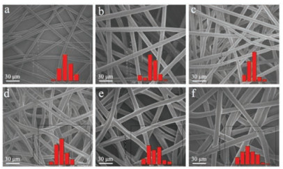
Electrospinning is a versatile method for the preparation of continuous fibers with diameters ranging from micrometers to nanometers [20, 21]. Coaxial electrostatic spinning can fabricate core-shell fibers in one step [22-25]. By using coaxial electrostatic spinning, we prepared healant-loaded core-shell PS fibers using epoxy resin or curing agent as the core material. The materials used in our contribution and the preparation and characterization methods are shown in Supportinginformation. A high content of healing agent in fibers is beneficial for the self-healing performance of composite, but it is limited by the formability of core-shell structure. We studied the influence of the epoxy content, controlled by the feed rate of the core material, on fiber morphology and diameter. Fig. 1 shows SEM images ofthe fibers fabricated at different epoxy feed rates. The mean diameter of the PS fibers without healing agent is about 5.7 ± 0.1 mm and the diameter is narrowly distributed, as shown in Fig. 1a. With the increase of the epoxy feed rate, the mean diameter of the fibers improves significantly (Figs. 1b-f), and the diameter distributions of the core-shell fibers fabricated with different feed rates are shown in Fig. 1. When the feed rate increased to 11 μL/min, fibers could not be formed.

|
Download:
|
| Fig. 1. SEM images and diameter distributions of the core-shell PS fibers fabricated at an epoxy feed rate of (a) 0, (b) 1, (c) 3, (d) 5, (e) 7 and (f) 9 μL/min, respectively. | |
FTIR spectroscopy was used for charactering the epoxy encapsulated in the fibers. The corresponding spectra are shown in Fig. S1 (Supporting information). The characteristic absorption peak of the epoxide ring at 1247, 912 and 832 cm-1 can be detected for the coaxial electrospun fibers. The absorption peak area of epoxide ring enhanced gradually with the epoxy feed rate increase.
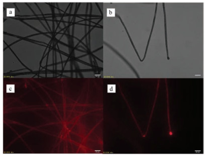
Fluorescent dye was added into epoxy to demonstrate the core-shell structure bythe microscope, and also to illustrate the liquid in the core can be released when the core-shell structure was broken. The optical and fluorescent microscope images are shown in Fig. 2. From the images, it can be clearly seen that the liquid epoxy is continuously and uniformly distributed in the PS shell. It can be found that the diameter in Fig. 2d is smaller than that in Fig. 2b, suggesting epoxy is mainly existing as the core. In addition, a large drop of epoxy at the fracture point of the fiber can be observed (Fig. 2d), indicating that epoxy can release from the broken fiber. The release of the healant encapsulated in the fiber is crucial for realizing the self-healing of the fibers filled composites.

|
Download:
|
| Fig. 2. (a, b) Optical images of the core-shell fibers and (c, d) fluorescent images of the core-shell fibers. | |
The core-shell structure was further confirmed by removing the core materials by using selective solvent. We immersed the core-shell fibers in ethanol at 60 ℃ for 30min to dissolve the healing agent. SEM images (Fig. 3) show that hollow PS fibers retaining a complete fiber structure were left after the core removing. No cracks or pores appeared on the pure PS fiber, as shown in Fig. 3a. Empty channels were formed for healant-loaded fibers and their diameters increased with the feed rate of core materials. The thickness of the fiber shell decreases gradually with the increase of feed rate of core material, since the flow rate of the shell material remains the same in the coaxial electrostatic spinning process. The shell thickness was very thin when the flow rate was 9 μL/min. The result explains why the core-shell structure could not be formed at a flow rate of 11 μL/min. In addition, residual shells are all intact with no cracks or holes, further indicating the core-shell fibers are stable. Also after heating the core-shell fibers at 60 ℃ for 5 min, we did not observe any leakage of the liquid from the core-shell fibers (Fig. S2 in Supporting information).

|
Download:
|
| Fig. 3. SEM images of the pure PS fibers (a) and the core-shell fibers obtained from the epoxy feed rate of (b) 1, (c) 3, (d) 5, (e) 7 and (f) 9 μL/min, respectively, after ethanol treatment. | |
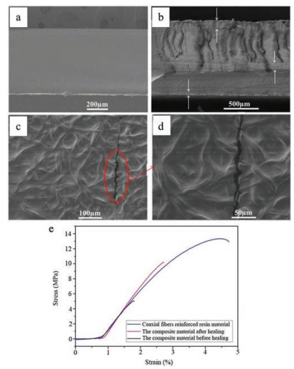
By the same method, PS core-shell fibers filled with curing agent were fabricated. Composite coatings were fabricated byembedding the core-shell fiber membranes in layers in acrylate resin. Different from the pure acrylate resin coating, the layered structure of the composite coating can be clearlyobserved bySEM(Figs. 4a and b). To characterize the self-healing property, a scratch was made on the coating surface. Fig. 4c shows the top view of the crack region which was left to heal by itself at room temperature for 24h. SEM image shows the liquid healing agents encapsulated in fibers released at the crack regiononce the core-shell structure of the fibers was destroyed, and ring-opening polymerization would be triggered when epoxy contact with the curing agent at the crack region. From the high magnificationview of the crack region after self-healing(Fig. 4d), we can see the original bumpy surface around the crack region was flattened due to the release of the core materials from the fibers. Fig. 4e shows after the sample was healed at room temperature for 24h and then at 60℃ for another 24h, the stress recovered from 13.3 MPa to 10.2 MPa, higher than the 5.0 MPa before healing, which indicates a good self-healing capacity.

|
Download:
|
| Fig. 4. SEM images of (a) the fracture surface of pure acrylate resin coating, (b) the composite coating, (c) the top view and (d) the high magnification view of the scratch on the coating surface after healing. (e) The mechanical property of the samples before and after healing. | |
In summary, we studied such an approach to make the polymer fibers with core-shell structures by means of coaxial electrostatic spinning technology, containing liquid healing agent as the core. The PS/healing agent core-shell fibers have been demonstrated effectively in self-healing process of polymer composites. Healing agents were successfully encapsulated in the fibers and the diameter of the fibers increased with the feed rate of the core materials. The core-shell structure and the release of healing materials from the mechanically ruptured fibers were confirmed by different techniques. Composite coatings embedded with the healant-loaded core-shell fibers have been prepared. Liquid healing agents encapsulated in fibers can be released and heal the scratch formed on the coatings by ring-opening polymeriza tion. Considered the rapid development of electrospinning technique and the availability of the highly effective healants, this simple and effective method may find practical application in many fields.
AcknowledgmentThis work was financially supported by the MOS of China (No. 2017YFB0703300), the National Natural Science Foundation of China (No. 51673117), the Science and Technology Innovation Commission of Shenzhen (Nos. JSGG20160226201833790, JCYJ20150625102750478).
Appendix A. Supplementary dataSupplementary data associated with this article can be found, in the online version, at https://doi.org/10.1016/j.cclet.2018.01.037.
| [1] |
S. Sinha-Ray, D.D. Pelot, Z.P. Zhou, et al., J. Mater. Chem. 22 (2012) 9138-9146. DOI:10.1039/c2jm15696b |
| [2] |
R.S. Trask, H. Rwilliams, I.P. Bond, Bioinspir. Biomim. 2 (2007) 1-9. DOI:10.1088/1748-3182/2/1/001 |
| [3] |
J.H. Park, P.V. Braun, Adv. Mater. 22 (2010) 496-499. DOI:10.1002/adma.v22:4 |
| [4] |
C.M. Dry, Proc. SPIE 1777 (1992) 367-371. |
| [5] |
C. Dry, N.R. Sottos, Proc. SPIE 1916 (1993) 438-444. DOI:10.1117/12.148501 |
| [6] |
X.F. Wu, A. Rahman, Z.P. Zhou, et al., J. Appl. Polym. Sci. 129 (2013) 1383-1393. DOI:10.1002/app.38838 |
| [7] |
S.R. White, N.R. Sottos, P.H. Geubelle, et al., Nature 409 (2001) 794-797. DOI:10.1038/35057232 |
| [8] |
R.P. Wool, Soft Matter 4 (2008) 400-418. DOI:10.1039/b711716g |
| [9] |
X.X. Liu, H.R. Zhang, J.X. Wang, Z. Wang, S.C. Wang, Surf. Coat. Technol. 206 (2012) 4976-4980. DOI:10.1016/j.surfcoat.2012.05.133 |
| [10] |
H.H. Jin, C.L. Mangun, A.S. Griffin, et al., Adv. Mater. 26 (2014) 282-287. DOI:10.1002/adma.201303179 |
| [11] |
H.R. Zhang, J.X. Wang, X.X. Liu, Z. Wang, S.C. Wang, Ind. Eng. Chem. Res. 52 (2013) 10172-10180. DOI:10.1021/ie400666a |
| [12] |
R.S. Trask, G.J. Williams, I.P. Bond, J. Roy. Soc. Interface 4 (2007) 363-371. DOI:10.1098/rsif.2006.0194 |
| [13] |
C.J. Hansen, W. Wu, K.S. Toohey, et al., Adv. Mater. 21 (2009) 4143-4147. DOI:10.1002/adma.v21:41 |
| [14] |
D. Therriault, S.R. White, J.A. Lewis, Nat. Mater. 2 (2003) 265-271. DOI:10.1038/nmat863 |
| [15] |
K.S. Toohey, N.R. Sottos, J.A. Lewis, J.S. Moore, S.R. White, Nat. Mater. 6 (2007) 581-585. DOI:10.1038/nmat1934 |
| [16] |
C.K. Gong, J.J. Liang, W. Hu, et al., Adv. Mater. 25 (2013) 4186-4191. DOI:10.1002/adma.201301069 |
| [17] |
X.X. Chen, M.A. Dam, K. Ono, et al., Science 295 (2002) 1698-1702. DOI:10.1126/science.1065879 |
| [18] |
X.X. Chen, F. Wudl, A.K. Mal, H.B. Shen, S.R. Nutt, Macromolecules 36 (2003) 1802-1807. DOI:10.1021/ma0210675 |
| [19] |
S. Gupta, Q.L. Zhang, T. Emrick, A.C. Balazs, T.P. Russell, Nat. Mater. 5 (2006) 229-233. DOI:10.1038/nmat1582 |
| [20] |
A. Greiner, J.H. Wendorff, Angew. Chem. Int. Ed. 46 (2007) 5670-5703. DOI:10.1002/(ISSN)1521-3773 |
| [21] |
D.M. Zhang, J. Chang, Adv. Mater. 19 (2007) 3664-3667. DOI:10.1002/(ISSN)1521-4095 |
| [22] |
J.F. Cooley, Patent, US 692631, 1902-02-04.
|
| [23] |
A. Formhals, Patent, US 1975504, 1934-10-2.
|
| [24] |
G. Taylor, Proc. R. Soc. A-Math. Phys. 313 (1969) 453-475. DOI:10.1098/rspa.1969.0205 |
| [25] |
Z.C. Sun, E. Zussman, A.L. Yarin, J.H. Wendorff, A. Greiner, Adv. Mater. 15 (2003) 1929-1932. DOI:10.1002/(ISSN)1521-4095 |