 2019, Vol. 30

 ,
Ke Pana,**
,
Ke Pana,**
b Affliated Hospital of Integrated Traditional Chinese and Western Medicine, Nanjing University of Chinese Medicine, Nanjing 210028, China;
c Laboratories of Translational Medicine, Jiangsu Province Academy of Traditional Chinese Medicine, Nanjing 210028, China;
d College of Pharmacy, Nanjing University of Chinese Medicine, Nanjing 210023, China;
e Department of Nuclear Medicine, The First Affiliated Hospital of Nanjing Medical University, Nanjing 210029, China;
f Theragnostic Laboratory, Campus Gasthuisberg, KU Leuven, 3000 Leuven, Belgium
Necrosis is described as the passive death of cells that subjected to illness or serious injury [1]. As a form of cell death, necrosis is widespread in various diseases, such as cancer, myocardial infarction (MI), neurodegenerative diseases, and stroke, etc. [2]. Necrosis avid agents (NAAs) can specifically accumulate in the necrotic area of tissues and show promising practical prospect in many fields, such as the diagnosis and therapy of necrosis-related diseases, as well as the evaluation of therapeutic efficacy [3, 4].
Previous studies have revealed that hypericin (Hyp), a naphthodianthrone compound, can specifically accumulate in necrotic tissues [5]. [123I]Hyp could visualize myocardial infarcts with small animal SPECT (single-photon emission computed tomography) imaging [6]. However, the self-aggregate property and high blood pool activity of Hyp have limited its clinical applications [6-8]. Recently, we found that monomeric anthraquinones (AQs) also exhibited peculiar affinity to necrotic tissues by splitting the chemical structure of Hyp [8]. Among them, rhein emerged as the most promising compound for imaging necrotic tissues due to its optimal pharmacokinetics properties and had been labeled with iodine-131 and Tc-99 m to visualize the necrotic myocardium [8, 9]. However, the hepatotoxicity and poor solubility of rhein limit to some extent the clinical applications of rheinbased NAAs [10]. Therefore, there is still a pressing need to seek NAAs with high safety and suitability for clinical applications.
It was reported that exposed DNA (E-DNA) is one of the most classical molecular targets for NAAs owning to its abundance and specificity in necrotic areas [11]. Recently, a series of NAAs targeted to E-DNA have been reported, such as Hoechst-IR [11], HoechstGemcitabine [12], and Gd-TO [13]. Besides, our previous studies also found that the necrosis avidity of Hyp derivatives and AQs might be related to the intercalations with E-DNA [9, 14, 15]. Hence, it might be practicable to develop potential NAAs from DNA binding agents.
With this in mind, 5-hydroxytryptophan (5-HTP), a kind of natural amino acids belonging to indole compounds, drew our attentions. Firstly, indole derivatives can interact with DNA. Both ellipticine and bis-indole alkaloid fascaplysin are reported as DNA intercalators [16]. Secondly, bis-indole derivative, ECIV-7, has been investigated to show extraordinary necrosisavidity [17]. Therefore, we speculated that 5-HTP might also feature necrosis avidity as a DNA binding agent. More importantly, 5-HTP is a kind of endogenous substances and has been used as over-the-counter drug or supplements with high safety [18].
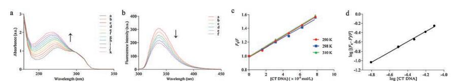
UV-vis absorption spectroscopy is one of the most effective techniques to investigate the interactions of small molecules with DNA. Generally, the absorbance and position of the peak show changes when small molecules interact with DNA. Hypochromic effect and red shift are associated with the intercalative interactions. For groove binding mode, hypochromic effect can be observed but the position of the absorption band changes hardly. However, hyperchromic effect can be caused when electrostatic attractions take place [19]. With the increasing concentration of calf thymus DNA (CT DNA), the absorption spectra of 5-HTP exhibited an apparent hyperchromism and blue shift (Fig. 1a). However, the CT DNA band at 260 nm obviously interfered with the 5-HTP absorption band at 275 nm. Therefore, there might be errors in the calculation of binding constant and binding mode via UV–vis absorption spectroscopy, and further experiments were required to understand the interactions between 5-HTP and DNA.

|
Download:
|
| Fig. 1. (a) UV–vis absorption spectra of 5-HTP with CT DNA. (b) Fluorescence emission spectra of 5-HTP with CT DNA at 298 K. (c) Stern-Volmer plot for interactions of 5-HTP with CT DNA at 290, 298, and 310 K. (d) Stern-Volmer plot of log[(F0-F)/F] versus log[CT DNA] at 298K to calculate the binding constants. Binding constant was obtained from the Y-intercept. | |
Fluorescence experiments were performed to clarify the interaction and binding constant of 5-HTP with CT DNA. The fluorescence intensity of small molecule reduces when interacts with DNA. Such a reduction is called fluorescence quenching. Stern-Volmer equation is used to assess the fluorescence quenching efficiency: F0/F =1 + KSV [Q] (1) where F0 and F are the fluorescence intensities in the absence and presence of the quencher respectively. KSV is the quenching constant and [Q] is the concentration of the quencher [19]. Besides, the fluorescence quenching could be classified as dynamic quenching and static quenching [20]. Dynamic quenching depends upon molecule diffusion. Higher temperatures will increase the diffusion coefficient and result in a larger KSV values, but lower the KSV values for static quenching due to the decrease of complexes stability [20]. Equation Kq = KSV/τ0 (2) was used to investigate the quenching process of small molecules and DNA, where Kq is the bimolecular quenching rate constant and τ0 is the fluorescence lifetime of the biomolecule in the absence of quencher (τ0 =10-8 s) [19].
The fluorescence intensity of 5-HTP decreased gradually with the addition of CT DNA at 298 K in Fig. 1b, suggesting that DNA interacted with 5-HTP and quenched the intrinsic fluorescence of 5-HTP. The KSV values calculated from the Stern-Volmer plots (Fig. 1c) showed a tinychange from7.42 ×103 to 7.45 ×10³ L/mol at different temperatures (290, 298 and 310 K) and were listed in Table S1 (Supporting information). The Kq values were found to be in the range of 7.42 ×10¹¹-7.45 ×10¹¹ L mol-1 s-1 at different temperatures (290, 298 and 310 K), higher than the limiting diffusion rate constant of biomolecules (2.0 ×10¹⁰). These results indicated that the fluorescence quenching process was initiated by static quenching.
Equation (3) can describe the relationship between fluorescence intensityand concentration of the quencher in the process of static quenching [20]. Log[(F0-F)/F] = logK + n log[Q] (3) where K is the binding constant, n is the number of binding sites and Q is the concentration of the quencher. From the plot of log (F0-F)/F versus log [CTDNA] in Fig. 1d, the K value of 5-HTPand DNA was estimated to be 1.69 ×10⁴ L/mol at 298K.
Competitive displacement assays, iodide quenching studies, comparison interactions of 5-HTP with double-stranded DNA and single-stranded DNA, circular dichroism studies and viscosity measurements were performed to further elucidate the binding mode of 5-HTP on CT DNA. All the results (Supporting information) indicated that 5-HTP bound to CT DNA in the groove binding mode.
The radiochemical purity of [131I]5-HTP measured by radioHPLC was 99.53% (Fig. S3a in Supporting infromation). 92.47% of the intact tracer remained in the rat serum at 12 h (Fig. S3b in Supporting infromation), indicating good stability of [131I]5-HTP in vitro.
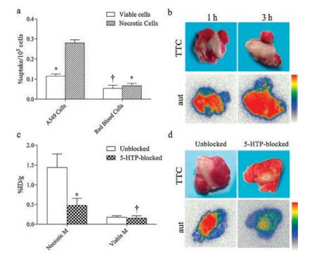
As shown in Fig. 2a, necrotic A549 cells (with cell nucleus) exhibited 2.43-fold increase in the uptake of [131I]5-HTP compared with viable A549 cells. It revealed that [131I]5-HTP features necrosis avidity in vitro. However, the uptake of [131I]5-HTP in necrotic red blood cells (without cell nucleus) showed no significant difference with that in viable red blood cells (P > 0.05), but was obviously lower than that in necrotic A549 cells (P < 0.01). These results indicated that the high accumulation of [131I]5- HTP in necrotic A549 cells might be related to cell nucleus.

|
Download:
|
| Fig. 2. (a) Uptake of [131I]5-HTP in A549 cells and red blood cells. Data were displayed as average percentage uptake per 105 cells plus or minus standard deviation (%uptake/105 cells ± SD). *P < 0.01 compared with necrotic A549 cells. †P > 0.05 compared with necrotic red blood cells. (b) TTC images and corresponding autoradiographs (aut) of partially necrotic muscle sections from muscular necrosis model mice at 1 h and 3 h after administrationwith [131I]5-HTP. (c) Uptake of [131I]5- HTP in unblocked and 20 mg/kg 5-HTP blocked (5-HTP-blocked) necrotic and viable muscle. Data are presented as average percentage of injected dose per gram (%ID/g) ± SD at 3 h after coinjection. Necrotic M = necrotic muscle; Viable M = viable muscle. *P < 0.01 compared with unblocked Necrotic M group. †P > 0.05 compared with unblocked Viable M group. (d) TTC images and corresponding aut of partially necrotic muscle sections from unblocked and 5-HTP-blocked groups. | |
The biologic distribution results in mice model of muscular necrosis are shown in Table 1. At each time point, the uptake of [131I]5-HTP in necrotic muscle was significantly higher than that in normal muscle (P < 0.01). The radioactivity ratios of necrotic and viable muscles increased from 3.59 ±0.47 at 1h to 6.95 ±0.68 at 3h. Lower uptake of [131I]5-HTP was detected in normal organs/ tissues such as viable muscle, heart and brain, except for kidney, bladder, and stomach at each time point. Besides, the biodistribution data displayed an obvious clearance of radioactivity from normal organs within 3 h except for the kidneys and bladder, indicating that [131I]5-HTP might prefer to metabolize in a renal metabolistic way.
|
|
Table 1 Biodistribution data of [131I]5-HTP in muscular necrosis models. |

Fig. 2b represents TTC staining and autoradiography outcomes. Higher tracer signals in the autoradiographs primarily appeared in necrotic regions of muscle, which were consistent with the data from biodistribution studies. These results also suggested that [131I]5-HTP could specifically localize in necrotic tissue.
Compared with the unblocked group, about 66.7% of the radioactivity in the necrotic muscle was blocked by unlabeled 5- HTP (Fig. 2c). However, the uptakes of [131I]5-HTP in the viable muscles had no significant differences between the unblocked and 5-HTP-blocked groups (P > 0.05). Results were consistent with the images of autoradiography (Fig. 2d). These results indicated that [131I]5-HTP may share the same specific targets with 5-HTP in vivo. Considering that the higher accumulation of [131I]5-HTP in necrotic A549 cells might relate to cell nucleus and that 5-HTP binds to DNA in groove binding mode, we speculate that the necrosis avidity mechanism of [131I]5-HTP might be attributable to its interactions with E-DNA in necrotic cells.
Fig. 3a shows the representative SPECT/CT images of control and MI model rats at 3 h post-injection (p.i.). Hotspot was visualized in the heart of rats with MI, while no obvious uptake was observed in the control rats, indicating that [131I]5-HTP could specifically accumulate in necrotic myocardium at 3 h p.i.

|
Download:
|
| Fig. 3. (a) SPECT/CT images of control rats and rat models with MI at 3 h p.i. of [131I] 5-HTP. (b) Biodistribution data after SPECT/CT imaging. Data are %ID/g, expressed as mean ± SD (n = 5). Viable M = viable myocardium; Necrotic M = necrotic myocardium. *P < 0.01 compared with necrotic myocardium. **P < 0.05 compared with necrotic myocardium. (c) Aut, corresponding H&E staining and micrograph (Micro) of 10 μm heart slices from rat models. N = necrotic area, V = viable area. Scale bar = 200 μm. | |
The biodistribution data after SPECT/CT imaging are illustrated in Fig. 3b. The radioactivity ratio of necrotic and viable myocardium was 4.87. Besides, the uptake of [131I]5-HTP in blood and lungs was less than that in necrotic myocardium significantly (P < 0.05). These results suggested that [131I]5-HTP may be a promising imaging probe of the necrotic myocardium. Prominent activity was detected in the kidney and liver, consistent with the SPECT/CT imaging. H&E staining and autoradiography of heart slices from rat models with MI are displayed in Fig. 3c. Higher tracer signals in autoradiographs primarily matched well with the pink parts in H&E staining. The result indicated that [131I]5-HTP could specifically localize in necrotic myocardium and evaluate myocardial viability.
In the present study, [131I]5-HTP (3 h p.i.) enabled the rapid imaging of necrotic myocardium compared with [123I]Hyp (9 h p.i.) [6] and [131I]Rhein (6 h p.i.) [8]. Meanwhile, [131I]5-HTP improved the necrotic myocardium-to-blood ratio (from 0.96 for[131I]Rhein to 1.99 for [131I]5-HTP), which greatly influences the image quality. It may be attributed to the better water solubility of [131I]5-HTP, which would result to the rapid distribution in necrotic myocardium and fast clearance from the circulation. Therefore, [131I]5-HTP may be more beneficial and potential for the early imaging and diagnosis of necrosis related diseases with high quality of image.
As a kind of endogenous substance, 5-HTP shows high safety and has been used as dietary supplements or neurotransmitters in clinic for over 30 years [18]. Moreover, the small dose of [131I]5-HTP (about 0.2 mg/kg of 5-HTP) for SPECT/CT imaging in this study is much less than the administration dosage of 5-HTP in clinic (≥ 3.3 mg kg-1 day-1) [18]. All the above facts indicate that [131I]5- HTP is a promising necrosis avid agent with high safety. Especially, the predominant necrosis avidity and necrotic myocardium imaging property of [131I]5-HTP were evaluated for the first time in the present paper. These results supplement the literature data on extending the applications of [131I]5-HTP in necrosis related pathologies.
Additionally, most of the NAAs are dark in color due to their big conjugate planar structures [3]. The unchangeable natures have largely limited their further commercial development. By contrast, colorless 5-HTP can be easily accepted by patients compared with the dark NAAs.
In conclusions, [131I]5-HTP is a promising necrosis avid agent with the advantages of high safety, good solubility and colorlessness. Furthermore, this study provides an effective way to develop novel NAAs from DNA binding agents.
AcknowledgmentsThis work was partially supported by the National Natural Science Foundation of China (Nos. 81473120, 81501536, 81473120). We appreciate Mr. Changwen Fu for his kindly work in SPECT/CT scanning.
Appendix A. Supplementary dataSupplementary material related to this article can be found, in the online version, at doi:https://doi.org/10.1016/j.cclet.2018.06.005.
| [1] |
T.V. Berghe, S. Grootjans, V. Goossens, et al., Methods 61 (2013) 117-129. DOI:10.1016/j.ymeth.2013.02.011 |
| [2] |
A.A. Neves, K.M. Brindle, J. Nucl. Med. 55 (2014) 1-4. DOI:10.2967/jnumed.112.114264 |
| [3] |
Y. Ni, G. Bormans, F. Chen, A. Verbruggen, G. Marchal, Invest. Radiol. 40 (2005) 526-535. DOI:10.1097/01.rli.0000171811.48991.5a |
| [4] |
M. Miranda Cona, R. Oyen, Y. Ni, Curr. Med. Chem. 22 (2015) 1829-1849. DOI:10.2174/0929867322666150227153550 |
| [5] |
M. van de Putte, H. Wang, F. Chen, P.A. de Witte, Y. Ni, Acad. Radiol. 15 (2008) 107-113. DOI:10.1016/j.acra.2007.08.008 |
| [6] |
H. Fonge, K. Vunckx, H. Wang, et al., Eur. Heart J. 29 (2007) 260-269. DOI:10.1093/eurheartj/ehm588 |
| [7] |
M. Pietrzak, M. Maciejczyk, M. Szabelski, A. Kasparek, Z. Wieczorek, Chem. Phys. Lett. 601 (2014) 39-44. DOI:10.1016/j.cplett.2014.03.076 |
| [8] |
Q. Wang, S. Yang, C. Jiang, et al., Sci. Rep 6 (2016) 21341. DOI:10.1038/srep21341 |
| [9] |
Q. Luo, Q. Jin, C. Su, et al., Anal. Chem. 89 (2017) 1260-1266. DOI:10.1021/acs.analchem.6b03959 |
| [10] |
Y. Yuan, J. Zheng, M. Wang, et al., J. Agric. Food Chem. 64 (2016) 5742-5750. DOI:10.1021/acs.jafc.6b01872 |
| [11] |
M. Dasari, S. Lee, J. Sy, et al., Org. Lett. 12 (2010) 3300-3303. DOI:10.1021/ol100923d |
| [12] |
M. Dasari, A.P. Acharya, D. Kim, et al., Bioconjugate Chem. 24 (2012) 4-8. |
| [13] |
S. Huang, H.H. Chen, H. Yuan, et al., J. Cardiov. Magn. Reson. 13 (2011) O23. DOI:10.1186/1532-429X-13-S1-O23 |
| [14] |
X. Duan, Z. Yin, C. Jiang, et al., European J. Pharm. Biop. 117 (2017) 151-159. DOI:10.1016/j.ejpb.2017.04.006 |
| [15] |
A.Y. Ji, Q.M. Jin, D.J. Zhang, et al., ACS Med. Chem. Lett. 8 (2017) 191-195. DOI:10.1021/acsmedchemlett.6b00398 |
| [16] |
N.K. Kaushik, N. Kaushik, P. Attri, et al., Molecules 18 (2013) 6620-6662. DOI:10.3390/molecules18066620 |
| [17] |
Y. Ni, MR contrast agents for cardiac imaging, in: J. Bogaert (Ed.), Clinical Cardiac MRI, Springer, Berlin, Heidelberg, 2011, pp. 31-51.
|
| [18] |
E.H. Turner, J.M. Loftis, A.D. Blackwell, Pharmacol. Therapeut. 109 (2006) 325-338. DOI:10.1016/j.pharmthera.2005.06.004 |
| [19] |
M. Sirajuddin, S. Ali, A. Badshah, J. Photoch. Photobio. B 124 (2013) 1-19. DOI:10.1016/j.jphotobiol.2013.03.013 |
| [20] |
S. Zhang, H. Yuan, L. Tian, J. Mol. Struct. 1130 (2017) 760-764. DOI:10.1016/j.molstruc.2016.11.003 |