 2019, Vol. 30


b State Key Laboratory of Microbial Technology, Shandong University, Ji'nan 250100, China
Palladium, an important transition metal, is widely distributed in the environment and displays extraordinary catalytic performances in coupling reactions [1]. In the past few decades, palladium has been extensively used in many areas including electronic industry [2], petroleum industry [3], automobile industry [3], glass manufacture [4] and fine chemicals engineering [1, 4], which has done harm to the environment in the meantime. Moreover, palladium is hard to be biodegraded and easy to be enriched through the food chain [5]. It is well known that palladium can coordinate with macromolecules such as DNA and proteins leading to DNA degradation, allergic reaction as well as enzyme inhibition [6, 7]. Therefore, specific and selective detection of palladium in environmental and biological samples is urgently desirable.
There are many traditional methods for palladium detection, including X-ray, atomic emission spectroscopy (AES), inductively coupled plasma atomic emission spectrometry (ICP-AES), inductively coupled plasma mass spectroscopy (ICP-MS), solid-phase microextraction high-performance liquid chromatography (SPMEHPLC) and atomic absorption spectroscopy (AAS), by which accurate detection can be achieved rapidly and sensitively. However, these methods require complicated sample-pretreatment procedures, well-controlled experimental conditions, expensive facilities and highly-trained individuals. As a result of high selectivity, sensitivity, rapidity, low cost and operational simplicity, optical probes [8, 9], such as colorimetric probes, fluorescent probes and luminescent probes, provide choices for determination of palladium species.
A highly sensitive readout can be provided by a triggered luminescence emission process, which is extremely favorable for bioimaging because of free interferences caused by light scattering and reduced background noise due to the absence of photonic excitation. For decades, bioluminescence has been widely applied in the preclinical analysis using genetically modified cells or organisms. While bioluminescent enzymes, for example, firefly luciferase and renilla luciferase, are necessary for bioimaging, chemiluminescence can be used with wild-type cells, organisms, and even animals and offers opportunities for clinical imaging.
Therefore, we decided to develop chemiluminescent imaging (CLI) agents that can be utilized in luciferase-null systems [10-12]. Currently, we chose 1, 2-dioxetane, which can be sterically stabilized by spiroadamantane, as our chemiluminescent platform [13-20]. Since the cleavage of a chemical bond triggers light emission of the chemiluminescence, it is significant to develop recognition moiety for detecting palladium ions sensitively and selectively. Benefitting from the capacity of palladium, "off-on" probes were explored, taking advantage of typical-metal binding [21], Pd-catalyzed Claisen rearrangement [22], Pd-catalyzed Tsuji-Trost reaction [23-25], Pdcatalyzed depropargylation [16, 26] and so on.
On account of these considerations and expanding the techniques of chemiluminescent imaging, we managed to introduce butynyl group to 1, 2-dioxetane moiety generating the palladium chemiluminescent probe (PCL) (Scheme 1). To be honest, our original plan was to design a mercury probe with alkynyl butylcarbonate as the recognition moiety [27, 28]. It was supposed that electronically excited m-oxybenzoate anion released once butynyl moiety is cleaved via metal-catalyzed reaction. Then, the excited intermediate undergoes an electron transfer process to return to the ground state, according to CIEEL (chemically initiated electron exchange luminescence) mechanism. Finally, the extra energy released in the form of light. However, according to the results of the preliminary experiment, our probe seems to be more sensitive to palladium cation than mercury at mild condition. Therefore, subsequent studies were centered on palladium(Ⅱ) imaging using PCL in the current report.

|
Download:
|
| Scheme 1. Structure and proposed chemiluminescent mechanism of palladium probe PCL. | |
The probe PCL was designed such that chemiluminescent emission would be initiated by the Pd-catalyzed cleavage of butynyl moiety and prepared conveniently according to the route illustrated in Scheme S1 (Supporting information). In brief, the synthesis begins with the preparation of the intermediate compound 4 which can convert to 1, 2-dioxetane by photocatalyzed oxidation. Assisted with N, N'-disuccinimidyl carbonate (DSC), but-3-yn-1-ol was connected to compound 4 to yield compound 6, the precursor of PCL. Finally, compound 6 was oxidized under visible light irradiation at 0 ℃ and the final probes were obtained.
After preparation, chemiluminescence intensity was measured with an IVIS kinetic imaging system (Caliper Life Sciences, Hopkinton, Massachusetts, U.S.A.) equipped with a cooled charge-coupled device (CCD) camera. More details about synthetic experiments and chemiluminescence imaging assays are well described in Supporting information.
Stability of PCL in an aqueous system with different pH was examined initially, since acid-base status in the environment may influence the carbonate moiety in PCL. The probe (100 μmol/L) was incubated in Tris-HCl with a wide pH range from 3 to 10 for approximately 60 min. After incubation, the chemiluminescence intensity was recorded with a living imaging system as depicted in Fig. 1. These results suggested that the palladium probe is stable in the aqueous system within a pH range of 3–7, although luminescence augmented remarkably in the system with pH more than 9.

|
Download:
|
| Fig. 1. Influence of pH on the stability of PCL. (A) Chemiluminescence imaging of PCL in Tris-HCl with various pH ranging from 3 to 10. (B) Quantification of chemiluminescence intensity of PCL for each pH. | |
Sensitivity of PCL (50 μmol/L) for palladium was evaluated in an aqueous system. As illustrated in Fig. 2 and Fig. S1 (Supporting information), optical signals of the probe fluctuated with time, when reacted with palladium species (25 μmol/L). As 60 min passed, a plateau occurred, and the luminescence intensity was almost invariant although declined slightly. Subsequently, after incubation with PdCl2 in PBS (1×, pH 7.4) of various concentrations (0 to 2.5 mmol/L) for 75 min, the probe's luminous flux was measured. It is indicated that PCL is sensitive to Pd2+ within a concentration range from 3.125 μmol/L to 500 μmol/L. Meanwhile, limit of detection (LOD) of PCL to Pd2+ can be calculated as less than 6.25 mmol/L. As for the decrease of photon emission when the concentration of palladium species reached a high level, we speculated that heavy metal species in higher concentration might depress the effect of Enhancer Emerald Ⅱ.

|
Download:
|
| Fig. 2. Sensitivity assay of PCL in vitro. (A) Time scans of the chemiluminescence intensity of PCL (50 μmol/L) reacted with PdCl2 (12.5 μmol/L) in PBS (1×, pH 7.4) containing 10% (v/v) Enhancer Emerald Ⅱ. (B) Quantification of chemiluminescence intensity of PCL (50 μmol/L) with Pd2+ (0–2.5 mmol/L) in PBS (1×, pH 7.4) containing 10% (v/v) Enhancer Emerald Ⅱ. (C) Quantification of chemiluminescence intensity of PCL with PdCl2 (100 μmol/L) and EDTA (0 to 2 mmol/L) in PBS (1×, pH 7.4) containing 10% (v/v) Enhancer Emerald Ⅱ. | |
It is also speculated that only free palladium ion can catalyze the cleavage of the butynyl moiety in PCL. Therefore, we commenced another experiment to confirm our conjecture. Previously, palladium chloride (100 μmol/L) was incubated with EDTA solution at various concentrations (0 to 2 mmol/L) at 37 ℃ for 60 min. Following the addition of PCL (50 μmol/L), the samples were incubated for an extra 60 min. Finally, the optical signals of PCL were measured by an IVIS kinetic imaging system to find that chemiluminescent intensity declined by the increasing amount of EDTA (Fig. 2 and Fig. S1). Accordingly, the probe displays an immediate concentration-dependent photon emission, which is an ideal property for imaging of free palladium ion in biological environment.
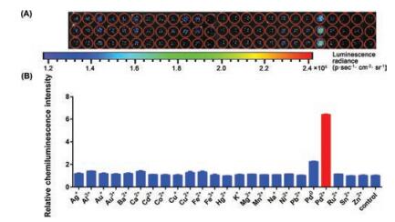
Selectivity of PCL (50 μmol/L) for Pd2+ (50 μmol/L) against a range of metal species, including heavy metals as well as microelements existing in the body, was assessed in an aqueous system. As illustrated in Fig. 3 and Fig. S2 (Supporting information), an evident enhancement in chemiluminescence intensity displayed when Pd2+ existing, compared with other metal cations. These response and selectivity data suggested that PCL with fine sensitivity and selectivity, combined with beneficial characteristics of the chemiluminescence technique, affording potential utility for detection of Pd2+ in cellulo and in vivo.

|
Download:
|
| Fig. 3. Selectivity assay of PCL in vitro. (A) Chemiluminescence imaging of selectivity of PCL towards various metal cations. (B) Quantification of the chemiluminescent intensity of PCL for each condition. | |
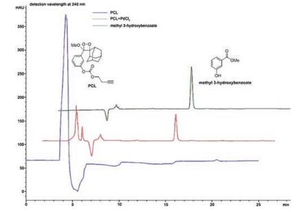
Experiments mainly focusing on proving the hypothesis of the probe's mechanism were also carried out. To evaluate the liability of PCL to palladium ion, the probe in an aqueous solution was reacted with palladium chloride at room temperature for 3 h. After microporous filtration, the sample was analyzed with HPLC by comparing the sample with PCL or methyl 3-hydroxybenzoate. According to the HPLC spectra exhibited in Fig. 4, when existing palladium ion, PCL decomposed, making absorbance at 4.3 min (the retention time of PCL) declined. At 13.0 min, which is the retention time of methyl 3-hydroxybenzoate, a new peak appeared simultaneously. The proposed mechanism of the palladium probe was confirmed by HPLC (Fig. 4).

|
Download:
|
| Fig. 4. HPLC spectra of PCL (100 μmol/L), PCL (100 μmol/L) incubated with PdCl2 (50 μmol/L) for 3 h and methyl 3-hydroxybenzoate (100 μmol/L). | |
Another palladium chemiluminescent probe with Pd-catalyzed depropargylation mechanism was designed developed previously [16]. It is confirmed that the chemiluminescence could be triggered via the catalytic depropargylation of Pd(0). Although light emission of the probe occurred when treated with Pd(Ⅱ), the responsibility still can be attributed to the reduction of Pd(Ⅱ) to Pd(0) by PPh3 actually. While the selectivity data of PCL manifested that this palladium probe is sensitive to Pd(Ⅱ) with specificity. Thus, PCL is a novel chemiluminescent probe for Pd(Ⅱ) detection despite it may not be the first reported one among its analogues.
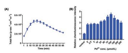
Afterwards, the ability of PCL to imaging palladium in living cells was investigated. To import palladium ion into living cells, human cervical cancer cell lines HeLa was cultivated with the PdCl2 solution in PBS (1×, pH 7.4) for 1 h before the test. As illustrated in Fig. 5 and Fig. S3 (Supporting information), chemiluminescence intensity continually enhanced along with the increasing concentrations (0–400 μmol/L) of palladium chloride. After 45-minreaction, luminescent flux reached the peak value, when the relative chemiluminescence intensity at 400 μmol/L of PdCl2 came up to approximately 130%. The results exposed that PCL was capable of palladium imaging at the cellular level with a series of dynamic changes. According to the results of cytotoxicity test accomplished by SRB cytotoxicity assay, the hypoxia chemiluminescent probe is low-toxic and fine-biocompatible (Fig. S4 in Supporting information).

|
Download:
|
| Fig. 5. Palladium imaging in HeLa cells using PCL. (A) Variation of chemiluminescence intensity in 60 min (400 μmol/L of PdCl2). (B) Quantification of the chemiluminescent intensity of PCL for each condition. | |
Additionally, bioactivity of PCL towards palladium ion in commercial rabbit plasma (20 folds diluted with saline) was investigated. For a 1-hour-incubation, as depicted in Fig. 6 and Fig. S5 (Supporting information), the chemiluminescence intensity augmented with the concentrations of PdCl2 from 25 μmol/L to 100 μmol/L. These results indicated that PCL could detect palladium quantitatively even in such complex biological samples. Unfortunately, 20-fold diluted rabbit plasma treated only with PCL released a chemiluminescence signal over 2.5 times stronger than PCL treated with saline, which drove down the LOD of PCL. The disadvantage may also be attributed to the instability of carbonate structure in complex biological samples. However, this study can still provide some valuable data and experience for establishing a chemiluminescent system for the animal model.

|
Download:
|
| Fig. 6. Palladium imaging in 20-fold diluted rabbit plasma using PCL. (A) Variation of chemiluminescence intensity in 60 min (100 μmol/L of PdCl2). (B) Quantification of the chemiluminescent intensity of PCL for each condition. | |
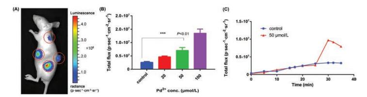
The capability of PCL for palladium(Ⅱ) imaging in living animals was interrogated. We attempted to subcutaneous inject the 50 mL of saline or palladium chloride (20 μmol/L, 50 μmol/L or 100 μmol/L) in saline. Following, 50 μL of PCL (1 mmol/L) in saline containing 10% (v/v) Enhancer Emerald Ⅱ was subcutaneous injected in situ respectively. Thereafter, subsequent chemiluminescence intensity was recorded with living imaging system. As shown in Fig. 7, after injection of PCL, obviously enhancing emission signals from injection regions in rude mice were observed, reaching peak values at about 30 min. The enhanced chemiluminescence intensity lasted for about 5 min. As was expected, distinct enhancement of luminescence intensity can be observed visually by living imaging, as injection amount of PdCl2 grew from 0 to 100 μmol/L.

|
Download:
|
| Fig. 7. Palladium imaging in vivo using PCL. (A) Chemiluminescence imaging of PCL towards Pd2+ with various concentrations (0–100 μmol/L). (B) Quantification of chemiluminescence intensity from the subcutaneous injection areas of the rude mice of triplicates (n = 4). (C) Variation of chemiluminescence intensity within 35 min. | |
In summary, herein we designed and prepared a palladium chemiluminescent probe (PCL) by using 1, 2-dioxetane as the chemiluminescence platform and butynyl moiety as the recognition unit. Exhibited desirable properties, including no requirement for excitation light, low background noise, high sensitivity, high selectivity and the ability to function well in aqueous media, PCL is capable of monitoring palladium ion visually in vitro, in cellulo and in vivo. Although the instability of carbonate structure in PCL may drive down the LODs in the basic system or in complex biological samples, the exploration brought experience and expand the palladium imaging toolkit and applications of chemiluminescence technology.
AcknowledgmentsThe present work was supported by grants from the Taishan Scholar Program at Shandong Province, the Qilu/Tang Scholar Program at Shandong University, the Key Research and Development Project of Shandong Province (No. 2017CXGC1401) and the Major Project of Science and Technology of Shandong Province (No. 2015ZDJS04001).
Appendix A. Supplementary dataSupplementary data associated with this article can be found, in the online version, at https://doi.org/10.1016/j.cclet.2018.03.028.
| [1] |
E. Negishi, Historical Background of Organopalladium Chemistry, Handbook of Organopalladium Chemistry for Organic Synthesis, John Wiley & Sons Inc., 2003, pp. 1-15.
|
| [2] |
J.L. Martin, M.P. Toben, K.J. Whitlaw, Met. Finish. 88 (1990) 39-41. |
| [3] |
M.S. Kamal, S.A. Razzak, M.M. Hossain, Atmos. Environ. 140 (2016) 117-134. DOI:10.1016/j.atmosenv.2016.05.031 |
| [4] |
B. Reinhard, Use and demand of palladium for the industry, in: F. Zereini, F. Alt (Eds.), Palladium Emissions in the Environment: Analytical Methods, Environmental Assessment and Health Effects, Springer Berlin Heidelberg, Berlin, Heidelberg, 2006, pp. 39-51.
|
| [5] |
K. Ravindra, L. Bencs, van Grieken R., Sci. Total Environ. 318 (2004) 1-43. DOI:10.1016/S0048-9697(03)00372-3 |
| [6] |
R. Merget, G. Rosner, Sci. Total Environ. 270 (2001) 165-173. DOI:10.1016/S0048-9697(00)00788-9 |
| [7] |
J. Kielhorn, C. Melber, D. Keller, I. Mangelsdorf, Int. J. Hyg. Environ. Health 205 (2002) 417-432. DOI:10.1078/1438-4639-00180 |
| [8] |
H. Li, J. Fan, X. Peng, Chem. Soc. Rev. 42 (2013) 7943-7962. DOI:10.1039/c3cs60123d |
| [9] |
P. Matthew, D. Pham, K. Koide, Chem. Soc. Rev. 44 (2015) 4769-4791. DOI:10.1039/C4CS00323C |
| [10] |
M. Matsumoto, J. Photochem. Photobiol. C 5 (2004) 27-53. DOI:10.1016/j.jphotochemrev.2004.02.001 |
| [11] |
M. Matsumoto, N. Watanabe, N. Hoshiya, H.K. Ijuin, Chem. Rec. 8 (2008) 213-228. DOI:10.1002/tcr.v8:4 |
| [12] |
N. Watanabe, N. Suga, M. Matsumoto, Luminescence 23 (2008) 344-349. DOI:10.1002/bio.v23:5 |
| [13] |
I.S. Turan, E.U. Akkaya, Org. Lett. 16 (2014) 1680-1683. DOI:10.1021/ol5003412 |
| [14] |
J. Cao, R. Lopez, J.M. Thacker, et al., Chem. Sci. 6 (2015) 1979-1985. DOI:10.1039/C4SC03516J |
| [15] |
J. Cao, J. Campbell, L. Liu, R.P. Mason, A.R. Lippert, Anal. Chem. 88 (2016) 4995-5002. DOI:10.1021/acs.analchem.6b01096 |
| [16] |
I.S. Turan, O. Yilmaz, B. Karatas, E.U. Akkaya, RSC Adv. 5 (2015) 34535-34540. DOI:10.1039/C5RA01551K |
| [17] |
I.S. Turan, O. Seven, S. Ayan, E.U. Akkaya, ACS Omega 2 (2017) 3291-3295. DOI:10.1021/acsomega.7b00537 |
| [18] |
O. Green, T. Eilon, N. Hananya, et al., ACS Cent. Sci. 3 (2017) 349-358. DOI:10.1021/acscentsci.7b00058 |
| [19] |
N. Hananya, A. Eldar-Boock, R. Satchi-Fainaro, D. Shabat, R. Bauer-Christoph, J. Am. Chem. Soc. 138 (2016) 13438-13446. DOI:10.1021/jacs.6b09173 |
| [20] |
O. Green, S. Gnaim, R. Blau, et al., J. Am. Chem. Soc. 139 (2017) 13243-13248. DOI:10.1021/jacs.7b08446 |
| [21] |
P. Kaur, N. Kaur, M. Kaur, et al., RSC Adv. 4 (2014) 16104-16108. |
| [22] |
A.L. Garner, K. Koide, J. Am. Chem. Soc. 130 (2008) 16472-16473. DOI:10.1021/ja8065539 |
| [23] |
X. Wang, Z. Guo, S. Zhu, H. Tian, W. Zhu, Chem. Commun. 50 (2014) 13525-13528. DOI:10.1039/C4CC05871B |
| [24] |
H. Tan, J. Liu, L. Zhou, et al., RSC Adv. 7 (2017) 6583-6586. DOI:10.1039/C6RA27502H |
| [25] |
T. Yu, G. Yin, P. Yin, et al., RSC Adv. 7 (2017) 24822-24827. DOI:10.1039/C7RA01731F |
| [26] |
M. Kumar, N. Kumar, V. Bhalla, RSC Adv. 3 (2013) 1097-1102. DOI:10.1039/C2RA22869F |
| [27] |
H. Yamamoto, M. Nishiyama, H. Imagawa, M. Nishizawa, Tetrahedron Lett. 47 (2006) 8369-8373. DOI:10.1016/j.tetlet.2006.09.067 |
| [28] |
F. Song, S. Watanabe, P.E. Floreancig, K. Koide, J. Am. Chem. Soc. 130 (2008) 16460-16461. DOI:10.1021/ja805678r |