 2018, Vol. 29


Cancer responsive nanoprobes with theranostic features have great attention in cancer diagnosis and therapy [1]. Cancer sensitive turn-on fluorescent probes have been attractive due to keeping an "off" state before reaching the target. These probes can be activated when specific reaction happened [2, 3]. For example, in nanocomposite Cy5.5-HANP/CuS (HANPC), which formed by loading copper sulfide (CuS) into Cy5.5-conjugated hyaluronic acid nanoparticles (HANP), Cy5.5 fluorescent signal is quenched by CuS inside the particle until the whole nanocomposites degraded by hyaluronidase present in tumors, giving strong fluorescence signals [4]. Moreover, multifunctional activatable theranostic nanoparticles, which composed of a photosensitizer and photothermal agent have been used for imaging-guided phototherapy [5]. Under irradiation, photosensitizers (PSs) generated reactive oxygen species (ROS), such as hydroxyl radicals, singlet oxygen, as well as peroxides, which irreversibly kill cancer cells via apoptosis or necrosis [6]. The NIR activated nanoparticles is of particular interest for cancer image in vivo since the human tissue is considered "transparent" to light in 700–1100 nm range [7]. The d–d energy band transition of Cu2+ ions in semiconductor CuS nanoparticles endows them with NIR light absorption. Therefore, the copper sulfide (CuS) nanoparticles (CuS NPs) have been NIR initiated photothermal therapy agents for the treatment of cancer [8]. On the other hand, proteins offer significant potential for templated synthesis of inorganic nanoparticles and protein nanocarriers have been developed as drug delivery devices due to their safety and biodegradability [9]. Bovine serum albumin (BSA) coated CuS NPs can increase the photothermal effect and biocompatibility. CuS NPs could also be coated directly with folic acid to target tumor cells that express folate receptors [10]. Generally, BSA coated nanospheres can be obtained by the assembly of BSA with dye in the presence of linker (such as, glutaraldehyde). Due to hydrophobic interaction between the dye and BSA, the emission of dye in dye@BSA was quenched, which can be recovered due to acid induced release of dyes [11, 12]. Although various techniques were successfully employed for development of hybrid protein-inorganic NPs, cancer activated protein-inorganic NPs have been challengeable. Recently, epithelial cell-adhesion molecule (EpCAM), a 40-kDa type I transmembrane glycoprotein, was identified as one of tumour stem cell markers and a potential target for cancer therapy [13]. EpCAM has been highly expressed in breast cancer cells. Therefore, the surface of cancer cells may be different from normal cells, and cancer cells activated nanoprobe can be cancer targeting imaging and therapy agents.
Herein, we report a cancer responsive nanoprobe (PDAMnCuS@BSA-FA, Scheme 1) for breast cancer cell detection and NIR activated attenuation. The quenching interaction of dyes (PDA) and central metal in NPs can be turn-on by CH3O-PEG-phosphatide or the cancer cells. First, Mn-CuS was formed by electrostatic interaction, and then Mn-CuS coordinated with PDA (8-[di(2- picolyl)amine-4-benzyl]-4, 4-difluoro-1, 3, 5, 7-tetramethyl-4-bora- 3a, 4a-diaza-s-indacene) producing PDAMn-CuS, which was assembled with folic acid (FA) modified BSA by hydrophobic interaction and electronic state interaction. PDAMn-CuS@BSA-FA showed cancer cell activated fluorescence emission, which was different from previous reported glutaraldehyde treated proteininorganic nanoparticles. Therefore, PDAMn-CuS@BSA-FA provided a convenient approach to be a breast cancer and CH3O-PEGphosphatide activated fluorescence nanosensor.

|
Download:
|
| Scheme 1. The synthesis route of PDAMn-CuS@BSA-FA NPs | |
Detailed experimental procedures are shown in the Supporting information. The synthesis procedure of PDAMn-CuS@BSA-FA NPs was shown in Scheme 1. Carboxylate group (COOH) modified supersmall nano-CuS was synthesized by using citric acid as template [14]. Secondly, the electrostatic interaction between the Mn(II) ion and COO- with on the surface of CuS resulting Mn-CuS NPs, which further linked with PDA forming PDAMn-CuS because of the coordination of Mn2+ with PDA (a fluorescence chelator of metal ions [15]). Finally, PDAMn-CuS (as core) assembled with HOOC-PEG-FA modified BSA (as shell) by hydrophobic and electronic state interaction forming PDAMn-CuS@BSA-FA nanoparticles [16]. The UV–vis absorbance spectra of PDA, CuS NPs and the assembly PDAMn-CuS@BSA-FA NPs is shown in Fig. 1A. PDA and CuS NPs exhibited high strong absorption at 494 nm and 900 nm. PDAMn-CuS@BSA-FA NPs showed absorption at 500 nm and 950 nm, assigned to the special absorption of PDA and CuS NPs, respectively. The two red shifted absorption indicates that there is the interaction between COO- on the surface of CuS NPs and the manganese (II) centre or the coordination of Mn(II) with PDA (Scheme 1).

|
Download:
|
| Fig. 1. (A) UV spectra of the ligand PDA (a, 44 mmol/L in acetonitrile), CuS NPs (b, 0.1 mg/mL in water) and PDAMn-CuS@BSA-FA NPs (c, 0.1 mg/mL in water). (B) Raman spectra of PDAMn-CuS@BSA-FA NPs | |
In IR spectra, the wide absorption peak at 3444 cm-1, which is assigned to stretching vibration of O-H bond indicating the existence of the COOH group in both CuS and PDAMn-CuS@BSA-FA NPs, respectively, and the absorption peak at 2900 ~ 2990 cm-1 is corresponded to stretching vibration of saturated C-H of PEG and BSA (Fig. S1 in Supporting information). In Raman spectra, the special peak of Cu-S bond appeared at 470 cm-1 (Fig. 1B, Figs. S2A and B in Supporting information). PDAMn-CuS@BSA-FA showed weak peak at 316 cm-1 of Mn-O bond. Two peaks occurred at 180 cm-1 and 385 cm-1 are attributed to the bending vibration and stretching vibration of Mn-N bond in PDAMn-CuS@BSA-FA, respectively, which indicates the successful bind of PDA toward Mn-CuS. For PDAMn-CuS@BSA-FA NPs, peaks at 586 cm-1, 955 cm-1 and 1185 cm-1 are due to vibrations form C-C bond and C-H bond of aromatic groups in PDA or BSA. Peaks at 755 cm-1, 1304 cm-1 and 1258 cm-1 indicate the existence of pyrrole and C-N bonds in PDA. Other peaks at 1428 cm-1, 1513 cm-1, 1539 cm-1 were attributed to the vibrations from C-C and N-H of BSA and PEGFA [17]. In the EPR spectra of PDAMn-CuS@BSA-FA NPs (Fig. S2C in Supporting information), g1 (4.11) and g2 (2.12) are two factors of the Mn(II) ion, representing the high and low spin of Mn(II), respectively. Therefore, there are Mn(II) species in PDAMnCuS@BSA-FA NPs.
The TEM image shows that the size of as-synthesized PDAMnCuS@BSA-FA nanoparticle is about 20 nm (Fig. 2A, Figs. S3A and B in Supporting information). Compared with the uniform size of CuS NPs (~8 nm), obvious protein layer can be seen on the surface of PDAMn-CuS@BSA-FA. Meanwhile, the DLS size of as-prepared PDAMn-CuS@BSA-FA NPs is about 15 ~ 25 nm, which agrees well with TEM image in size (Fig. 2B). The molecular weight values of PDAMn-CuS@BSA-FA NPs were measured by method of GPC (Gel Permeation Chromatography), then the PDI value was calculated to be 1.03, which confirmed the size of PDAMn-CuS@BSA-FA NPs was almost uniform. Furthermore, zeta potential of NPs in PB solution at different pH was further tested, and the highest zeta potential indicated NPs were more stable and well dispersed at pH 5.0 (Fig. S3C in Supporting information). The stability of nanoparticle was carried out by measuring released free Mn2+ ions of nanoparticles in different PB buffer solution (Fig. S4 in Supporting information) [18]. Less 5%–8% free Mn2+ can be found. Fluorescence titration results also demonstrated that the nanoparticle PDAMnCuS@BSA-FA showed no obvious emission change when DMEM, PB buffer solution and Bovine Serum Album was added. Results indicate that the nanoparticle is stable in PB buffer solution and cell medium.

|
Download:
|
| Fig. 2. HRTEM image (A) and DLS size distribution (B) of PDAMn-CuS@BSA-FA NPs | |
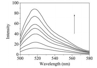
PDA has emission at 520 nm when excited 485 nm [14], however, nearly no emission can be detected for PDAMnCuS@BSA-FA in the same condition. It demonstrates that there is charge transfer interaction between PDA and CuS, which quenched the emission of PDA. The quenched emission cannot be recovered with the titration of BSA (Fig. S5 in Supporting information). However, the emission of the nanoparticle at 520 nm increased with the titration of CH3O-PEG-phosphatide (Fig. 3). These demonstrated that PDAMn-CuS@BSA-FA was different from previously reported glutaraldehyde linked protein-inorganic nanoparticle. The detection limit for nanoparticles (100 mg/mL) to CH3O-PEG-phosphatide is 1.68 mmol/L. Because the nanoparticle PDAMn-CuS@BSA-FA showed no obvious emission change when DMEM, PB buffer solution and BSA was added, we deduced that the PDAMn-CuS@BSA-FA was loosed and the quenched interaction between dye and metal central was decreased because of the hydrophobic and electrostatic interactions between CH3OPEG-phosphatide and BSA. Because the nanoparticle was sensitive to CH3O-PEG-phosphatide, it may sense the cell membrane or adhesion molecules. To investigate the nanoparticle can be cancer turn-on nanoprobe, the fluorescence response of PDAMnCuS@BSA-FA to cancer cells lysis was studied (Fig. S6 in Supporting information, Fig. 4). MCF-7 cells, AGS cells, PaTu8988 cells and SW1990 cells can switch on the emission greatly, in contrast, weak emission can be found when normal cells (HUEVC cells) was added. The detection limit for nanoparticle (50 mg/mL) to breast cancer cell lysis is 0.06 mL. Results demonstrate PDAMn-CuS@BSA-FA NPs can be a breast cancer cell activated nanosensor.

|
Download:
|
| Fig. 3. Fluorescence emission spectra of PDAMn-CuS@BSA-FA NPs aqueous solution (100 mg/mL) without NIR laser irradiation in the presence of CH3O-PEGphosphatide aqueous solution (the concentration of CH3O-PEG-phosphatide from the bottom to top is 0, 0.24, 0.48, 0.72, 0.96, 1.2 and 1.68 mmol/L, respectively), (λex = 485 nm, λem = 520 nm; Slit = 5) | |

|
Download:
|
| Fig. 4. (A) The fluorescence intensity of PDAMn-CuS@BSA-FA NPs aqueous solution (200 mg/mL) cultivated with different cells lysis (100 mL). (Normal cells: 1, HUEVC cells; Cancer cells: 2, MCF-7 cells; 3, AGS cells; 4, PaTu8988 cells; 5, SW1990 cells). (B) The fluorescence intensity of PDAMn-CuS@BSA-FA NPs aqueous solution (50 mg/mL, 3 mL) mixed with different concentrations of MC-7 cell lysis. (The density of cell lysis solution is 50000/mL). Data are expressed as means ± S.D. (n = 3), ***P < 0.001 vs. controls | |
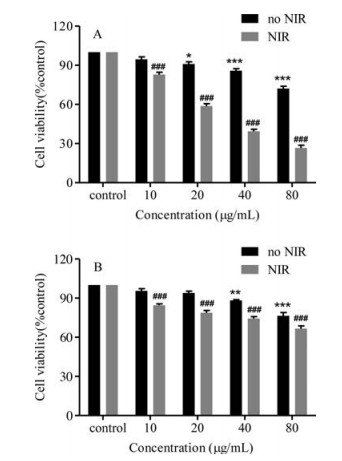
The cytotoxicity of PDAMn-CuS@BSA-FA to MCF-7 cells and SW1990 cells was evaluated by the MTT method (Fig. 5). It shows less toxicity to MCF-7 cells without irradiation. However, PDAMnCuS@BSA-FA can inhibit the proliferation of MCF-7 in dose dependent manner in 10–80 mg/mL under irradiation. This demonstrates the nanoparticle can be a NIR responsive photothermal effect agent. The different cytotoxicity of PDAMnCuS@BSA-FA to MCF-7 cells and SW1990 cells indicate that the nanoparticle shows high accumulation in MCF-7 cells due to the highly expression of folic acid acceptor.

|
Download:
|
| Fig. 5. Cytotoxicity of the PDAMn-CuS@BSA-FA NPs. Cell viability of MCF-7 cells (A) and SW1990 cells (B) treated with five different concentrations of NPs with NIR light (808 nm, 0.5 W/cm2) and without light for 24 h was determined by MTT assay. (Control: no NPs in the cells). Data are expressed as means ± S.D. (n = 3), *P < 0.05, **P < 0.01 and ***P < 0.001 vs. controls, ###P < 0.001 vs. controls | |
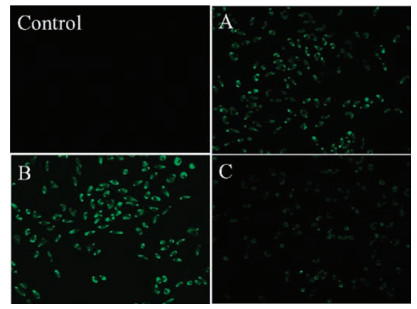
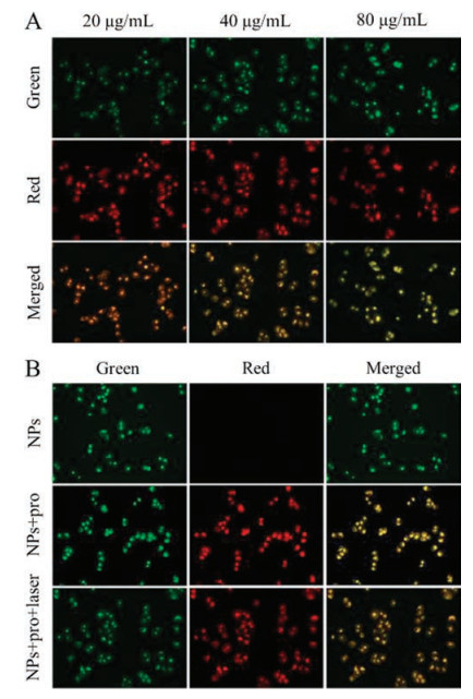
Folic acid is highly expressed in breast cancer, and nanoparticle shows high fluorescence sensitivity to cancer cells, so it may be breast cancer cell (MCF-7) turn-on fluorescence imaging agents. PDAMn-CuS@BSA-FA was cultivated with MCF-7 cells at 37 ℃ for 6 h, the cell imaging was recorded (Fig. 6). The green fluorescence images of MCF-7 cells show that PDAMn-CuS@BSA-FA can permeate through cell membranes and enter MCF-7 cells. These cell experiments show that the adhesion molecules in MCF-7 cells weaken the interaction of PDA and central metal in PDAMnCuS@BSA-FA, which can be the MCF-7 cells turn-on nanoprobe. Moreover, strong green fluorescence image was observed when the MCF-7 cells were irradiated with NIR light (808 nm, 0.5 W/cm2) for 5 s at 37 ℃ (Fig. 6B). It indicates that PDA could be released from PDAMn-CuS@BSA-FA under NIR irradiation. Moreover, the release of PDA was further confirmed by NIR enhanced emission of PDAMn-CuS@BSA-FA in BSA solution (Fig. S7 in Supporting information). To confirm the mitochondria target imaging, the imaging of PDAMn-CuS@BSA-FA was assessed by comparison with the well-known MitoTracker red FM. When the MCF-7 cells were treated with both MitoTracker red FM and nanoparticle, yellow fluorescent image was observed due to the overlapped image of MitoTracker red FM and green image of nanoparticles in the cell (Fig. 7 and Fig. S8 in Supporting information).

|
Download:
|
| Fig. 6. Fluorescence images of MCF-7 cells (scale bar, 100 mm; λex: 485 nm). Control: MCF-7 cells without nanoparticle; (A) MCF-7 cells after cultivated with PDAMn-CuS@BSA-FA NPs (40 mg/mL) for 6 h; (B) MCF-7 cells after cultivated with PDAMn-CuS@BSA-FA NPs (40 mg/mL) for 2 h, and then irradiated with a nearinfrared laser (808 nm, 0.5 W/cm2) for 5 s; (C) MCF-7 cells after cultivated with PDAMn-CuS@BSA NPs (40 mg/mL) for 6 h | |

|
Download:
|
| Fig. 7. Mitochondria target imaging of PDAMn-CuS@BSA-FA NPs cultivated with MCF-7 cells (scale bar, 100 mm; λex: 485 nm). (A) PDAMn-CuS@BSA-FA NPs with different concentrations (20, 40, 80 mg/mL) with NIR laser irradiation (Green), treated with MitoTracker red FM (Red) and overlapped images (Merged); (B) PDAMn-CuS@BSA-FA NPs (40 mg/mL) treated with MCF-7 cells without NIR laser irradiation (NPs), with MitoTracker red FM (NPs + pro), and with both NIR laser irradiation and MitoTracker red FM (NPs + pro+ laser), (NIR lase irradiation, 808 nm, 0.5 W/cm2) | |
In summary, a breast cancer sensitive nanoparticle PDAMnCuS@BSA-FA (NPs) was constructed by the electronic state interaction, coordination and hydrophobic interaction based layer-layer assembly strategy. The photoinitiated cytotoxicity indicates that NPs can be employed as effective NIR light initiated attenuators. In particular, the emission of the nanoparticle at 520 nm increased with the titration of CH3O-PEG-phosphatide. Moreover, MCF-7 cells lysis switched on the green emission of NPs greatly, in contrast, weak emission can be found in normal cells lysis. It indicates that the interaction of PDA and central metal in PDAMn-CuS@BSA-FA can be adjusted by CH3O-PEG-phosphatide or the cancer cells. Results demonstrate PDAMn-CuS@BSA-FA can be a breast cancer activated nanosensor. Our results provide a convenient method for the development of non-aptamer cancer activated nanoprobes or drug carriers based on the CH3O-PEGphosphatide recognized nanopartices. MCF-7 cell turn-on fluorescence images demonstrate the NPs can enter MCF-7 cells. Therefore, NPs can be a breast cancer activated fluorescence nanoprobe for breast cancer diagnosis.
AcknowledgmentThis work was financially supported by the National Natural Science Foundation of China (No. 21571085).
Appendix A. Supplementary dataSupplementary data associated with this article can be found, in the online version, at https://doi.org/10.1016/j.cclet.2018.02.011.
| [1] |
(a) Q. Chen, H.T. Ke, Z.F. Dai, Z. Liu, Biomaterials 73 (2015) 214-230; (b) A. Palma, L.A. Alvarez, D. Scholz, et al., J. Am. Chem. Soc. 133 (2011) 19618-19621. |
| [2] |
X. Zhao, C.X. Yang, L.G. Chen, X.P. Yan, Nature Commun. 8 (2017) 14998-14999. DOI:10.1038/ncomms14998 |
| [3] |
B.B. Li, P. Zhang, J.W. Du, X. Zhao, Y.X. Wang, Colloids Surf. B:Biointerf. 154 (2017) 133-141. DOI:10.1016/j.colsurfb.2017.03.020 |
| [4] |
L.W. Zhang, S. Gao, F. Zhang, et al., ACS Nano 8 (2014) 11250-12258. |
| [5] |
N. Gao, W. Yang, H.L. Nie, et al., Biosens. Bioelectron. 96 (2017) 300-307. DOI:10.1016/j.bios.2017.05.019 |
| [6] |
(a) S.H. Wang, A. Riedinger, H.B. Li, et al., ACS Nano 9 (2015) 1788-1800; (b) L.R. Guo, D.D. Yan, D.F. Yang, et al., ACS Nano 8 (2014) 5670-5681. |
| [7] |
(a) M. Zhou, M. Tian, C. Li, Bioconjugate Chem. 27 (2016) 1188-1199; (b) R.C. Lv, P.P. Yang, B. Hu, et al., ACS Nano 11 (2017) 1064-1072. |
| [8] |
(a) X.X. Yao, Z.F. Tian, J.X. Liu, Y.F. Zhu, N. Hanagata, Langmuir 33 (2017) 591-599; (b) M. Zhou, J.J. Li, S. Liang, et al., ACS Nano 9 (2015) 7085-7096; (c) J. Shen, G.Y. Chen, A.M. Vu, et al., Adv. Optical Mater. 1 (2013) 644-650. |
| [9] |
A.O. Elzoghby, W.M. Samy, N.A. Elgindy, J. Control. Release 161 (2012) 38-49. DOI:10.1016/j.jconrel.2012.04.036 |
| [10] |
(a) W.L. Lu, Y.Q. Lan, K.J. Xiao, et al., J. Mater. Chem. B 5 (2017) 1275-1283; (b) L. Han, Y. Zhang, X.W. Chen, Y. Shu, J.H. Wang, J. Mater. Chem. B 4 (2016) 105-112. |
| [11] |
J. Bai, Y.W. Liu, X. Jiang, Biomater 35 (2014) 5805-5813. DOI:10.1016/j.biomaterials.2014.04.008 |
| [12] |
(a) Z.T. Wang, P. Huang, O. Jacobson, et al., ACS Nano 10 (2016) 3453-3460; (b) W.T. Yang, W.S. Guo, W.J. Le, et al., ACS Nano 10 (2016) 10245-10257. |
| [13] |
(a) P.A. Baeuerle, O. Gires, Br. J. Cancer 96 (2007) 417-423; (b) S.D. Soysal, S. Muenst, T. Barbie, et al., Br. J. Cancer 108 (2013) 1480-1487. |
| [14] |
X.J. Liu, Q.L. Ren, F.F. Fu, et al., Dalton Trans. 44 (2015) 10343-10351. DOI:10.1039/C5DT00198F |
| [15] |
Z. Li, Q.Y. Chen, P.D. Wang, Y. Wu, RSC Adv. 3 (2013) 5524-5528. DOI:10.1039/c3ra22907f |
| [16] |
A. Topete, M. Alatorre-Meda, P. Iglesias, et al., ACS Nano 8 (2014) 2725-2738. DOI:10.1021/nn406425h |
| [17] |
G.S. Saini, Acta Part A 64 (2006) 981-986. DOI:10.1016/j.saa.2005.09.008 |
| [18] |
M. Lópezlázaro, Mol. Med. 16 (2010) 144-153. |