 2018, Vol. 29


Endonucleases are enzymes that can hydrolyze the phosphodiester bond within a polynucleotide chain, which catch much attention because they play an important role in lots of biological processes including recombination, DNA replication, molecular cloning, genetyping, etc. [1-5]. The detection of endonuclease thus is of importance in biosensing and clinical diagnosis. Some conventional techniques, such as polyacrylamide gel electrophoresis, high performance liquid chromatography and enzymelinked immunosorbent assay, have been applied for the detection of endonuclease activity [6-8]. However, they are time-consuming and laborious. Recently, colorimetric, electrochemical, and fluorescent assays have been employed to develop sensitive and selective assays for endonuclease activities [5, 9-12]. It is noted that fluorometric detection has become an attractive means due to its high sensitivity, rapid response, and simple manipulation [13-17]. Especially, label-free and fluorescence turn-on method is desirable for endonuclease detection because it is economic and can avoid false positive response.
Recently, perylene diimide derivatives (PDI) have attracted attention due to high thermal stability and photostability [18, 19]. A cationic PDI can associated with single-stranded DNA (ssDNA) resulting in strong tendency to form a self-aggregated or π-stacked structure [20-22], which can efficiently quench a variety of adjacent fluorophores that emit over a wide wavelength range from the visible to NIR region. Thus the PDI with quenching abilitiy brings a favorable application in biological sensing system [23, 24].
Cationic conjugated polymers (CCPs) possessing strong lightharvesting ability, fluorescent signal amplification and good watersolubility, have received intense research interest [25-29]. CCPs have exhibited the broad applications in biological detection, cell imaging, drug transport and release, and disease diagnosis, anticancer, antibacterial, etc. [30-39]. In this paper, by taking advantage of the excellent photoelectric properties of CCPs, we designed a new label-free biosensor for endonuclease detection based on cationic conjugated poly(9, 9-bis(60-N, N, N-trimethylammonium)hexyl)fluorine phenylene) (PFP) and PDI derivatives by using fluorescence "sign-on" mechanism. In the biosensor, CCPs, single-stranded nucleic acid and PDI were used as signal reporter, probe and fluorescence quencher, respectively. Owing to the aggregation of PDI and its quenching ability being related closely to the length of nucleic acids, it is reasonable to design a biosensor for endonuclease. S1 nuclease was chosen as model enzyme for the proof-of-concept application of the method. When ssDNA was present in the PFP and PDI mixture solution, PFP/ssDNA/PDI complex can form through electrostatic attraction interactions, resulting in the fluorescence of PFP being quenched due to the adjacence between PDI and PFP. Upon addition of S1 nuclease, ssDNA is hydrolyzed into small fragment. The PFP/ssDNA/PDI complex thus is broken and the aggregation of PDI is decreased, resulting in PFP fluorescence recovering. In addition, nucleic acid aptamer, as single-stranded nucleic acid species, can transfer its configuration when binding to a specific target molecule, which weakens the aggregation of PDI and motivates the fluorescence of PFP recovering. This method is thus potential to detect target molecules, such as bisphenol A. Moreover, the method provides a new platform for other enzymes and small molecules.
The proposed principle of the detection of S1 nuclease and BPA is illustrated in Scheme 1. PDI itself could not quench the fluorescence of PFP because of the electrostatic repulsion between the two (Fig. 1a). When ssDNA was added into the mixture solution (Scheme 1a), PFP/ssDNA/PDI complex can form due to stronger electrostatic attraction interactions. The PDI thus aggregates on the ssDNA and quenches the fluorescence of PFP due to the adjacence between PDI and PFP. So in the absence of S1 nucleas, the fluorescence of PFP is weak when PFP is excited. On the contrary, upon addition of S1 nuclease, ssDNA is hydrolyzed into small fragment by S1 nuclease, the PFP/ssDNA/PDI complex is broken down and the aggregation of PDI on small ssDNA fragment is uneffective. Correspondingly, the fluorescence of PFP recovers dramatically due to the weaker quenching ability of PDI and the farther distance between PFP and PDI. In virtue of target-mediated aggregation of PDI quencher, we also construct a label-free assay for small biomolecules based on the aptamer configuration changing. As shown in Scheme 1b, in the absence of BPA, PFP/ ssDNA/PDI complex can form due to stronger electrostatic interactions, resulting in the fluorescence of PFP quenching. In the presence of BPA, the BPA aptamer can bind BPA specifically and switch its configuration, which weakens the effective aggregation of PDI. Correspondingly, the fluorescence of PFP recovers when PFP is excited at 380 nm. This fluorescence turn-on assay thus can be used to detect S1 nuclease and BPA without the requirement of labelling probe.

|
Download:
|
| Scheme 1. Proposed principle for the detection of S1 nuclease and BPA using cationic conjugated polymers and PDI. | |

|
Download:
|
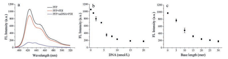
| Fig. 1. (a) Fluorescence emission spectra of PFP, PFP/PDI, PFP/ssDNA/PDI in Tris-HCl buffer solution (pH 7.5). (b, c) Relationship between fluorescence intensity of PFP/DNA/PDI and ssDNA concentration or length. [PFP] = 1.0 ×10⁻⁶ mol/L in RUs, [ssDNA] = 1.5 ×10⁻⁸ mol/L, [PDI] = 5.0 × 10⁻⁷ mol/L. The excitation wavelength is 380 nm. | |
Firstly, the concentration of PFP and PDI were optimized by measuring the fluorescence emission spectra. The solution containing S1 nuclease and ssDNA was incubated at 37 ℃ for 30 min in advance. Then, PFP in various concentration and the mixture solution were added into Tris-HCl (pH 7.5), followed by adding PDI (5.0 × 10⁻⁷ mol/L) to incubate for 5 min. As shown in Fig. S1a (Supporting information), when the concentration of PFP is 1.0 × 10⁻⁶ mol/L, the ratio of fluorescence intensity of PFP reaches the maxium. Thus, 1.0 × 10⁻⁶ mol/L was chosen as the optimal concentration of PFP. Furthermore, to optimize the concentration of PDI, the concentration of PFP and ssDNA were fixed to 1.0 × 10⁻⁶ mol/L and 20 nmol/L, respectively. As shown in Fig. S1b (Supporting information), the intensity of PFP/ssDNA/ PDI reduced gradually with the increase of PDI concentration. When the concentration of PDI is added to 5.0 × 10⁻⁷ mol/L, the quenching effeciency reaches the plateau. 5.0 × 10⁻⁷ mol/L PDI was thus selected for the following analytical purposes.
To study the effect of the concentration of the nucleotide acids on the fluorescence quenching, we measured the fluorescence intensity of PFP in the presence of the ssDNA at different concentrations. As shown in Fig. 1b, PFP intensity decreases with the increase of ssDNA concentration. When the concentration of ssDNA is added to 1.5 ×10⁻⁸ mol/L, the quenching efficiency remains nearly unchanged. Thus, 15 nmol/L was used in following experiments. To investigate the effect of the length of nucleotide acid on the PDI aggregation, ssDNA with different base lengths varying from 5 to 30 mer, were tested. As shown in Fig. 1c, the fluorescence intensity of PFP gradually decreased with the increase of the ssDNA length due to the stronger electrostatic interactions between ssDNA with longer length and PDI or PFP. When the length of ssDNA was 30 mer, the quenching efficiency of PDI reaches the plateau. Accordingly, 30 mer ssDNA at a concentration of 15 nmol/L was used in the following research.
To investigate the response of label-free assay for S1 nuclease, we measured PFP fluorescence intensity in Tris-HCl buffer at pH 7.4 at various concentrations of S1 nuclease. As shown in Fig. 2a, the fluorescence intensity of PFP increased with the increasing concentration of S1 from 0 to 0.02 U/mL. Since S1 nuclease can cleave ssDNA into small fragment, the aggregation of PDI is destroyed and then the quenching ability of PDI decreases. The relationship between the fluorescence intensity ratio (I/I0) at 425 nm and S1 nuclease concentration is shown in Fig. 2b. The ratio increases dramatically with the increasing concentration of S1 nuclease. When the concentration of S1 nuclease is 2.0 × 10⁻² U/mL, the ratio almost reaches a plateau. The detection limit (LOD) was determined to be 1.0 × 10⁻⁶ U/mL (S/N = 3). These results represent that this method is sensitive to S1 nuclease detection, As shown in Table S1 (Supporting information), this LOD is lower than that from previous biosensors reported by Dai et al. (5.9 × 10⁻⁴ U/mL) [1], Wang et al. (0.04 U/mL) [5], Zhang et al. (5 ×10⁻⁵ U/mL) [40], and Tang et al. (5.8 × 10⁻⁴ U/mL) [41], etc.

|
Download:
|
| Fig. 2. (a) Fluorescence emission spectra of PFP in Tris-HCl buffer (pH 7.5) with addition of S1. (b) The ratio of PFP intensity with various concentrations of S1 nuclease. (c) Inhibition efficiency of S1 nuclease by ATP in Tris-HCl (pH 7.5) buffer solution. [PFP] = 1.0 × 10⁻⁶ mol/L, [PDI] = 5.0 × 10⁻⁷ mol/L, [ssDNA] = 1.5 ×10⁻⁸ mol/L. The error bars represent standard deviations three parallel measurements. The excitation wavelength is 380 nm. | |
S1 nuclease reaction time was investigated by fixing the concentration of S1 nuclease at 0.1 U/mL. As shown in Fig. S2 (Supporting information), the ratio increases to the plateau once incubation for 25 min. Thus, to ensure S1 nuclease digesting ssDNA adequately, the incubation time was set for 30 min in our experiments, which demonstrates that the S1 nuclease detection can be completed in a short time.
In addition, the inhibition of enzyme activity was studied by incubating ssDNA with different concentrations of ATP which is a well-known inhibitor of S1 nuclease. The concentration of S1 nuclease is fixed at 0.1 U/mL. Fig. 2c shows that the fluorescence intensity of PFP decreases with the increasing concentration of ATP in the range of 0–5 μmol/L. It is observed that 5 μmol/L ATP can inhibit S1 nuclease completely. These results confirm that our strategy is potential for screening S1 nuclease inhibitors.
As a result of the quenching ability of PDI being dependent on the length of ssDNA, we construct a label-free aptasensor based on aptamer configuration changing for small molecules. As shown in Fig. 3a, in the absence of BPA, the fluorescence of PFP is weak when PFP is excited at 380 nm, because PDI aggregates on the aptamer and quenching the fluorescence of PFP. In the presence of BPA, the aptamer can recognize targets and switch its configuration, which decreases the aggregation of PDI, resulting in the fluorescence of PFP recovers. The fluorescence increases with the increase of BPA concentration. The relationship between the fluorescence intensity ratio (I/I0) at 425 nm and BPA concentration is shown in Fig. 3b, where I0 and I are the fluorescence intensities of the sensor solution in the absence and presence of BPA, respectively. The LOD was determined to be 0.05 ng/mL (S/N = 3). By changing the sequence of aptamer, this platform also is potential to sense other targets.

|
Download:
|
| Fig. 3. (a) Fluorescence emission spectra of PFP in Tris-HCl buffer (pH 7.5) with addition of BPA. (b) The ratio of PFP intensity with various concentrations of BPA. (c) Response of the assay toward other BPA analogues. [PFP] = 1.0 × 10⁻⁶ mol/L, [aptamer] = 2.0 ×10⁻⁸ mol/L, [PDI] = 5.0 × 10⁻⁷ mol/L, [BPA] = 50 ng/mL, [BPB] = [BPC] = [BPE] = [DES] = [DPA] = 100 ng/mL. The excitation wavelength is 380 nm. | |
We also investigated the selectivity of the proposed method by measuring the fluorescence intensity after aptamer incubating with other BPA analogues, such as BPB, BPC, BPE, DPA and DES. As shown in Fig. 3c, none of these analogues caused obvious enhancement in the fluorescence intensity, however, a significant intensity increase was observed for BPA. These results show the assay demonstrates an advantageous specificity for BPA detection when using BPA aptamer as probe.
In summary, we designed a new lable-free biosensor based on cationic conjugated polymer and perylene diimide derivatives. In the biosensor, the targets can tune the length of nucleic acids to mediate the aggregation of PDI, which can be used for endonuclease and biomolecules detection such as S1 nuclease and BPA. The detection limit is determined to be 1.0 × 10⁻⁶ U/mL for S1 nuclease, which is far lower than reported in the literature. In other words, this strategy has the advantages as high sensitivity, simplicity and cost efficiency by taking advantage of the fluorescence signal amplification of conjugated polymers, and provides new insight in the area of biosensors for the detection of enzyme and other biomolecules.
AcknowledgmentsThe authors are grateful for the financial support from the National Natural Science Foundation of China (No. 21675106), the 111 Project (No. B14041), Natural Science Basic Research Plan in Shaanxi Province of China (No. 2017JM2019), the Program for Changjiang Scholars and Innovative Research Team in University (No. 14R33), and the Program for Innovative Research Team in Shaanxi Province (No. 2014KCT-28).
Appendix A. Supplementary dataSuppleme2 ntary data associated with this article can be found, in the online version, at http://dx.doi.org/10.1016/j.cclet.2017.08.032.
| [1] |
Z. Wang, J. Zhao, J. Bao, Z. Dai, ACS Appl. Mater. Interfaces 8(2016) 827-833. DOI:10.1021/acsami.5b10165 |
| [2] |
N.D. Grindley, K.L. Whiteson, P.A. Rice, Annu. Rev. Biochem. 75(2006) 567-605. DOI:10.1146/annurev.biochem.73.011303.073908 |
| [3] |
P. Norberg, T. Bergstrom, J.A. Liljeqvist, J. Clin. Microbiol. 44(2006) 4511-4514. DOI:10.1128/JCM.00421-06 |
| [4] |
S.C. West, Nat. Rev. Mol. Cell Biol. 4(2003) 435-445. |
| [5] |
Z. Zhou, J. Zhu, L. Zhang, Y. Du, S. Dong, E. Wang, Anal. Chem. 85(2013) 2431-2435. DOI:10.1021/ac303440d |
| [6] |
A. Jeltsch, A. Fritz, J. Alves, H. Wolfes, A. Pingoud, Anal. Biochem. 213(1993) 234-240. DOI:10.1006/abio.1993.1415 |
| [7] |
L.W. McLaughlin, F. Benseler, E. Graeser, N. Piel, S. Scholtissek, Biochemistry 26(1987) 7238-7245. DOI:10.1021/bi00397a007 |
| [8] |
S. Spitzer, F. Eckstein, Nucleic Acids Res. 16(1988) 11691-11704. DOI:10.1093/nar/16.24.11691 |
| [9] |
J. Ding, W. Qin, Biosens. Bioelectron. 47(2013) 559-565. DOI:10.1016/j.bios.2013.03.066 |
| [10] |
Y. Tang, F. Feng, F. He, et al., J. Am. Chem. Soc. 128(2006) 14972-14976. DOI:10.1021/ja065159b |
| [11] |
X. Yang, F. Pu, J. Ren, X. Qu, Chem. Commun. 47(2011) 8133-8135. DOI:10.1039/c1cc12216a |
| [12] |
R. Cao, B. Li, Y. Zhang, Z. Zhang, Chem. Commun. 47(2011) 12301-12303. DOI:10.1039/c1cc15994a |
| [13] |
Y. Huang, S. Zhao, M. Shi, et al., Anal. Chem. 83(2011) 8913-8918. DOI:10.1021/ac2013114 |
| [14] |
Y. Huang, S. Zhao, Z.F. Chen, Y.C. Liu, H. Liang, Chem. Commun. 47(2011) 4763-4765. DOI:10.1039/c1cc10325c |
| [15] |
W. Li, Z. Liu, H. Lin, et al., Anal. Chem. 82(2010) 1935-1941. DOI:10.1021/ac902670c |
| [16] |
J. Lee, Y.K. Kim, D.H. Min, Anal. Chem. 83(2011) 8906-8912. DOI:10.1021/ac201298r |
| [17] |
L.H. Wang, K.K. Ma, Y.D. Zhang, Anal. Biochem. 468(2015) 34-38. DOI:10.1016/j.ab.2014.09.011 |
| [18] |
Y.Y. Gao, H.Z. Li, S.W. Yin, et al., New J. Chem. 38(2014) 5647-5653. DOI:10.1039/C4NJ01083C |
| [19] |
B. Wang, C. Yu, Angew. Chem. Int. Ed. 49(2010) 1485-1488. DOI:10.1002/anie.200905237 |
| [20] |
R. Hu, T. Liu, X.B. Zhang, et al., Anal. Chem. 86(2014) 5009-5016. DOI:10.1021/ac500618v |
| [21] |
R. Hu, X. Zhang, Q. Xu, et al., Biosens. Bioelectron. 92(2017) 40-46. DOI:10.1016/j.bios.2017.01.051 |
| [22] |
T. Takada, M. Ido, A. Ashida, et al., Chem. Eur. J. 21(2015) 6846-6851. DOI:10.1002/chem.201406592 |
| [23] |
H.J. Ben, X.K. Ren, B. Song, et al., J. Mater. Chem. C 5(2017) 2566-2576. DOI:10.1039/C6TC05171E |
| [24] |
J. Schill, L.G. Milroy, J.A.M. Lugger, A. Schenning, L. Brunsveld, ChemistryOpen 6(2017) 266-272. DOI:10.1002/open.v6.2 |
| [25] |
X.L. Feng, L.B. Liu, S. Wang, D.B. Zhu, Chem. Soc. Rev. 39(2010) 2411-2419. DOI:10.1039/b909065g |
| [26] |
H.A. Ho, A. Najari, M. Leclerc, Acc. Chem. Res. 41(2008) 168-178. DOI:10.1021/ar700115t |
| [27] |
L.H. Wang, K.Y. Pu, J. Li, et al., Adv. Mater. 23(2011) 4386-4391. DOI:10.1002/adma.201102227 |
| [28] |
L. Feng, C. Zhu, H. Yuan, et al., Chem. Soc. Rev. 42(2013) 6620-6633. DOI:10.1039/c3cs60036j |
| [29] |
X.Q. Zhang, Q. Zhao, Y.R. Li, X.R. Duan, Y.L. Tang, Anal. Chem. 89(2017) 5503-5510. DOI:10.1021/acs.analchem.7b00477 |
| [30] |
H. Bai, H. Yuan, C. Nie, et al., Angew. Chem. Int. Ed. 54(2015) 13208-13213. DOI:10.1002/anie.201504566 |
| [31] |
X. Liu, Q. Fan, W. Huang, Biosens. Bioelectron. 26(2011) 2154-2164. DOI:10.1016/j.bios.2010.09.025 |
| [32] |
M.R. Pinto, K.S. Schanze, Proc. Natl. Acad. Sci. USA 101(2004) 7505-7510. DOI:10.1073/pnas.0402280101 |
| [33] |
Y. Tang, F. Feng, F. He, et al., J. Am. Chem. Soc. 128(2006) 14972-14976. DOI:10.1021/ja065159b |
| [34] |
Y. Tang, Y. Liu, A. Cao, Anal. Chem. 85(2013) 825-830. DOI:10.1021/ac302840t |
| [35] |
C. Zhu, L. Liu, Q. Yang, F. Lv, S. Wang, Chem. Rev. 112(2012) 4687-4735. DOI:10.1021/cr200263w |
| [36] |
H. Yuan, C. Xing, Y. Fan, et al., Macromol. Rapid. Commun. 38(2017). DOI:10.1002/marc.201600726 |
| [37] |
H. Yuan, B. Wang, F. Lv, L. Liu, S. Wang, Adv. Mater. 26(2014) 6978-6982. DOI:10.1002/adma.v26.40 |
| [38] |
J.T. Li, Q. Zhao, F. Shi, C.H. Liu, Y.L. Tang, Adv. Healthc. Mater. 5(2016) 2967-2971. DOI:10.1002/adhm.201600868 |
| [39] |
R. Hu, S.L. Li, H.T. Bai, et al., Chin. Chem. Lett. 27(2016) 545-549. DOI:10.1016/j.cclet.2016.02.001 |
| [40] |
L. Wang, K. Ma, Y. Zhang, Anal. Biochem. 468(2015) 34-38. DOI:10.1016/j.ab.2014.09.011 |
| [41] |
Y. He, B.N. Jiao, H.W. Tang, RSC Adv. 4(2014) 18294-18300. DOI:10.1039/C4RA01102C |