 2017, Vol. 28


 , Yue-Wei Guob
b State Key Laboratory of Drug Research, Shanghai Institute of Materia Medica, Chinese Academy of Sciences, Shanghai 201203, China;
c Jiangsu Kanion Pharmaceutical Co., Ltd., Lianyungang 222002, China
Soft corals belonging to the genus Clavularia (class Octocorallia, order Alcyonacea, family Clavulariidea), inhabiting abundantly in the South China Sea, have been proved to be a rich source of structurally diverse and biologically active terpenoids, prostanoids and steroids [1–4]. Remarkably, the genus Clavularia is really a productive factory of dolabellane type diterpenoids and many of them showed significant cytotoxic activities [5, 6]. The complex and unique structures of dolabellane diterpenoid have also attracted the attention of synthetic chemists for their total synthesis [7, 8].
In the course of our ongoing research aiming at searching for the biologically active substances from South China Sea soft corals [9–12], we made a collection of the title sample C. viridis off the Xisha Islands, Hainan Province, China. Chemical investigation of the Et2O-soluble fraction of the acetone extract of this animal resulted in the isolation of a new dolabellane-type diterpenoid, clavirolide G (1), and a related one, clauvdiol A (3) [13] (Fig. 1). This paper describes the isolation and structure elucidation of these diterpenoids.

|
Download:
|
| Figure 1. Structures of compounds 1–3. | |
2. Results and discussion
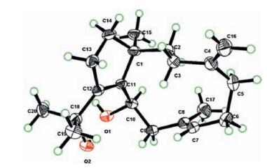
The structural determination of the isolate 3 is straightforward since it is a crystal suitable for X-ray diffraction analysis allowing to unambiguously recognize its structure as depicted in Fig. 2, the same as clauvdiol A [13], a dolabellane-type diterpenoid previously isolated from the same species by Su et al. However, it may be worth to point out that in the literature [13], the absolute configuration (AC) of 3 was tentatively assigned based mainly on the interpretation of CD profile of 3 and biogenetic consideration. In our case, the AC of 3 was, for the first time, unambiguously elucidated by using X-ray crystallography with Cu Kα radiation on a single crystal of 3.

|
Download:
|
| Figure 2. Perspective drawing of X-ray structure of compound 3. | |
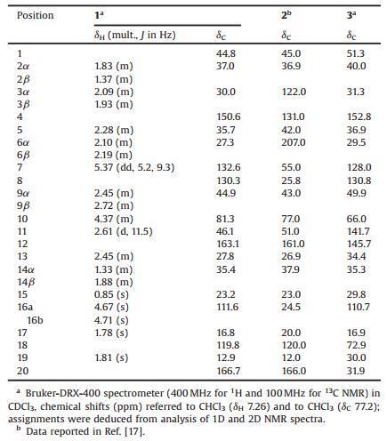
Clavirolide G (1) was obtained as an optically active, colorless oil. The molecular formula, C20H28O2, consistent with seven degrees of unsaturation, was determined by HREIMS (m/z 300.2091 [M]+, calcd. 300.2089) suggesting the diterpenoid nature of the metabolite. Further, 1H NMR and 13C NMR data (Table 1) showed similarities with those of the co-occurring 3 indicating that 1 is also a dolabellane-type diterpenoid, rare among octacorals. The UV absorption of 1 at 230 nm (log ε = 4.95), together with IR absorptions at 1712, and 1094 cm-1 and 13C NMR signals at δ 166.7 (C-20), 163.1 (C-12), and 119.8 (C-18), suggested the presence of a conjugated lactone system. In addition, the 13C NMR signals at δ 150.6 (C-4) and 111.6 (C-16) indicated an exocyclic methylene function. Signals in the 13C NMR spectrum at δ 130.3 (C-8) and 132.6 (C-7), together with a signal accounting for one vinylic hydrogen in the 1H NMR spectrum at δ 5.37 (dd, lH, J = 5.2, 9.3 Hz, H-7), established the presence of one trisubstituted double bond. The E configuration of this double bond was indicated by the 13C NMR chemical shift of the vinyl methyl group (δ 16.8, CH3) [14]. These data, coupled with the degrees of unsaturation (seven), suggested that compound 1 to be tricyclic. Detailed analysis of the 1D and 2D NMR spectra (1H-1H COSY, HSQC, HMBC, NOESY) of 1 allowed us to propose the structure of clavirolide G as 1 (Fig. 1).
|
|
Table 1 1H NMR and 13C NMR spectral data for compound 1 and 13C NMR spectral data for compounds 2 and 3. |
Literature checking revealed that the NMR data of 1 were strongly reminiscent of the structurally related dolabellane-type diterpenoids, e.g. clavulactone (2) [2, 15] and 3 [13]. A comparison the overall 13C NMR data of 1 and 2, 3 (Table 1) readily recognized that 1 and 2 shared the common partial structures for the rings a and b (Fig. 1), while 1, like 3, possessed the same olefins at △7 and △4(16) for the ring c. These spectroscopic evidences give further support that compound 1 is an analogue of the model compound 2 [15]. In fact, 1 differs from 2 only by the isomerization of the olefin at △3, dehydrogenation between C-7 to C-8 and reduction of the C-6 ketone. Finally, the AC at C-1, C-10, and C-11 of 1 were assigned as R, R and S, respectively, the same as those of already determined for clavulactone (2) [2, 15] and other related analogues [2, 13, 16], by both direct CD spectra comparison [15, 17, 18] and biogenetic consideration.
It is interesting to note that although the structures of 1 and the co-occurring 3 are formally quite different, they are biogenetically related. As outlined in Scheme 1, the △11 olefin migration accompanying the hydroxyl group shifting from C-18 to C-20 will generate the intermediate 4, of which further oxidation at C-20 gives 5 and successive esterification between the C-20 carboxyl group and C-10 hydroxyl group will yield the compound 1.

|
Download:
|
| Scheme 1. Possible biosynthetic pathway of compound 1 from its parent precursor 3. | |
In light of the observation that many dolabellane diterpenoids displayed promising anti-tumoral activities, the cytotoxicity of compounds 1 and 3 were tested. In the bioassay, only the new compound 1 exhibited moderate cytotoxic activity against KB and HL-60 cell lines with IC50 values of 15.4 μmol/L and 17.8 μmol/L, respectively, with adriamycin as the positive control (IC50 = 0.04 μmol/L and 0.02 μmol/L).
3. ConclusionThe Xisha soft coral C. viridis has been proved to be able to produce various dolabellane diterpenoids. To our knowledge, till now, nine such diterpenoids, namely clavulactone (2) [2], clavirolides A–F [2, 13, 16], clavudiol A (3) and B [13, 16], were isolated from the title species. The present work is the second chemical study on the same species from the same location. The discovery of clavirolide G has added a new member of this class of diterpenes, of which the numbers are expanding. The real producer responsible for these unique metabolites is a matter worth to discuss. It is reported that they were initially found either from sea hare [19, 20] or algae [21], latter found in seawhips [22] and Aplysia [23]. Further studies should be conducted to verify the true origin of these metabolites and to understand their real ecological role played in the life cycle of the title animal.
4. Experimental 4.1. General experimental proceduresECD spectrum was recorded on a J-810 spectropolarimeter. UV spectrum was recorded on a Shimadzu UV-2550 spectrophotometer. IR spectrum was recorded on a Nicolet Magna FT-IR 750 spectrometer. Optical rotations were measured on a PerkinElmer 241MC polarimeter. NMR spectra were measured on a Bruker DRX-400 (400 MHz for 1H and 100 MHz for 13C NMR) spectrometer (Bruker Biospin AG, Fällanden, Germany) in CDCl3, chemical shifts are referenced to the residual solvent signal (δH 7.26 ppm; δC 77.2 ppm). EIMS and HREIMS spectra were recorded on a FinniganMAT-95 mass spectrometer (Finnigan MAT, San Jose, CA, USA). Commercial silica gel (Qingdao Haiyang Chemical Group Co., Ltd., Qingdao, China, 200–300 and 400–500 mesh) was used for column chromatography, and precoated silica gel GF254 plates (Sinopharm Chemical Reagent Co., Shanghai, China) were used for TLC. Sephadex LH-20 (Pharmacia, USA) was also used for column chromatography. All solvents were of analytical grade. KB, HL-60 and A549 cells were bought from AmericanType Culture Collection (ATCC, Rockville, MD).
4.2. Collection of biological materialsSpecimens of the Clavularia sp. (class Octocorallia, order Alcyonacea, family Clavulariidea) were collected from Xisha Island, Hainan Province, P.R. China, in March 2013. The soft coral was identified as C. viridis by Prof. H. Hang of South China Sea Institute of Oceanology, Chinese Academy of Sciences. Freshly collected coral tissue was frozen on-site and stored at -20 ℃ until processed. The voucher specimen (No. 13XS-49) is available for inspection at the Shanghai Institute of Materia Medica, CAS.
4.3. Extraction and isolationThe frozen specimens of C. viridis (dry weight, 60 g) were cut into pieces and exhaustively ultrasonic extracted with acetone at room temperature (1.0 L × 4). The solvent-free acetone extract was partitioned between Et2O and H2O. The organic phase was evaporated under reduced pressure to give a dark-red residue (0.9 g), which was subjected to silica gel CC (200–300 mesh) and eluted with PE in Et2O (0–100%, gradient) to yield six fractions (A–F). Fraction D was first purified by Sephadex LH-20 (PE/CH2Cl2/ MeOH, 2:1:1), and then fraction B2 (41.0 mg) repeatedly purified by silica gel CC (400–500 mesh) using a PE/Et2O gradient as eluent (98:2–90:10) affording compound 1 (20.4 mg) and 3(25.9 mg). All the spectra are deposited in Supporting information.
Clavirolide G (1): Colorless oil; αD20 -96.8 (c 0.20, CHCl3); UV (MeOH) λmax (log ε): 206 (5.28), 230 (4.95) nm; CD (MeOH): 206 (∆ε -36.5), 227 (∆ε +14.5) nm, 255 (∆ε -46.0) nm; IR (KBr, cm-1) νmax 2932, 1712, 1441, 1309, 1094, 1001; 1H NMR (CDCl3, 400 MHz) and 13C NMR(CDCl3, 100 MHz) spectral data see Table 1; HREIMS m/z 300.2091 [M]+ (calcd for C20H28O2: 300.2091).
Clauvdiol A (3): Colorless crystals; αD20 -62.3 (c 0.13, CHCl3); 1H NMR (CDCl3, 400 MHz): δ 1.09 (3H, s, Me-15), 1.39 (3H, s, Me-20), 1.44 (3H, s, Me-19), 1.69 (3H, s, Me-17), 3.25 (1H, t, J = 11.8 Hz, H-9), 4.12 (1H, dd, J = 11.4, 2.5 Hz, H-10), 4.63 (1H, s, H-16), 4.71 (1H, s, H-16), 5.33 (1H, dd, J = 9.4, 5.2 Hz, H-7); 13C NMR (CDCl3, 100 MHz) see Table 1; EIMS m/z 304 [M]+.
4.4. Single-crystal X-ray diffraction analysis of clauvdiol A (3)A colorless block crystal of 3 was obtained by recrystallization from PE-CH2Cl2. A single crystal with dimensions of 0.32 mm × 0.18 mm × 0.15 mm was used for X-ray diffraction studies on a Bruker APEX-II CCD diffractometer employing graphite-monochromated Cu Kα radiation (l.54178 Å). The structure was solved by direct methods (SHELXS-97) and refined using full-matrix least squares difference Fourier techniques. All non-hydrogen atoms were refined anisotropically, and all hydrogen atoms were placed in idealized positions and refined as riding atoms with the relative isotropic parameters.
Crystal data: C20H32O2 (Mr = 304.45), orthorhombic with space group P212121, with a = 8.5414(3) Å, b = 10.4689(3) Å, c = 20.7809 (7) Å, V = 1858.21(10) Å3, T = 170 K, Z = 4. Dx = 1.0088 mg/m3, F(000) = 672. Independent reflections 3281 [Rint = 0.039], Flack parameter = 0.03(7).
4.5. Cytotoxicity assaysThe cytotoxicity of compounds 1 and 3 against KB cell lines has been tested by using the sulforhodamine B (SRB) method [24] and the cytotoxicity against HL-60 and A549 cell lines has been tested by using the Cell Counting Kit-8 (CCK-8) method [25], according to the protocols described in previous literatures. Cells were seeded into 96-well plates and grown for 24 h and then treated with increasing concentrations of compounds and cultured for further 72 h. Inhibition rates of cell proliferation after compounds treatments were determined by the SRB and CCK-8 methods. ADR (adriamycin) was used as the positive control with IC50 values of 0.04 μmol/L, 0.02 μmol/L and 0.13 μmol/L on KB, HL-60 and A549 cancer cells, respectively.
AcknowledgmentsThis research was financially supported by the National Natural Science Foundation of China (Nos. 81520108028, 21672230, 41506187, 816013016) and the SKLDR/SIMM Project (No. SIMM1501ZZ-03).
Appendix A. Supplementary dataSupplementary data associated with this article can be found, in the online version, at http://dx.doi.org/10.1016/j.cclet.2017.01.004.
| [1] | K. Iguchi, H. Sawai, H. Nishimura, M. Fujita, T Yamori, New dolabellane-type diterpenoids from the Okinawan soft coral of the genus Clavularia. Bull.Chem. Soc.Jpn. 75 (2002) 131–136. DOI:10.1246/bcsj.75.131 |
| [2] | J.Y. Su, Y.L. Zhong, L.M Zeng, Four novel diterpenoids:clavirolides B, C, D, and E from the Chinese soft coral Clavularia viridis. J.Nat.Prod. 54 (1991) 380–385. DOI:10.1021/np50074a005 |
| [3] | C.Y. Duh, A.A.H. El-Gamal, C.J. Chu, S.K. Wang, C.F Dai, New cytotoxic constituents from the Formosan soft corals Clavularia viridis and Clavularia violacea. J.Nat.Prod. 65 (2002) 1535–1539. DOI:10.1021/np0201873 |
| [4] | Y.C. Shen, Y.B. Cheng, Y.C. Lin, New prostanoids with cytotoxic activity from Taiwanese octocoral Clavularia viridis. J.Nat.Prod. 67 (2004) 542–546. DOI:10.1021/np030435a |
| [5] | C.Y. Duh, M.C. Chia, S.K. Wang, Cytotoxic dolabellane diterpenes from the Formosan soft coral Clavularia inflata. J.Nat.Prod. 64 (2001) 1028–1031. DOI:10.1021/np010106n |
| [6] | K. Mori, K. Iguchi, N. Yamada, Y. Yamada, Inouye Y.Bioactive marine diterpenoids from Japanese soft coral of Clavularia sp. Chem.Pharm.Bull 36 (1988) 2840–2852. DOI:10.1248/cpb.36.2840 |
| [7] | H. Miyaoka, Y. Isaji, H. Mitome, Y Yamada, Total synthesis of the dolabellane marine diterpenoids claenone, palominol and dolabellatrienone. Tetrahedron 59 (2003) 61–75. DOI:10.1016/S0040-4020(02)01474-6 |
| [8] | H. Miyaoka, T. Baba, H. Mitome, Y Yamada, Total synthesis of marine diterpenoid stolonidiol. Tetrahedron Lett. 42 (2001) 9233–9236. DOI:10.1016/S0040-4039(01)02032-9 |
| [9] | W.T. Chen, J. Li, J.R. Wang, X.W. Li, Y.W Guo, Structural diversity of terpenoids in the soft coral Sinularia flexibilis, evidenced by a collection from the South China Sea. RSC Adv. 5 (2015) 23973–23980. DOI:10.1039/C5RA01151E |
| [10] | L.F. Liang, L.X. Gao, J. Li, O. Taglialatela-Scafati, Y.W Guo, Cembrane diterpenoids from the soft coral Sarcophyton trocheliophorum Marenzeller as a new class of PTP1B inhibitors. Bioorg.Med.Chem. 21 (2013) 5076–5080. DOI:10.1016/j.bmc.2013.06.043 |
| [11] | L.F. Liang, T. Kurtan, Y.W. Guo, Unprecedented diterpenoids as a PTP1B inhibitor from the Hainan soft coral Sarcophyton trocheliophorum Marenzeller. Org.Lett. 15 (2013) 274–277. DOI:10.1021/ol303110d |
| [12] | W.T. Chen, L.G. Yao, X.W. Li, Y.W Guo, Sarcophytrols A-C, new capnosane diterpenoids from the South China Sea soft coral Sarcophyton trocheliophorum. Tetrahedron Lett. 56 (2015) 1348–1352. DOI:10.1016/j.tetlet.2015.01.157 |
| [13] | J.Y. Su, Y.L. Zhong, K.L. Shi, Clavudiol A and clavirolide A, two marine dolabellane diterpenes from the soft coral Clavularia viridis. J.Org.Chem. 56 (1991) 2337–2344. DOI:10.1021/jo00007a019 |
| [14] | D.E. Dorman, M. Jautelat, J.D Roberts, Carbon-13 nuclear magnetic resonance spectroscopy.Quantitative correlations of the carbon chemical shifts of acyclic alkenes. J.Org.Chem. 36 (1971) 2757–2766. DOI:10.1021/jo00818a007 |
| [15] | J.C. Li, Z.M. Zhang, Z.X. Xia, C.Z. Ni, Y.L Wu, New diterpene from soft coral qunzhuchong(Clavularia sp.)—isolation and structural elucidation of clavulactone. Acta Chim.Sin. 45 (1987) 558–563. |
| [16] | J.Y. Su, Y.L. Zhong, L.M Zeng, Two new diterpenoids from the marine soft coral Clavularia viridis. Chin.J.Chem. 10 (1992) 155–160. |
| [17] | G Snatzke, Circular dichroism and optical rotatory dispersion—principles and application to the investigation of the stereochemistry of natural products. Angew.Chem.Int.Ed. 7 (1968) 14–25. DOI:10.1002/(ISSN)1521-3773 |
| [18] | A.F Beecham, The CD of α, β-unsaturated lactones. Tetrahedron 28 (1972) 5543–5554. DOI:10.1016/S0040-4020(01)93618-X |
| [19] | C. Ireland, D.J. Faulkner, J. Finer, J Clardy, A novel diterpene from Dollabella californica. J.Am.Chem.Soc. 98 (1976) 4664–4665. DOI:10.1021/ja00431a063 |
| [20] | G.R. Pettit, R.H. Ode, C.L. Herald, Dreele R.B.Von, C. Michel, Antineoplastic agents 46.The isolation and structure of dolatriol. J.Am.Chem.Soc. 98 (1976) 4677–4678. DOI:10.1021/ja00431a072 |
| [21] | H.H. Sun, W Fenical, Diterpenoids of the brown seaweed Glossophora galapagensis. Phytochemistry 18 (1979) 340–341. DOI:10.1016/0031-9422(79)80095-3 |
| [22] | S.A. Look, W Fenical, New bicyclic diterpenoids from the Caribbean gorgonian octocoral Eunicea calyculata. J.Org.Chem. 47 (1982) 4129–4134. DOI:10.1021/jo00142a024 |
| [23] | A.G. Gonzalez, J.D. Martin, M. Norte, Marine natural products from Atlantic zone.Part 35.A new diterpene from Aplysia dactylomela. Tetrahedron Lett. 24 (1983) 1075–1076. DOI:10.1016/S0040-4039(00)81608-1 |
| [24] | P. Skehan, R. Storeng, D. Scudiero, New colorimetric cytotoxicity assay for anticancer-drug screening. J.Natl.Cancer Inst. 82 (1990) 1107–1112. DOI:10.1093/jnci/82.13.1107 |
| [25] | H. Tominaga, M. Ishiyama, F. Ohseto, A water-soluble tetrazolium salt useful for colorimetric cell viability assay. Anal.Commun. 36 (1999) 47–50. DOI:10.1039/a809656b |