 2017, Vol. 28


Dopamine (DA) is a monoamine neural transmitter, a simple organic chemical in the catecholamine family associated with activities including the central nervous system, movement control and immune functions. Low levels of DA would result in diseases such as Parkinson's disease and attention deficit hyperactivity disorder [1]. Therefore, the accurate determination of DA is crucial. As DA can be oxidized easily, electrochemical methods are generally employed to detect it [2]. However, because of the overlapping electrochemical oxidation potentials of DA, ascorbic acid (AA) and uric acid (UA), it is difficult to simultaneously detect each species in a mixture with well selectivity and high sensitivity [3, 4]. Therefore, it is desired to develop a novel electrode material to achieve the highly selective and sensitive detection of DA.
Current years, more and more attentions have been paid to fabricating metal oxide nanostructures based p-n junction, due to their unique optical and electronic properties in the applications of sensors, optoelectronics and catalysis [5-9]. It offers an important pathway to combine the different physical and chemical properties of individual component into one system [6]. For example, the heavily doped Si p-n junction nanowire exhibited a highly localized sensing region at the p-n interface and used for intracellular recording [10]. Introducing the p-n interface into single nanowire based probe can be effectively employed for hemoglobin detection [11]. The superior functional performances of the p-n junction in comparison to the corresponding singlephase metal oxides are mainly ascribed to the build-up of potential barriers at the p-n interface. The change of the potential barrier can be used as a sensing factor [12]. Therefore, it may be an important way to achieve novel sensing applications by developing the p-n junction based on nanostructures.
Ni foam with low weight and cross-linked grid structure allows providing high porosity, large surface area and well mechanical stability [13-15]. This 3D porous architecture would not only reduce the diffusion resistance of electrolyte but also maintain integrated electron transport pathways in the rapid electrochemical reactions. However, Ni, as a metal, has poor catalytic and sensing properties. NiO, a p-type semiconductor material, is widely studied in catalysts and sensors due to its well catalytic and sensing performance [16]. Therefore, it is desired to fabricate NiO foam for electrochemical sensing applications. ZnO, an n-type semiconductor material with direct wide band-gap (3.37 eV), is studied widely in catalysts, sensors, solar cells and UV lasing devices [17-20]. Varieties of ZnO nanostructures, such as nanowires, nanorods and nanotubes, have been successfully synthesized [21-23]. Recent studies demonstrated the crystal facet exposing and defect distributions of ZnO played important roles in its functional performances. As ZnO nanopyramids (ZnO NPys) have particular crystal facets exposing of the {1011} facet, it exhibits special performance in dye-sensitized solar cells, gas sensing and photocatalysis [24-26]. However, as the {1011} facet has a high surface energy and a fast growth rate, it is difficult to be synthesized [24].
In this work, NiO foam with rough nanostructured surface was fabricated by the surface treatment of Ni foam and used for electrochemical detection of DA. The n-type ZnO NPys were then decorated on the p-type NiO foam to construct the 3D p-n junction foam. Because of the introduction of the p-n junction interface and the high catalytic activity of ZnO NPys, the sensitivity to DA was enhanced significantly.
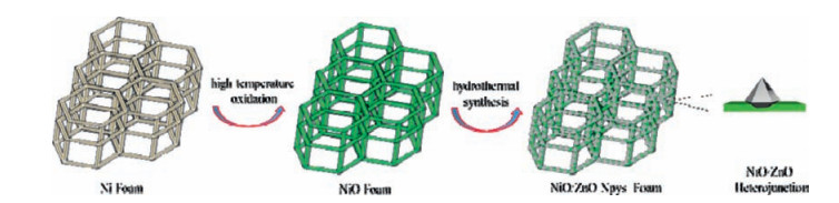
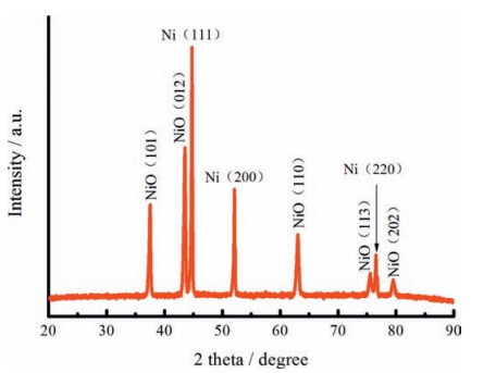
2. Results and discussion 2.1. Characterization of the prepared ZnO/NiO p-n junction foamThe schematic illustration for the fabrication of the ZnO/NiO p-n junction foam is shown in Fig. 1. Ni foam was first heat treated at high-temperature to form NiO foam with rough nanostructured surface. Then, ZnO NPys were in situ grown on the NiO foam under the hydrothermal condition to construct the p-n junction interfaces. Fig. 2 shows the XRD pattern of NiO/Ni foam. The diffraction peaks at 37.2°, 43.3°, 62.8°, 75.4° and 79.4° can be assigned to (1 0 1), (0 1 2), (11 0), (11 3) and (2 0 2) reflections, respectively. They are indexed to the face-centered cubic (JCPDS No. 47-1049) structure of NiO crystal. In addition, the three peaks locating at 2θ=44.5°, 51.8° and 76.3° assigned to (111), (2 0 0) and (2 2 0) planes reflection of the face-centered cubic (JCPDS No. 04-0850) structure of Ni crystal. Therefore, it was confirmed that the formation of crystalline NiO on the Ni substrate. Fig. 3a shows the SEM image of the prepared NiO foam, it is observed that a 3D interconnected framework with pores from 50 μm to 300 μm. Fig. 3b reveals that the surface of the NiO foam is rough with the widely distributed nano-wrinkles and nano-pores, which increases the specific surface area and facilitates the adsorption of reactants. The surface of the NiO foam is uniformly decorated with ZnO NPys after the hydrothermal growth (Fig. 3c-d). The ZnO NPys have regular shape and a stable incorporation with NiO wrinkles, ensuring the effective formation of the p-n junction interface [11]. The specific structure of the single ZnO NPy is revealed by the TEM images (Fig. 4). It is observed that the single ZnO NPy has a hexagonal base (~100 nm in diameter and~20 nm in height) and a pyramid (~150 nm in height) on the top of the hexagonal base. The high-resolution TEM image (Fig. 4b insert) of the ZnO NPys shows a lattice spacing of 0.28 nm, which corresponds to the distance between (100) planes. This special structure is determined to consist of six O-terminated {1011} facets and one O-terminated {0001} facet and exhibits excellent catalytic oxidation and sensing ability [24, 25], so it will help to enhance the catalytic oxidation of DA.

|
Download:
|
| Figure 1. Schematic illustration of the fabricating procedures of the ZnO NPys/NiO foam. | |

|
Download:
|
| Figure 2. XRD pattern of the NiO/Ni foam. | |

|
Download:
|
| Figure 3. The morphology and structure of the NiO foam and ZnO NPys/NiO foam. (a) The SEM image of the NiO foam. (b) Rough surface of the NiO foam. (c) The SEM image of the ZnO NPys/NiO foam. (d) The high-resolution image of the ZnO NPys and the structure diagram. | |

|
Download:
|
| Figure 4. TEM images of the single ZnO NPy. (a) The image of the side face. (b) The image of the bottom, insert is the high-resolution image. | |
2.2. Electrochemical characteristics of the prepared biosensor
The biosensor was characterized by cyclic voltammetry (CV) between the potentials of 0.1 V and 1.0 V in PBS (0.1 mol/L, pH 7.4) at the scan rate of 50 mV/s. Fig. 5a shows the CV curves of the 3D ZnO/NiO foam based electrode in the presence and absence of DA. It is observed that the addition of 5 mmol/L DA caused a significant increase in the oxidative peak current, indicating the 3D ZnO/NiO foam had fine electro-catalytic activity toward DA. To further investigate the electrochemical sensing performance, the amperometric measurements were carried out. As shown in Fig. 5b, the typical current-time is performed at 0.46 V that responses for the successive injection of DA into PBS step by step under stirring. It is observed that the biosensor exhibited rapid and sensitive response to the change of DA concentration. The current increased with the increase of DA concentration and achieved 95% of the steadystated current within 2 s. It indicates that the as-prepared biosensor can well catalyze the oxidation of DA. Fig. 5c shows the calibration curve of DA concentrations versus current, a well linear range from 10 to 100 mmol/L is found and the linear equation is as follows: I (μA)=171.35 C (mmol/L) + 0.4779 (R=0.9998). The sensitivity of the biosensor is 171.35μA/mmol/L, which is much higher than the measured sensitivity of the NiO foam (101.47 μA/mmol/L) (Fig. 5e-f) and other reported ZnO nanostructures (Table 1). The detection limit is as low as 3.15 μmol/L (based on the signal-to-noise of 3). This result may arise from several factors: (ⅰ) The 3D network and porous nanostructured surface possess a large surface area and are beneficial to the biomolecules' approach; (ⅱ) The NiO foam decorated with ZnO NPys exposes numerous active sites; (ⅲ) The formed amount of p-n junctions on the surface can accelerate electron transport. Hence, the results demonstrate the combined effect of ZnO and NiO results in the enhanced biosensing performance.

|
Download:
|
| Figure 5. (a) CVs of the ZnO NPys/NiO foam in 0.1 mol/L PBS (pH 7.4) in the presence and absence of 5 mmol/L DA. (b) Current-time curves of the ZnO NPys/NiO foam to DA by increasing the concentration gradually with a step of 0.01 mmol/L. (c) The calibration curve for DA concentrations versus current of the ZnO NPys/NiO foam. (d) The selectivity of the ZnO NPys/NiO foam to DA under 0.46 V by adding 0.01 mmol/L AA, DA, UA successively. (e) Current–time curves of the NiO foam to DA by increasing concentrations gradually. (f) The calibration curve for DA concentrations versus current of the NiO foam. | |
|
|
Table 1 Comparison of the analytical performance of various biosensors. |

In physiological conditions, the UA, AA and DA are coexisted. As it is well known the oxidation peak potentials of AA, UA and DA are generally overlapped, so it is hard to determine DA selectively. Interference tests were thus carried out (Fig. 5d), it is found that addition of AA and UA only caused the negligible current fluctuations. The results demonstrate the prepared biosensor has excellent selectivity to DA.
It is known that the potential barrier is formed at the p-n junction interface, which has important effects on the conductivity of the material. According to the previous report [11], when the positively charged molecules contact the p-n junction, the induced charges (Q > 0) will cause the height of the p-n junction interfacial potential barrier decrease, so the conductivity increases. As DA (pKa=8.8) is positively charged [33], but AA (pKa=4.1) and UA (pKa=3.7) are negatively charged [34] at pH 7.4, the introduction of the p-n junction interface will cause the enhanced response to DA but the depressed response to AA and UA, correspondingly. Therefore, the sensitivity and selectivity to DA was enhanced. In addition, the decorated ZnO NPys with particular crystal facets have strong catalytic ability, which further boosts the response to DA. The 3D porous architecture affords porous reactants diffusion channels and integrated electron transport pathway, so the response is fast. Therefore, introducing p-n junction interface into the 3D foam may be a new way to improve the biosensing performance.
The stability of the fabricated biosensor is measured by successively cycling the electrode, negligible change of the current response was found after 10 cycles. Further, the long-term stability was investigated by cycling the electrode every two days for two weeks. It is observed that 90% of the initial response was remained, suggesting the fine reproducibility and repeatability of the biosensors based on 3D ZnO/NiO foam.
3. ConclusionIn conclusion, the ZnO/NiO p-n junction foam was fabricated by decorating the ZnO NPys on NiO foam and used for the electrochemical detection of DA. The enhanced sensing performance was achieved compared with the single ZnO and NiO, including the high sensitivity (171.35μA/mmol/L), excellent selectivity and rapid response (2 s). These excellent results are attributed to the formation of the p-n junction, the interfacial potential barrier played an important role in enhancing the electrochemical response to DA. The fabrication of the 3D p-n junction based framework opens a new avenue to prepare the excellent electrochemical biosensors.
4. Experimental 4.1. Reagents and apparatusNi foam was purchased from alibaba website, DA was purchased from Alfa Aesar. L-AA was purchased from Tianjin Bodi Chemical Co., Ltd. Humiseal 948-06G silver conductive paint was manufactured by Loka chemicals laboratory. Other reagents were purchased from Sinopharm Chemical Reagent Co., Ltd. (Shanghai, China) and all of them were analytically pure. All solutions were prepared using double-distilled water.
All the electrochemical experiments were performed on a CHI660E Electrochemical Workstation (Shanghai Chenhua Instrument Corporation, China). The obtained sample was characterized by PHI-5702 X-ray powder diffraction (XRD). An S-4800 scanning electron microscope (SEM) and an FEI Tecnai G20 high-resolution transmission electron microscope (HRTEM) were used to determine the morphology, and structure of products.
4.2. Synthesis of NiO foamThe number of meshes in the Ni foam mesh substrates was 110. First, we cut out small pieces (~1 cm2) of Ni foam and cleaned it by sonication in acetone, ethanol and deionized (DI) water for 10 min each case, successively. Then, dried them with N2 flow and transferred into an open pipe furnace immediately (without using any gas flow), the furnace temperature was raised to 700 ℃ for 300 min, samples were kept at this temperature for 4 h. Finally, the samples were removed after the furnace was gradually cooled down without opening the furnace.
4.3. Synthesis of the ZnO/NiO p-n junction foamThe ZnO NPys were decorated on NiO foam using a hydrothermal synthesis. Primitively, 0.01 mol/L Zn (CH3COO)2 ·2H2O was dispersed in methanol, and then NiO foam was dipped in it and annealed at 130 ℃ to prepare the ZnO seeding layer. A mixed solution consisting of Zn (NO3)2 ·6H2O (0.05 mol/L), hexamethylenetetramine (HMTA, 0.05 mol/L), NH3·H2O (0.05 mol/L) and PEI (2 mmol/L) was prepared under stirring for 5 min. Then, the solution and the prepared ZnO-seeded/NiO foam were transferred to a Teflon-lined stainless steel autoclave with a volume of 100 mL. A hydrothermal treatment was conducted at 100 ℃ for 12 h. After that, the samples were removed after the autoclave cooled down to ambient temperature. Finally, the ZnO/NiO p-n junction foam was rinsed with deionized water and annealed in air at 450 ℃ for 1 h to remove any residual organics.
4.4. Electrochemical measurementsThe electrochemical sensing properties of the samples were evaluated with DA, AA and UA. Firstly, 0.3 μm and 0.05 μm sized alumina powder was used to polish the bare glassy carbon electrode (GCE, 3 mm in diameter), and the polished GCE was rinsed by ethanol and DI water. After the GCE was dried, a piece of the prepared ZnO/NiO p-n junction foam was cut and immobilized onto the surface of GCE by silver conductive paint. Then the electrode was cleaned and was dried at 4 ℃ overnight in a refrigerator. Three electrodes system was adopted for measurement, consisting of the as-prepared ZnO/NiO p-n junction foam modified electrode as working electrode, a Pt electrode as counter electrode, and an Ag/AgCl electrode as reference electrode.
AcknowledgmentsThis work was sponsored by Qingdao City Programs for Science and Technology Plan Projects (No.15-9-1-82-jch), National Natural Science Foundation of China (No. 51572249), Fundamental Research Funds for the Central University (No. 201513008), Natural Science Foundation of Shandong Province (No. ZR2014EMM021).
| [1] | H.Y. Yue, S. Huang, J. Chang, ZnO nanowire arrays on 3D hierachical graphenefoam:biomarkerdetectionofParkinson's disease. ACS Nano 8 (2014) 1639–1646. DOI:10.1021/nn405961p |
| [2] | L. Liu, J.M. Du, S.J. Li, Amplified voltammetric detection of dopamine using ferrocene-capped gold nanoparticle/streptavidin conjugates. Biosens. Bioelectron. 41 (2013) 730–735. DOI:10.1016/j.bios.2012.09.061 |
| [3] | L. Yang, D. Liu, J.S. Huang, T.Y. You, Simultaneous determination of dopamine, ascorbic acid and uric acid at electrochemically reduced graphene oxide modified electrode. Sens. Actuators B 193 (2014) 166–172. DOI:10.1016/j.snb.2013.11.104 |
| [4] | Y.Z. Zhou, L.J. Zhang, S.L. Chen, S.Y. Dong, X.H. Zheng, Electroanalysis and simultaneous determination of dopamine and epinephrine at poly (isonicotinic acid)-modified carbon paste electrode in the presence of ascorbic acid. Chin. Chem. Lett. 20 (2009) 217–220. DOI:10.1016/j.cclet.2008.10.026 |
| [5] | H.W. Lin, H.B. Liu, X.M. Qian, Synthesizing axial inserting p-n heterojunction nanowire arrays for realizing synergistic performance. Inorg. Chem. 52 (2013) 6969–6974. DOI:10.1021/ic400302e |
| [6] | Y.L. Liu, G.Z. Li, R.D. Mi, C.K. Deng, P.Z. Gao, An environment-benign method for the synthesis of p-NiO/n-ZnO heterostructure with excellent performance for gas sensing and photocatalysis. Sens. Actuators B 191 (2014) 537–544. DOI:10.1016/j.snb.2013.10.068 |
| [7] | Y.J. Chen, L. Yu, D.D. Feng, Superior ethanol-sensing properties based on Ni-doped SnO2 p-n heterojunction hollow spheres. Sens. Actuators B 166- 167 (2012) 61–67. |
| [8] | H.T. Wang, H.T. Yuan, S.S. Hong, Y.B. Li, Y. Cui, Physical and chemical tuning of two-dimensional transition metal dichalcogenides. Chem. Soc. Rev. 44 (2015) 2664–2680. DOI:10.1039/C4CS00287C |
| [9] | X.Y. Ren, L.H. Lu, Luminescent nanoscale metal-organic frameworks for chemical sensing. Chin. Chem. Lett. 26 (2015) 1439–1445. DOI:10.1016/j.cclet.2015.10.014 |
| [10] | B.Z. Tian, T. Cohen-Karni, Q. Qing, Three-dimensional, flexible nanoscale field-effect transistors as localized bioprobes. Science 329 (2010) 830–834. DOI:10.1126/science.1192033 |
| [11] | M.G. Zhao, B. Cai, Y. Ma, Introducing heterojunction barriers into single kinked nanowires for the probe-free detection of proteins and intracellular recording. Nanoscale 6 (2014) 4052–4057. DOI:10.1039/c3nr06159k |
| [12] | P.H. Yeh, Z. Li, Z.L. Wang, Schottky-gated probe-free ZnO nanowire biosensor. Adv. Mater. 21 (2009) 4975–4978. DOI:10.1002/adma.v21:48 |
| [13] | B. Pierozynski, T. Mikolajczyk, M. Turemko, On the temperature performance of ethanol oxidation reaction at palladium-activated nickel foam. Electrocatalysis 6 (2015) 173–178. DOI:10.1007/s12678-014-0231-0 |
| [14] | G.W. Yang, C.L. Xu, H.L. Li, Electrodeposited nickel hydroxide on nickel foam with ultrahigh capacitance. Chem. Commun. (2008) 6537–6539. |
| [15] | Y.L. Wang, Y.Q. Zhao, C.L. Xu, Improved performance of Pd electrocatalyst supported on three-dimensional nickel foam for direct ethanol fuel cells. J. Power Sources 195 (2010) 6496–6499. DOI:10.1016/j.jpowsour.2010.04.025 |
| [16] | L.N. Jin, Q. Liu, W.Y. Sun, Room temperature solution-phase synthesis of flower-like nanostructures of[Ni3(BTC)2·12H2O] and their conversion to porous NiO. Chin. Chem. Lett. 24 (2013) 663–667. DOI:10.1016/j.cclet.2013.05.001 |
| [17] | A. Ohtomo, M. Kawasaki, Y. Sakurai, Room temperature ultraviolet laser emission from ZnO nanocrystal thin films grown by laser MBE. Mater. Sci. Eng. B 54 (1998) 24–28. DOI:10.1016/S0921-5107(98)00120-2 |
| [18] | Ü. Özgür, Y.I. Alivov, C. Liu, A comprehensive review of ZnO materials and devices. J. Appl. Phys. 98 (2005) 041301. DOI:10.1063/1.1992666 |
| [19] | G.Q. Wan, D.X. Li, C.F. Li, J. Xu, W.G. Hou, From Zn-Al layered double hydroxide to ZnO nanostructure:gradually etching by sodium hydroxide. Chin. Chem. Lett. 23 (2012) 1415–1418. DOI:10.1016/j.cclet.2012.10.020 |
| [20] | Y.M. Dong, G.L. Wang, P.P. Jiang, Simple preparation and catalytic properties of ZnO for ozonation degradation of phenol in water. Chin. Chem. Lett. 22 (2011) 209–212. DOI:10.1016/j.cclet.2010.10.010 |
| [21] | Y.R. Li, C.Y. Wan, C.T. Chang, Thickness effect of NiO on the performance of ultraviolet sensors with p-NiO/n-ZnO nanowire heterojunction structure. Vacuum 118 (2015) 48–54. DOI:10.1016/j.vacuum.2015.01.018 |
| [22] | L. Vayssieres, Growth of arrayed nanorods and nanowires of ZnO from aqueous solutions. Adv. Mater. 15 (2003) 464–466. DOI:10.1002/adma.200390108 |
| [23] | G.W. She, X.H. Zhang, W.S. Shi, Controlled synthesis of oriented singlecrystal ZnO nanotube arrays on transparent conductive substrates. Appl. Phys. Lett. 92 (2008) –053111. |
| [24] | Z.H. Wang, J. Xue, D.M. Han, F.B. Gu, Controllable defect redistribution of ZnO nanopyramids with exposed facets for enhanced1011 gas sensing performance. ACS Appl. Mater. Interfaces 7 (2015) 308–317. DOI:10.1021/am506206c |
| [25] | P. Li, Z. Wei, T. Wu, Q. Peng, Y.D. Li, Au-ZnO hybrid nanopyramids and their photocatalytic properties. J. Am. Chem. Soc. 133 (2011) 5660–5663. DOI:10.1021/ja111102u |
| [26] | J. Chang, R. Ahmed, H.X. Wang, ZnO nanocones with high-index facets for enhanced energy conversion efficiency of dye-sensitized solar cells. J. Phys. Chem. C 117 (2013) 13836–13844. DOI:10.1021/jp402742n |
| [27] | C. Xia, N. Wang, L. Wang, Optical and electro-catalytic properties of bundled ZnO nanowires grown on a ITO substrate. J. Nanopart. Res. 12 (2010) 1869–1875. DOI:10.1007/s11051-009-9748-1 |
| [28] | X.X. Dong, Y.X. Liu, Y.M. Sun, C. Yang, Z.L. Xu, In situ growth ofmicroporous ZnO nanorods on ITO for dopamine oxidization. Mater. Lett. 162 (2016) 246–249. DOI:10.1016/j.matlet.2015.10.021 |
| [29] | J.P. Wu, F. Yin, Studies on the electrocatalytic oxidation of dopamine at phosphotungstic acid-ZnO spun fiber-modified electrode. Sens. Actuators B 185 (2013) 651–657. DOI:10.1016/j.snb.2013.05.052 |
| [30] | S. Reddy, B.E.K. Swamy, H.N. Vasan, H. Jayadevappa, ZnO and ZnO/polyglycine modified carbon paste electrode for electrochemical investigation of dopamine. Anal. Methods 4 (2012) 2778–2783. DOI:10.1039/c2ay25203a |
| [31] | K. Pandiselvi, S. Thambidurai, Chitosan-ZnO/polyanilne nanocomposite modified glassy carbon electrode for selective detection of dopamine. Int. J. Biol. Macromol. 67 (2014) 270–278. DOI:10.1016/j.ijbiomac.2014.03.028 |
| [32] | L.X. Fang, K.J. Huang, B.L. Zhang, Nanosheet-based 3D hierarchical ZnO structure decorated with Au nanoparticles for enhanced electrochemical detection of dopamine. RSC Adv. 4 (2014) 48986–48993. DOI:10.1039/C4RA06090C |
| [33] | Y. Bao, J.X. Song, Y. Mao, Graphene oxide-templated polyaniline microsheets toward simultaneous electrochemical determination of AA/DA/UA. Electroanalysis 23 (2011) 878–884. DOI:10.1002/elan.201000607 |
| [34] | P. Manivel, M. Dhakshnamoorthy, A. Balamurugan, Conducting polyaniline-graphene oxide fibrous nanocomposites:preparation, characterization and simultaneous electrochemical detection of ascorbic acid, dopamine and uric acid. RSC Adv. 3 (2013) 14428–14437. DOI:10.1039/c3ra42322k |