 2016, Vol. 27


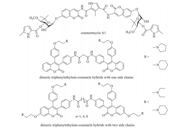
Coumarins, also known as benzopyran-2-ones, are a family of naturally occurring lactones and have attracted intense interest in recent years because of their diverse pharmacological activities such as anti-cancer, anti-HIV, antibacterial, and anti-inflammatory [1-7]. As the anti-tumor agents, they can act on the cancer formation [8-11] by blocking cell cycle [12], inducing cell apoptosis [13-15], modulating estrogen receptor (ER) [16], or inhibiting the DNA-associated enzymes, such as topoisomerase or gryase [17, 18]. Due to their potential applications in cancer therapy, extensive studies have been carried out on the design and synthesis of coumarin derivatives with improved anticancer activity [19-26]. Among them, oligomerization (di/tri) of coumarin is one of the effective ways [27, 28]. For example, coumermycin A1 (IC50~70 μmol/L, Fig. 1), the dimeric natural product, which was shown to be more effective than the monomeric species (IC50~700 μmol/L) [29, 30]. Recently, the concept of molecular oligomerization led us to discover two novel series of dimeric derivatives of triphenylethylene-coumarin hybrid [31, 32] (Fig. 1). The dimeric compounds had potent anti-tumor activity possibly by acting on DNA via the intercalative mode, and higher than their corresponding monomeric compounds [33, 34], respectively. The positive results inspired our interests to explore the trimeric variants of the triphenylethylene-coumarin hybrid in an effort to produce more efficient anti-tumor agents. Herein we would like to report the synthesis of the novel trimeric triphenylethylene-coumarin hybrids with two side chains. Such trimers were preliminarily evaluated for their anti-proliferatic activity against tumor cells, and their interactions with Ct-DNA by UV-vis, fluorescence (lifetime) and thermal denaturation experiment.

|
Download:
|
| Figure 1. Coumermycin A1 and the diemric triphenylethylene-coumarin hydrids. | |
2. Experimental
Melting points were measured in an open capillary on a SGW X-4 melting point apparatus and are uncorrected. Element analysis was performed using a Heraeus (CHNO, rapid) elemental analyzer. 1H NMR and 13C NMR spectra were measured on a RT-NMR Bruker AVANCE 600M, NMR spectrometer using tetramethylsilane (Me4Si) as an internal standard. ESI-MS was determined using an Agilent G6300 ion trap mass spectrometer, and signals were recorded in m/z. High resolution mass spectra (HRMS) were carried out on a FTICR-MS (Ionspec 7.0 T) mass spectrometer with electrospray ionization (ESI) and MALTI-TOF mass spectrometer. The optical densities for examining the anti-proliferative activity were measured on a BioRad Model 3550 microplate spectrophotometer. Absorption spectra were recorded on a Shimadzu UV-3600 spectrophotometer. Emission spectra were recorded on a Hitachi F-7000 fluorescence spectrophotometer. The fluorescence lifetime was measured on a Fluorescence Lifetime and Steady State Spectroscopy FLS 920. The silica gel (300-400 mesh) for flash column chromatography was from Qingdao Marine Chemical (China). Ct-DNA was purchased from Sigma. Other reagents were from commercial sources.
2.1. General procedure for the synthesis of compound 3To a solution of 2a [32, 34] (0.5 mmol) in dry DCM (5.0 mL) was added Boc-6-aminohexanoic acid (0.8 mmol) and 2-(7-aza-1Hbenzotriazole-1-yl)-1, 1, 3, 3-tetramethyluronium hexafluorophosphate (HATU, 0.8 mmol). After all the solids were dissolved in the solution by stirring at room temperature, N-ethyldiisopropylamine (DIPEA, 0.8 mmol) was added by stirring until the completion of reaction (monitored by TLC). The mixture was neutralized with 0.1 mol/L HCl solution, and the residue was extracted with EtOAc (15 mL × 2). The organic layer was then sequentially washed with water, satd. NaCl solution and dried over anhydrous Na2SO4, evaporated to dryness under reduced pressure. Finally the residue was purified by column chromatography of silica gel (DCM-MeOH-NH3·H2O=100:2:0.1) to afford the product 3a in good yield. Under the same conditions, compounds 3(b-d) were obtained.
tert-Butyl(6-oxo-6-((4-(2-oxo-7-(2-(piperidin-1-yl)ethoxy)-4-(4-(2-(piperidin-1-yl)ethoxy)phenyl)-2H-chromen-3-yl)phenyl) amino)hexyl)carbamate (3a): Pale yellow solid, yield 83%, mp 48-51 ℃; 1H NMR (600 MHz, CD3OD): δ 7.39 (d, 2H, J=8.4 Hz, Ar-H), 7.13 (d, 1H, J=9.0 Hz, Ar-H), 7.04-7.02 (m, 4H, Ar-H), 6.95 (d, 1H, J=2.4 Hz, Ar-H), 6.90 (d, 2H, J=2.4 Hz, Ar-H), 6.85 (dd, 1H, J=9.0 Hz, Ar-H), 4.25 (t, 2H, J=5.4 Hz, CH2), 4.18 (t, 2H, J=5.4 Hz, CH2), 3.03-2.99 (m, 6H, CH2), 2.81-2.76 (m, 8H, CH2), 2.33 (t, 2H, J=7.8 Hz, CH2), 1.72-1.64 (m, 10H, CH2), 1.54 (t, 4H, J=3.6 Hz, CH2), 1.50-1.46 (m, 2H, CH2), 1.42 (s, 9H, CH3), 1.38-1.35 (m, 2H, CH2); 13C NMR (150 MHz, CD3OD): δ 173.1, 162.2, 161.3, 158.3, 157.2, 154.5, 152.2, 137.7, 131.1, 130.8, 129.9, 128.7, 127.4, 123.1, 118.6, 114.4, 114.1, 112.4, 101.1, 78.5, 64.4, 63.4, 56.6, 54.2, 54.1, 53.4, 39.8, 36.5, 29.3, 27.4, 26.1, 25.2, 24.2, 23.9, 22.6, 22.4; MS(ESI), m/z 781 ([M+H]+); HRMS(ESI): Calcd. for C46H61N4O7 ([M+H]+), 781.4540, found 781.4546.
tert-Butyl(6-((4-(7-(2-morpholinoethoxy)-4-(4-(2-morpholinoethoxy)phenyl)-2-oxo-2H-chromen-3-yl)phenyl)amino)-6-oxohexyl)carbamate (3b): Pale yellow solid, yield 81%, mp 51-54 ℃; 1H NMR (600 MHz, CDCl3): δ 8.13 (s, 1H, NH), 7.45 (d, 2H, J=9.0 Hz, Ar-H), 7.16 (d, 1H, J=9.0 Hz, Ar-H), 7.03 (t, 4H, J=7.8 Hz, Ar-H), 6.90 (d, 1H, J=2.4 Hz, Ar-H), 6.83 (d, 2H, J=8.4 Hz, Ar-H), 6.78 (d, 1H, J=9.0 Hz, Ar-H), 4.25 (s, 4H, CH2), 3.87-3.81 (m, 8H, CH2), 3.04 (s, 2H, CH2), 2.94-2.85 (m, 8H, CH2), 2.71 (brs, 4H, CH2), 2.36 (t, 2H, J=7.2 Hz, CH2), 1.74-1.69 (m, 2H, CH2), 1.54-1.49 (m, 2H, CH2), 1.44 (s, 9H, CH3), 1.37-1.34 (m, 2H, CH2); 13C NMR (150 MHz, CDCl3): δ 161.8, 156.1, 154.7, 137.7, 131.3, 130.8, 129.9, 129.6, 128.9, 123.7, 123.4, 119.0, 114.9, 114.5, 112.5, 101.4, 77.2, 57.2, 57.2, 53.9, 53.6, 45.8, 38.6, 37.3, 29.7, 28.4, 26.3, 25.1; MS(ESI), m/z 785 ([M+H]+); HRMS(ESI): Calcd. for C44H57N4O9([M+H]+), 785.4125, found: 785.4133.
tert-Butyl(6-oxo-6-((4-(2-oxo-7-(2-(pyrrolidin-1-yl)ethoxy)-4-(4-(2-(pyrrolidin-1-yl)ethoxy)phenyl)-2H-chromen-3-yl)phenyl) amino)hexyl)carbamate (3c): Pale yellow solid, yield 79%, mp 57-59 ℃; 1H NMR (600 MHz, CD3OD): δ 7.42 (d, 2H, J=8.4 Hz, ArH), 7.20 (d, 1H, J=9.0 Hz, Ar-H), 7.11-7.06 (m, 4H, Ar-H), 7.04 (d, 1H, J=2.4 Hz, Ar-H), 6.94 (d, 2H, J=9.0 Hz, Ar-H), 6.91 (d, 1H, J=9.0 Hz, Ar-H), 4.27 (t, 2H, J=5.4 Hz, CH2), 4.16 (t, 2H, J=5.4 Hz, CH2), 3.06-3.02 (m, 6H, CH2), 2.80-2.78 (m, 8H, CH2), 2.36 (t, 2H, J=7.8 Hz, CH2), 1.91-1.88 (m, 8H, CH2), 1.71-1.67 (m, 2H, CH2), 1.53-1.49 (m, 2H, CH2), 1.44 (s, 9H, CH3), 1.39-1.36(m, 2H, CH2); 13C NMR (150 MHz, CD3OD): δ 173.1, 162.2, 162.7, 158.7, 154.6, 152.3, 137.7, 131.1, 130.7, 129.9, 128.7, 127.1, 123.0, 118.6, 114.3, 114.0, 112.4, 100.9, 78.4, 66.7, 65.8, 54.4, 54.3, 54.2, 39.8, 36.5, 29.3, 27.4, 26.1, 25.2, 22.8, 22.8; MS(ESI), m/z 753 ([M+H]+); HRMS(ESI): Calcd. for C44H57N4O7 ([M+H]+), 753.4227, found: 753.4241.
tert-Butyl(6-((4-(7-(2-(diethylamino)ethoxy)-4-(4-(2-(diethylamino)ethoxy)phenyl)-2-oxo-2H-chromen-3-yl)phenyl)amino)-6-oxohexyl)carbamate (3d): Pale yellow solid, yield 85%, mp 60-62 ℃; 1H NMR (600 MHz, CDCl3): δ 7.77 (s, 1H, NH), 7.41 (d, 2H, J=8.4 Hz, Ar-H), 7.17 (d, 1H, J=9.0 Hz, Ar-H), 7.05-7.01 (m, 4H, ArH), 6.91 (d, 1H, J=2.4 Hz, Ar-H), 6.83 (d, 2H, J=9.0 Hz, Ar-H), 6.78 (d, 1H, J=9.0 Hz, Ar-H), 4.18-4.16 (m, 4H, CH2), 3.12 (d, 2H, J=6.0 Hz, CH2), 3.04 (t, 2H, J=6.0 Hz, CH2), 2.97 (t, 2H, J=5.4 Hz, CH2), 2.82 (d, 4H, J=5.4 Hz, CH2), 2.71 (t, 4H, J=8.4 Hz, CH2), 2.35 (t, 2H, J=7.2 Hz, CH2), 1.73-1.70 (m, 2H, CH2), 1.54-1.50 (m, 2H, CH2), 1.45 (s, 9H, CH3), 1.35-1.32 (m, 2H, CH2), 1.18 (t, 6H, J=7.2 Hz, CH3), 1.13 (t, 6H, J=7.2 Hz, CH3); 13C NMR (150 MHz, CDCl3): δ 161.9, 161.5, 156.1, 154.7, 151.5, 131.4, 130.8, 128.8, 118.9, 114.4, 112.5, 101.3, 77.2, 51.5, 47.9, 47.8, 45.9, 37.4, 29.7, 28.4, 26.3, 25.1, 11.5, 11.0; MS(ESI), m/z 757 ([M+H]+); HRMS(ESI): Calcd. for C44H61N61O7 ([M+H]+), 757.4540, found: 757.4529.
2.2. General procedure for the synthesis of compound 4Compound 3a (0.3 mmol) was dissolved in 90% CF3COOH/H2O (2.0 mL), then the solution was stirred at room temperature until the completion of reaction (monitored by TLC). CF3COOH was removed under reduced pressure. The residue was dissolved in DCM and was purified by column chromatography of silica gel (DCM-MeOH-NH3·H2O=50:2:0.5) to afford the product 4a in good yield. Under the same conditions, compounds 4(b-d) were obtained.
6-Amino-N-(4-(2-oxo-7-(2-(piperidin-1-yl)ethoxy)-4-(4-(2-(piperidin-1-yl)ethoxy)phenyl)-2H-chromen-3-yl)phenyl)hexanamide (4a): Pale yellow solid, yield 90%, mp 73-76 ℃; 1H NMR (600 MHz, CDCl3): δ 7.42 (d, 2H, J=9.6 Hz, Ar-H), 7.18 (d, 1H, J=9.0 Hz, Ar-H), 7.07 (d, 4H, J=8.4 Hz, Ar-H), 7.00 (d, 1H, J=2.4 Hz, Ar-H), 6.92 (d, 2H, J=8.4 Hz, Ar-H), 6.88 (d, 1H, J=9.0 Hz, Ar-H), 4.29 (t, 2H, J=5.4 Hz, CH2), 4.16 (t, 2H, J=5.4 Hz, CH2), 2.95-2.89 (m, 6H, CH2), 2.68 (brs, 8H, CH2), 2.39 (d, 2H, J=9.0 Hz, CH2), 1.76-1.66 (m, 12H, CH2), 1.53 (brs, 4H, CH2), 1.49-1.43 (m, 2H, CH2); 13C NMR (150 MHz, CD3OD): δ 172.7, 162.1, 161.6, 161.5, 158.6, 154.5, 152.3, 137.6, 131.1, 130.7, 128.7, 127.1, 122.9, 118.6, 114.2, 114.0, 112.4, 100.9, 65.4, 64.5, 57.2, 54.4, 39.2, 36.1, 27.1, 25.6, 24.8, 24.6, 23.3, 23.2; MS(ESI), m/z 681 ([M+H]+), 703 ([M+Na]+); HRMS(ESI): Calcd. for C41H52N4O5Na ([M+Na]+), 703.3835, found: 703.3842.
6-Amino-N-(4-(7-(2-morpholinoethoxy)-4-(4-(2-morpholinoethoxy)phenyl)-2-oxo-2H-chromen-3-yl)phenyl)hexanamide (4b): Pale yellow solid, yield 85%, mp 74-76 ℃; 1H NMR (600 MHz, CD3OD): δ 7.45 (d, 2H, J=8.4 Hz, Ar-H), 7.15 (d, 1H, J=8.4 Hz, ArH), 7.05-7.04 (m, 4H, Ar-H), 6.98 (d, 1H, J=2.4 Hz, Ar-H), 6.92-6.90 (m, 2H, Ar-H), 6.85 (d, 1H, J=9.0 Hz, Ar-H), 4.24 (t, 2H, J=5.4 Hz, CH2), 4.15 (t, 2H, J=5.4 Hz, CH2), 3.73 (t, 8H, J=4.8 Hz, CH2), 2.95 (t, 2H, J=7.8 Hz, CH2), 2.86-2.82 (m, 4H, CH2), 2.62 (d, 8H, J=4.2 Hz, CH2), 2.4 (t, 2H, J=7.8 Hz, CH2), 1.75-1.70 (m, 4H, CH2), 1.49-1.44 (m, 2H, CH2); 13C NMR (150 MHz, CD3OD): δ 172.8, 162.1, 161.7, 158.7, 154.6, 152.2, 137.8, 131.1, 130.7, 129.9, 128.7, 127.1, 122.9, 118.6, 114.2, 114.1, 112.4, 101.0, 66.2, 66.1, 65.8, 65.0, 57.2, 57.0, 52.2, 39.3, 36.2, 26.8, 25.5, 24.7; MS(ESI), m/z 685 ([M+H]+), 707 ([M+Na]+); HRMS(ESI): Calcd. for C39H48N48O7Na ([M+Na]+), 707.3420, found: 707.3418.
6-Amino-N-(4-(2-oxo-7-(2-(pyrrolidin-1-yl)ethoxy)-4-(4-(2-(pyrrolidin-1-yl)ethoxy)phenyl)-2H-chromen-3-yl)phenyl) hexanamide (4c): Pale yellow solid, yield 91%, mp 77-79 ℃; 1H NMR (600 MHz, CD3OD): δ 7.33 (d, 2H, J=9.0 Hz, Ar-H), 7.06 (d, 1H, J=9.0 Hz, Ar-H), 6.96 (d, 4H, J=9.0 Hz, Ar-H), 6.86 (d, 1H, J=2.4 Hz, Ar-H), 6.81 (d, 2H, J=8.4 Hz, Ar-H), 6.76 (d, 1H, J=9.0 Hz, Ar-H), 4.10 (t, 2H, J=6.6 Hz, CH2), 4.02 (t, 2H, J=4.8 Hz, CH2), 2.85 (t, 2H, J=6.0 Hz, CH2), 2.82 (t, 2H, J=6.0 Hz, CH2), 2.72-2.69 (m, 2H, CH2), 2.59-2.57 (m, 8H, CH2), 2.28 (t, 2H, J=7.8 Hz, CH2), 1.78-1.74 (m, 8H, CH2), 1.62 (brs, 2H, CH2), 1.50 (brs, 2H, CH2), 1.28 (s, 2H, CH3); 13C NMR (150 MHz, CDCl3): δ 172.3, 162.3, 161.7, 158.8, 154.7, 152.0, 138.1, 131.3, 130.8, 129.5, 129.0, 126.8, 123.1, 119.0, 114.5, 114.4, 112.8, 101.3, 67.8, 67.0, 55.1, 54.9, 54.8, 54.8, 40.1, 36.8, 28.8, 25.8, 25.0, 23.6, 23.5; MS(ESI), m/z 653 ([M+H]+), 675 ([M+Na]+); HRMS(ESI): Calcd. for C39H48N48O5Na ([M+Na]+), 675.3522, found: 675.3526.
6-Amino-N-(4-(7-(2-(diethylamino)ethoxy)-4-(4-(2-(diethylamino)ethoxy)phenyl)-2-oxo-2H-chromen-3-yl)phenyl)hexanamide (4d): Pale yellow solid, yield 87%, mp 82-84 ℃; 1H NMR (600 MHz, CD3OD): δ 7.42 (d, 2H, J=8.4 Hz, Ar-H), 7.17 (d, 1H, J=9.0 Hz, Ar-H), 7.07-7.05 (m, 4H, Ar-H), 6.98 (d, 1H, J=2.4 Hz, ArH), 6.90 (d, 2H, J=3.0 Hz, Ar-H), 6.85 (d, 1H, J=9.0 Hz, Ar-H), 4.19 (t, 2H, J=5.4 Hz, CH2), 4.08 (t, 2H, J=5.4 Hz, CH2), 2.94 (t, 2H, J=5.4 Hz, CH2), 2.90 (t, 2H, J=5.4 Hz, CH2), 2.75 (t, 2H, J=7.2 Hz, CH2), 2.71-2.65 (m, 8H, CH2), 2.37 (t, 2H, J=7.8 Hz, CH2), 1.73-1.68 (m, 2H, CH2), 1.61-1.56 (m, 2H, CH2), 1.45-1.40 (m, 2H, CH2), 1.14-1.07 (m, 12H, CH3); 13C NMR (150 MHz, CD3OD): δ 172.9, 162.2, 161.8, 158.8, 154.6, 152.3, 137.7, 131.2, 130.7, 130.0, 128.7, 126.9, 122.8, 118.5, 114.1, 114.0, 100.9, 66.4, 65.6, 51.1, 51.0, 40.4, 36.4, 30.4, 26.0, 25.1, 10.2, 10.1; MS(ESI), m/z 657 ([M+H]+), 679 ([M+Na]+); HRMS(ESI): Calcd. for C39H52N52O5Na ([M+Na]+), 679.3835, found: 679.3846.
2.3. General procedure for the synthesis of compounds 5 and 6To a solution of 4a (0.3 mmol) in dry DMF (3.0 mL) was added trimesic acid (0.1 mmol) and HATU (0.4 mmol). After all the solids were dissolved in the solution by stirring at room temperature for 15 min, DIPEA (0.5 mmol) was added by stirring until the completion of reaction (monitored by TLC). The mixture was neutralized with 0.1 mol/L HCl solution, and the residue was extracted with DCM (10 mL × 2). The organic layer was then sequentially washed with water, satd. NaCl solution, and dried over anhydrous Na2SO4, evaporated to dryness under reduced pressure. Finally the residue was purified by column chromatography of silica gel (DCM-MeOH-NH3·H2O=80:2:0.5) to afford the product 5a. Under the same conditions, compounds 5(b-d) and 6(a-d) were obtained.
N1, N3, N5-Tris(4-(2-oxo-7-(2-(piperidin-1-yl)ethoxy)-4-(4-(2-(piperidin-1-yl)ethoxy)phenyl)-2H-chromen-3-yl)phenyl)benzene-1, 3, 5-tricarboxamide (5a): Light yellow solid, yield 45%, mp 142-145 ℃; 1H NMR (600 MHz, CDCl3): δ 9.83 (s, 1H, NH), 8.43 (s, 1H, Ar-H), 7.62 (s, 2H, Ar-H), 7.12 (d, 1H, J=7.8 Hz, Ar-H), 7.02 (s, 4H, Ar-H), 6.84-6.73 (m, 4H, Ar-H), 4.18-4.12 (m, 4H, CH2), 2.96-2.10 (m, 4H, CH2), 2.67-2.59 (m, 8H, CH2), 1.64-1.61 (m, 8H, CH2), 1.47-1.42 (m, 4H, CH2); 13C NMR (150 MHz, CDCl3): δ 164.5, 162.2, 161.7, 158.8, 154.7, 152.0, 137.4, 135.4, 131.2, 130.7, 130.2, 129.2, 128.9, 126.8, 123.1, 119.5, 114.5, 114.4, 112.7, 101.3, 66.7, 65.8, 57.9, 57.6, 55.1, 55.0, 25.9, 25.8, 24.2, 24.1; MS(MALDI-TOF), m/z 1858 ([M+H]+).
N1, N3, N5-Tris(4-(7-(2-morpholinoethoxy)-4-(4-(2-morpholinoethoxy)phenyl)-2-oxo-2H-chromen-3-yl)phenyl)benzene-1, 3, 5-tricarboxamide (5b): Light yellow solid, yield 42%, mp 147-149 ℃; 1H NMR (600 MHz, CDCl3): δ 9.59 (s, 1H, NH), 8.36 (s, 1H, Ar-H), 7.62 (d, 2H, J=7.8 Hz, Ar-H), 7.01 (d, 2H, J=7.8 Hz, Ar-H), 6.91 (s, 1H, Ar-H), 6.84 (d, 2H, J=8.4 Hz, Ar-H), 6.78 (d, 1H, J=9.6 Hz, Ar-H), 4.20 (t, 2H, J=4.8 Hz, CH2), 4.09 (t, 2H, J=4.8 Hz, CH2), 3.75-3.74 (m, 4H, CH2), 3.68-3.66 (m, 4H, CH2), 2.85 (t, 4H, J=5.4 Hz, CH2), 2.76 (t, 4H, J=5.4 Hz, CH2), 2.59 (brs, 4H, CH2), 2.53 (brs, 4H, CH2); 13C NMR (150 MHz, CDCl3): δ 164.8, 162.1, 161.6, 158.7, 154.7, 151.9, 137.7, 135.2, 131.1, 130.7, 130.3, 130.2, 129.6, 129.0, 126.8, 123.1, 119.7, 114.5, 112.7, 101.2, 66.9, 66.5, 65.7, 57.6, 57.3, 54.1, 54.1; MS(MALDI-TOF), m/z 1870 ([M+H]+).
N1, N3, N5-Tris(4-(2-oxo-7-(2-(pyrrolidin-1-yl)ethoxy)-4-(4-(2-(pyrrolidin-1-yl)ethoxy)phenyl)-2H-chromen-3-yl)phenyl) benzene-1, 3, 5-tricarboxamide (5c): Light yellow solid, yield 39%, mp 153-156 ℃; 1H NMR (600 MHz, CDCl3): δ 9.26 (s, 1H, NH), 8.30 (s, 1H, Ar-H), 7.16 (d, 1H, J=9.0 Hz, Ar-H), 6.99 (d, 4H, J=8.4 Hz, ArH), 6.88 (d, 1H, J=3.6 Hz, Ar-H), 6.83 (s, 2H, J=7.8 Hz, Ar-H), 6.78 (d, 1H, J=2.4 Hz, Ar-H), 4.17 (t, 2H, J=4.8 Hz, CH2), 4.08 (s, 2H, CH2), 2.93 (t, 2H, J=5.4 Hz, CH2), 2.85 (t, 2H, J=5.4 Hz, CH2), 2.63 (brs, 4H, CH2), 2.53 (brs, 4H, CH2), 1.81 (brs, 4H, CH2), 1.70 (brs, 4H, CH2); 13C NMR (150 MHz, CDCl3): δ 164.5, 162.2, 161.7, 158.8, 154.7, 152.1, 137.4, 135.5, 131.2, 130.7, 130.2, 129.2, 128.9, 126.8, 123.1, 119.6, 114.5, 114.4, 112.7, 101.3, 67.8, 67.0, 55.0, 54.8, 54.7, 54.7, 53.4, 23.56, 23.5; MS(MALDI-TOF), m/z 1775 ([M+H]+).
N1, N3, N5-Tris(4-(7-(2-(diethylamino)ethoxy)-4-(4-(2-(diethylamino)ethoxy)phenyl)-2-oxo-2H-chromen-3-yl)phenyl)benzene-1, 3, 5-tricarboxamide (5d): Light yellow solid, yield 40%, mp 159-161 ℃; 1H NMR (600 MHz, CDCl3): δ 10.27 (s, 1H, NH), 8.70 (s, 1H, Ar-H), 7.82 (d, 2H, J=8.4 Hz, Ar-H), 7.15 (d, 1H, J=6.0 Hz, ArH), 7.04-7.00 (m, 4H, Ar-H), 6.88 (s, 1H, Ar-H), 6.82 (d, 2H, J=7.8 Hz, Ar-H), 6.76 (d, 1H, J=9.0 Hz, Ar-H), 4.15 (d, 4H, J=5.4 Hz, CH2), 3.32-3.28 (m, 4H, CH2), 2.79 (d, 4H, J=7.2 Hz, CH2), 2.70 (d, 4H, J=7.2 Hz, CH2), 1.22 (t, 4H, J=7.2 Hz, CH2), 1.14-1.10 (m, 12H, CH3); 13C NMR (150 MHz, CDCl3): δ 164.8, 162.1, 161.6, 158.6, 154.7, 151.8, 137.9, 135.2, 135.2, 131.3, 131.1, 130.8, 130.1, 129.6, 128.9, 126.8, 123.2, 119.6, 119.5, 114.5, 114.4, 112.6, 101.2, 67.2, 65.9, 54.8, 54.7, 52.8, 51.5, 51.5, 47.9, 47.8, 11.8, 11.5; MS(MALDI-TOF), m/z 1787 ([M+H]+).
N1, N3, N5-Tris(6-oxo-6-((4-(2-oxo-7-(2-(piperidin-1-yl)ethoxy)-4-(4-(2-(piperidin-1-yl)ethoxy)phenyl)-2H-chromen-3-yl)phenyl) amino)hexyl)benzene-1, 3, 5-tricarboxamide (6a): Pale yellow solid, yield 41%, mp 115-119 ℃; 1H NMR (600 MHz, CD3OD): δ 9.95 (s, 1H, NH), 8.80 (s, 1H, Ar-H), 8.64 (s, 1H, Ar-H), 7.60 (d, 2H, J=7.2 Hz, Ar-H), 7.12 (d, 1H, J=9.0 Hz, Ar-H), 6.97-6.94 (m, 4H, Ar-H), 6.86 (s, 1H, Ar-H), 6.79 (d, 2H, J=8.4 Hz, Ar-H), 6.75 (d, 1H, J=10.8 Hz, Ar-H), 4.19 (d, 4H, J=5.4 Hz, CH2), 3.36 (s, 2H, CH2), 3.24 (d, 4H, J=6.6 Hz, CH2), 2.98-2.94 (m, 4H, CH2), 2.58 (brs, 4H, CH2), 2.32 (brs, 2H, CH2), 1.71 (brs, 4H, CH2), 1.64-1.48 (m, 14H, CH2); 13C NMR (150 MHz, CDCl3): δ 172.5, 166.8, 162.1, 161.5, 158.2, 154.6, 151.7, 138.2, 131.1, 130.7, 129.3, 128.9, 127.1, 114.4, 114.4, 112.6, 112.6, 101.3, 66.2, 64.6, 57.4, 57.1, 54.9, 54.6, 45.9, 39.9, 37.1, 29.7, 28.9, 26.4, 25.5, 25.3, 24.7, 23.8, 23.2; MS(MALDITOF), m/z 2197 ([M+H]+).
N1, N3, N5-Tris(6-((4-(7-(2-morpholinoethoxy)-4-(4-(2-morpholinoethoxy)phenyl)-2-oxo-2H-chromen-3-yl)phenyl)amino)-6-oxohexyl)benzene-1, 3, 5-tricarboxamide (6b): Pale yellow solid, yield 46%, mp 118-121 ℃; 1H NMR (600 MHz, CD3OD): d 9.19 (s, 1H, NH), 8.12 (s, 1H, Ar-H), 7.52 (s, 1H, Ar-H), 7.32 (s, 2H, Ar-H), 7.13 (d, J=8.4 Hz, 1H, Ar-H), 6.96 (s, 2H, J=7.8 Hz, ArH), 6.86-6.84 (m, 3H, Ar-H), 6.78 (d, 2H, J=7.8 Hz, Ar-H), 6.74 (d, 1H, J=9.0 Hz, Ar-H), 4.16 (s, 2H, CH2), 4.05 (s, 2H, CH2), 3.74-3.71(m, 8H, CH2), 3.28 (brs, 2H, CH2), 2.82 (brs, 2H, CH2), 2.76 (brs, 2H, CH2), 2.57 (brs, 8H, CH2), 2.33 (brs, 2H, CH2), 1.67 (brs, 2H, CH2), 1.53 (brs, 2H, CH2), 1.33 (brs, 2H, CH2); 13C NMR (150 MHz, CDCl3): δ 172.5, 162.2, 161.5, 158.6, 154.6, 151.8, 138.0, 135.6, 131.1, 130.7, 129.3, 129.2, 128.9, 126.8, 123.1, 118.7, 114.4, 114.4, 112.7, 101.2, 66.8, 61.3, 65.6, 57.5, 57.3, 54.1, 54.0, 45.7, 40.0, 37.3, 29.0, 26.5, 25.5; MS(MALDI-TOF), m/z 2210 ([M+H]+).
N1, N3, N5-Tris(6-oxo-6-((4-(2-oxo-7-(2-(pyrrolidin-1-yl)ethoxy)-4-(4-(2-(pyrrolidin-1-yl)ethoxy)phenyl)-2H-chromen-3-yl)phenyl)amino)hexyl)benzene-1, 3, 5-tricarboxamide (6c): Pale yellow solid, yield 36%, mp 125-129 ℃; 1H NMR (600 MHz, CD3OD): δ 9.30 (s, 1H, NH), 8.19 (s, 1H, Ar-H), 7.88 (s, 1H, Ar-H), 7.39 (d, 2H, J=6.6 Hz, Ar-H), 7.13 (d, 1H, J=8.4 Hz, Ar-H), 6.97 (d, 2H, J=8.4 Hz, Ar-H), 6.90-6.87 (m, 3H, Ar-H), 6.80-6.76 (m, 3H, ArH), 4.18 (brs, 2H, CH2), 4.08 (brs, 2H, CH2), 3.27 (brs, 2H, CH2), 2.97-2.95 (m, 4H, CH2), 2.71-2.67 (m, 8H, CH2), 2.29 (brs, 2H, CH2), 1.83-1.81 (m, 8H, CH2), 1.62 (brs, 2H, CH2), 1.50 (brs, 2H, CH2), 1.28 (brs, 2H, CH2); 13C NMR (150 MHz, CDCl3): δ 172.5, 162.3, 161.6, 158.6, 154.6, 151.9, 138.0, 131.2, 131.1, 131.1, 130.7, 129.3, 128.9, 128.9, 126.8, 123.0, 118.9, 118.8, 114.4, 112.7, 101.2, 67.6, 66.5, 60.4, 54.8, 54.7, 54.6, 40.0, 39.9, 37.2, 37.2, 29.0, 26.5, 25.4, 25.4, 23.5, 23.4; MS(MALDI-TOF), m/z 2114 ([M+H]+).
N1, N3, N5-Tris(6-((4-(7-(2-(diethylamino)ethoxy)-4-(4-(2-(diethylamino)ethoxy)phenyl)-2-oxo-2H-chromen-3-yl)phenyl)amino)-6-oxohexyl)benzene-1, 3, 5-tricarboxamide (6d): Pale yellow solid, yield 37%, mp 130-132 ℃; 1H NMR (600 MHz, CD3OD): d 9.20 (s, 1H, NH), 8.12 (s, 1H, Ar-H), 7.66 (s, 1H, Ar-H), 7.36 (d, 2H, J=7.2 Hz, Ar-H), 6.96 (d, 2H, J=8.4 Hz, Ar-H), 6.88-6.87 (m, 3H, ArH), 6.77 (d, 2H, J=8.4 Hz, Ar-H), 6.74 (d, 1H, J=8.4 Hz, Ar-H), 4.11 (t, 2H, J=6.0 Hz, CH2), 4.01 (t, 2H, J=6.0 Hz, CH2), 3.28 (s, 2H, CH2), 2.92-2.89 (m, 4H, CH2), 2.67-2.64 (m, 8H, CH2), 2.32 (brs, 2H, CH2), 1.65 (brs, 2H, CH2), 1.51 (brs, 2H, CH2), 1.30 (brs, 2H, CH2), 1.10-1.05 (m, 12H, CH3); 13C NMR (150 MHz, CDCl3): δ 172.5, 162.2, 161.6, 158.3, 154.6, 151.8, 138.0, 135.5, 131.1, 131.1, 130.7, 129.3, 128.9, 128.9, 126.9, 123.1, 118.8, 114.3, 112.6, 101.2, 66.9, 65.4, 51.3, 51.1, 47.7, 47.6, 40.0, 37.2, 29.0, 26.5, 25.4, 23.0, 11.5; MS(MALDI-TOF), m/z 2126 ([M+H]+).
2.4. Anti-proliferative activities assayThe anti-proliferative activities of the compounds against Hela, A549, K562 and MCF-7 cell lines were examined by the modified Mosmann's protocol [35]. Briefly, cancer cells (104 cells per well) were seeded in 96-well culture microplates and cultured overnight in the DMEM (Dulbecco's modified eagle medium) with 10% NBS (new bovine serum) at 37 ℃ in a 5% CO2 humidified incubator. Compounds at desired concentrations in culture medium were added to the wells in six replicates. Wells containing cells without compounds were used as blanks. After the plates were incubated for 48 h, the MTT (3-(4, 5-dimethylthiazol-2-yl)-2, 5-diphenyl tetrazolium bromide) dye stock solution (10 μL, 5 mg/mL) was added to each well. After 4 h, the supernatant was removed and DMSO (100 μL) was added to solubilize the MTT. The optical density was measured on a microplate spectrophotometer at a wavelength of 570 nm. The inhibition rate was calculated according to the formula: (ODcontrol -ODtreated)/ODcontrol × 100%.
2.5. DNA binding assayAliquots of a micromolar stock solution of Ct-DNA in phosphate/NaCl buffer (0-50 μmol/L) were added into the tested compounds (5 μmol/L) in phosphate buffer (10 mmol/L, pH 7.4) containing 50 mmol/L NaCl and 4% DMSO. Absorption spectra were recorded in the wavelength range of 250-450 nm after equilibration at 25 ℃ for 10 min.
Aliquots of a micromolar stock solution of Ct-DNA in phosphate/NaCl buffer (0-50 μmol/L) were added to the tested compounds (5 μmol/L) in phosphate buffer (10 mmol/L, pH 7.4) containing 50 mmol/L NaCl and 2% DMSO. The emission spectra were recorded at the wavelength range of 350-600 nm after equilibration at 25 ℃ for 10 min. The binding efficiency was studied following the Stern-Volmer equation [36]: F0/F=1 + KSV[Q], where F0 and F are the fluorescence intensities in the absence and presence of the Ct-DNA [Q] and KSV is the Stern-Volmer binding constant.
Ct-DNA in phosphate/NaCl buffer (50 μmol/L) was added to the dimeric derivatives (5 μmol/L) in phosphate buffer (1 mmol/L, pH 7.4) containing 5 mmol/L NaCl and 4% DMSO. The absorption at 260 nm was recorded in the temperature range of 25-85 ℃ after equilibration for 10 min.
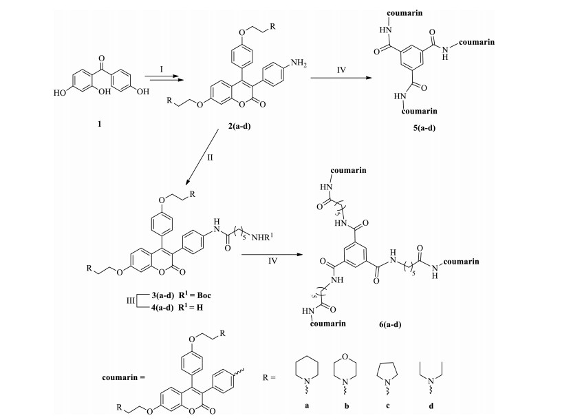
3. Results and discussionThe monomeric amino 3, 4-diphenyl coumarins 2(a-d) were prepared via Perkin reaction route from benzophenone (1) according to the previous procedures [32-34]. 2 reacted with Boc-6-aminohexanoic acid to mainly give 3, then the t-butyloxy carbonyl (Boc) group was removed in 90% CF3COOH to yield another monomeric coumarins 4(a-d) (Scheme 1). The target trimeric compounds (5 or 6) were easily synthesized by condensation of 2 or 4 with trimesic acid using tetramethyluronium hexafluorophosphate (HATU) and N-ethyldiisopropyl amine (DIPEA) as catalysts at room temperature in the middle yields of 36%-46%. The structures of all the new compounds (4-6) were determined by NMR, and MS (HRMS). Both analytical and spectral data of compounds are in agreement with the proposed structures.

|
Download:
|
| Scheme. 1. Synthesis of compounds 2-6. Reagents and conditions: (Ⅰ) See Refs. [32-34]; (Ⅱ) Boc-6-aminohexanoic acid, HATU, DCM, DIPEA, r.t.; (Ⅲ) 90% CF3COOH/H2O, r.t.; two (Ⅱ and Ⅲ) steps: 69%-75%; (Ⅳ) Trimesic acid, HATU, DIPEA, DMF, r.t., 36%-46%. | |
Compounds 5 and 6 were subjected to anti-proliferative tests against the following cancer cell lines, MCF-7 (human breast cancer), A549 (human lung cancer) and Hela (cervical carcinoma). Cisplatin was used as positive control to preliminarily estimate the antitumor activity of the coumarins. As shown in Table 1, the trimer 6a (R=piperidinyl) exhibited significant anti-proliferative activities against three cancer cells at IC50 of near 10 mmol/L, and especially against A-549, better than the positive control. Compounds 6c and 6d (R=pyrrolinyl, NEt2, respectively) expressed medium anti-tumor activities. The trimers 5a, 5c and 5d showed weak or no anti-tumor activities possible due to their overcrowded structures which hampered the activities. Additionally, neither 5b nor 6b (R=morpholinyl) expressed anti-proliferative activities, which was similar with the previous observation [34] that the morpholinyl demonstrated a detrimental effect on the anti-proliferative activity. The results implied that the basic amino group had certain effect on their activities, and among them, piperidinyl group was the best.
|
|
Table 1 Cytotoxicity of compounds against the cancer cell lines. |

In order to evaluate the interactions with DNA, UV-vis, fluorescence (lifetime) and DNA thermal denaturation experiment were used to ascertain the DNA binding properties of compounds 5a, 6a, 5d and 6d with calf thymus (Ct) DNA.
The DNA binding properties of compounds 5a, 6a, 5d and 6d with Ct-DNA were investigated by UV-vis spectra in phosphate buffer (10 mmol/L, pH 7.4) containing 50 mmol/L NaCl at 25 ℃. As shown in Fig. 2 and Table 2, in the presence of increasing concentration of Ct-DNA, the absorption intensities of these four compounds enhanced in middle hyperchromities of 14%, 25%, 13%, and 17%, respectively, without the obvious shift of the maximum absorption (λmax) except 5d (3.0 nm). The observed hypsochromism implied that the trimeric compounds or part of the structures would insert into the base pairs of DNA as DNAintercalating agents [37, 38]. By the double inverse method [39], the binding constants of compounds 5a, 6a, 5d and 6d with Ct-DNA were calculated to be 2.87 × 10³ (mol/L)-1, 4.30 × 10³ (mol/L)-1, 2.44 × 10³ (mol/L)-1, and 3.20 × 10³ (mol/L)-1, respectively. The Kb values showed that compounds 6a and 6d have stronger binding properties with Ct-DNA than those of 5a and 5d. Furthermore, the Kb values and the rate of hyperchromities of the trimers followed the order (5-6)a > (5-6)d, suggested that the extended linker (six carbons) and piperidinyl group on the side chains are beneficial to DNA binding. These results were in agreement with the SAR analysis of their anti-proliferative activities.

|
Download:
|
| Figure 2. UV-vis spectral changes of compounds 5a, 6a, 5d and 6d at the concentration of 5.0 × 10⁻⁶ mol/L upon addition of Ct-DNA (0-50 μmol/L) in phosphate buffer (10 mmol/L, pH 7.4) containing 50 mmol/L NaCl and 4% DMSO at 25 ℃. Inset: the fitting plots for 5a, 6a, 5d and 6d with Ct-DNA obtained at the maximum absorption. | |
|
|
Table 2 Photometric properties of the trimeric coumarin derivatives 5a, 6a, 5d and 6d with Ct-DNA investigated by UV-vis spectra and binding constants (K) by UV-vis and fluorescence spectra. |

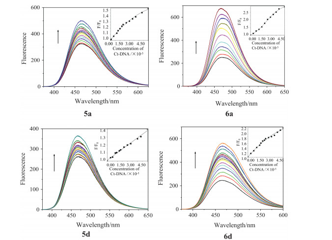
The fluorescence properties were performed to investigate the interactions between compounds 5a, 6a, 5d and 6d and Ct-DNA in phosphate buffer (10 mmol/L, pH 7.4) containing 50 mmol/L NaCl at 25 ℃. As shown in Fig. 3, the trimeric compounds showed similar binding properties with Ct-DNA around 345 nm in the fluorescence spectra. Upon addition of DNA, the maximum emission bands red shift obviously, and the fluorescence intensities increased markedly except 5a and 5d. The observed fluorescence intensity increment (6a and 6d) points to binding interaction between the compounds and Ct-DNA and makes these novel coumarins possess potential application as DNA staining agent. Binding of the coumarins with the Ct-DNA hinders the rotations around various bonds and thereby decreases the possible nonradiative process through the twisted intramolecular charge transfer (TICT) state [36, 40, 41]. The fluorescence intensities were quantified by plotting F/F0 as a function of Ct-DNA concentrations, where F0 and F are the fluorescence intensity without and with Ct-DNA, respectively. Stern-Volmer analysis gave deep insight into the binding efficiency of fluorescence enhancement of the trimers with increasing concentrations of Ct-DNA [36]. The calculated binding efficiency (Table 2) were 8.63 × 10³ (mol/L)-1, 3.38 × 10⁴ (mol/L)-1, 6.16 × 10³ (mol/L)-1 and 2.02 × 10⁴ (mol/L)-1 for compounds 5a, 6a, 5d and 6d, respectively [42, 43]. The Stern-Volmer plots indicate that the fluorescence of compound 6a with piperidinyl group is higher sensitive to the Ct-DNA concentrations than those of the others. These results were consistent with the observations by UV and their anti-proliferative activity, that is, the order of the Kb values was same with the order of the IC50 values.

|
Download:
|
| Figure 3. Fluorescence spectral changes of 5a and 5d (λex=350.5 nm), 6a and 6d (λex=345.5 nm) at the concentration of 5.0 × 10⁻⁶ mol/L upon addition of Ct-DNA (0-50 μmol/L) in phosphate buffer (10 mmol/L, pH 7.4) containing 50 mmol/L NaCl and 4% DMSO at 25 ℃. The excitation and emission slit widths were 5.0 nm for other compounds. Inset: Stern-Volmer plots for the observed fluorescence enhancement upon addition of Ct-DNA to the dimeric coumarin derivatives. | |
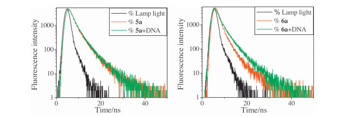
Additionally, the fluorescence lifetime was also performed to investigate the interactions between the trimers 5a, 6a and Ct-DNA (Fig. 4 and Table 3). Both compounds 5a and 6a had two fluorescence lifetimes, τ1=1.34 ns, τ2=4.26 ns; and τ1=0.97 ns, τ2=3.87 ns, respectively. In the presence of Ct-DNA, the lifetimes were τ1=1.35 ns, τ2=4.28 ns for 5a, and τ1=1.40 ns, τ2=4.41 ns for 6a. The △τ data (increased lifetime) suggested that the trimer 6a had the stronger binding with DNA than 5a due to the obviously prolonged fluorescence lifetime of 6a.

|
Download:
|
| Figure 4. The fluorescence lifetime of 5a and 6a (5.0 × 10⁻⁶ mol/L) in the absence and presence of Ct-DNA (5.0 × 10⁻⁵ mol/L) in phosphate buffer (10 mmol/L, pH 7.4) containing 50 mmol/L NaCl and 4% DMSO at 25 ℃. | |
|
|
Table 3 The fluorescence lifetimes of 5a and 6a. |

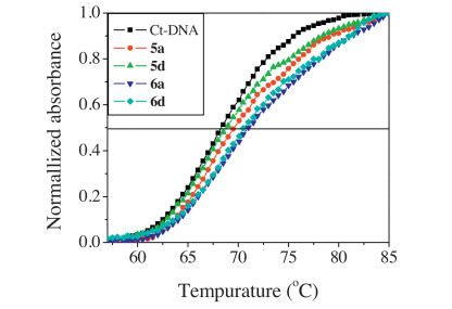
It is well known that the temperature at which a half of a DNA sample melts is known as the melting temperature (Tm). A change of Tm may be observed if a molecule binds with DNA [44]. Thus the thermal behavior of DNA in the presence of the triphenylethylene-coumarin hybrids provides useful information on the conformational changes and the strength of the DNA-compound complexes. The melting curves of Ct-DNA in the absence and presence of 5a, 5d, 6a and 6d are illustrated in Fig. 5 and Table 4, respectively. The Tm value for the free Ct-DNA was 68.2 ℃. Upon addition of 6a, obvious changes in the DNA melting temperature were observed. The Tm value increased to 71.0 ℃, and the level of the increased melting temperature (△Tm) induced by DNA-compound interactions was 2.8 ℃. The others possessed lower DNA melting temperature than 6a, and the △Tm values were 1.2, 0.8 and 2.6 ℃, respectively. These results further illuminated that trimer 6a with piperidinyl group exhibited strongest affinity with Ct-DNA and the extended trimer 6 had more binding with Ct-DNA than 5, consistent with the results from the fluorescence data.

|
Download:
|
| Figure 5. DNA melting curves for Ct-DNA (5.0 × 10⁻⁵ mol/L) in the absence and presence of 5a, 6a, 5d and 6d with concentration of 5.0 × 10⁻⁶ mol/L in phosphate buffer (1 mmol/L, pH 7.4) containing 5 mmol/L NaCl and 4% DMSO at 25 ℃. | |
|
|
Table 4 Average Tm and △Tm for Ct-DNA in the absence and presence of 5a, 6a, 5d and 6d. |

4. Conclusion
A series of novel trimeric triphenylethylene-coumarin hybrids were designed and synthesized by the condensation of trimesic acid with the amino monomeric hybrids. The extended trimer 6a (six carbons linker and R=piperidinyl) showed the best broadspectrum anti-tumor activities and binding affinity with DNA in intercalative mode. Both the length of the linker and the basic amino group had important effects on the anti-proliferative activity and DNA binding capacity of the compounds. The agreement of the DNA binding constants and the anti-tumor activities of 5 and 6 suggested that DNA might be one of the potential targets for such novel trimeric coumarin hybrids as antitumor drug candidates.
AcknowledgmentThe financial support was from the National Natural Science Foundation of China (NSFC, No. 20902016).
| [1] | K.M. Amin, N.M. Abdel Gawad, D.E. Abdel Rahman, M.K.M. El Ashry, New series of 6-substituted coumarin derivatives as effective factor Xa inhibitors: synthesis, in vivo antithrombotic evaluation and molecular docking. Bioorg. Chem. 52 (2014) 31–43. DOI:10.1016/j.bioorg.2013.11.002 |
| [2] | V. Rempel, N. Volz, F. Gläser, Antagonists for the orphan G-protein-coupled receptor GPR55 based on a coumarin scaffold. J. Med. Chem. 56 (2013) 4798–4810. DOI:10.1021/jm4005175 |
| [3] | Y. Chen, S.L. Wang, X.Q. Xu, Synthesis and biological investigation of coumarin piperazine (Piperidine) derivatives as potential multireceptor atypical antipsychotics. J. Med. Chem. 56 (2013) 4671–4690. DOI:10.1021/jm400408r |
| [4] | Q.L. Zou, Y.Y. Fang, Y.X. Zhao, Synthesis and in vitro photocytotoxicity of coumarin derivatives for one-and two-photon excited photodynamic therapy. J. Med. Chem. 56 (2013) 5288–5294. DOI:10.1021/jm400025g |
| [5] | R.S. Keri, B.S. Sasidhar, B.M. Nagaraja, M.A. Santos, Recent progress in the drug development of coumarin derivatives as potent antituberculosis agents. Eur. J. Med. Chem. 100 (2015) 257–269. DOI:10.1016/j.ejmech.2015.06.017 |
| [6] | B. Li, R. Pai, M. Di, Coumarin-based inhibitors of Bacillus anthracis and Staphylococcus aureus replicative DNA helicase: chemical optimization, biological evaluation, and antibacterial activities. J. Med. Chem. 55 (2012) 10896–10908. DOI:10.1021/jm300922h |
| [7] | S. Hamulakova, L. Janovec, M. Hrabinova, Synthesis and biological evaluation of novel tacrine derivatives and tacrine-coumarin hybrids as cholinesterase inhibitors. J. Med. Chem. 57 (2014) 7073–7084. DOI:10.1021/jm5008648 |
| [8] | A. Thakur, R. Singla, V. Jaitak, Coumarins as anticancer agents: a review on synthetic strategies, mechanism of action and SAR studies. Eur. J. Med. Chem. 101 (2015) 476–495. DOI:10.1016/j.ejmech.2015.07.010 |
| [9] | M. Basanagouda, V.B. Jambagi, N.N. Barigidad, S.S. Laxmeshwar, V.D. Narayanachar, Synthesis, structure-activity relationship of iodinated-4-aryloxymethylcoumarins as potential anti-cancer and anti-mycobacterial agents. Eur. J. Med. Chem. 74 (2014) 225–233. DOI:10.1016/j.ejmech.2013.12.061 |
| [10] | J. Dandriyal, R. Singla, M. Kumar, V. Jaitak, Recent developments of C-4 substituted coumarin derivatives as anticancer agents. Eur. J. Med. Chem. 119 (2016) 141–168. DOI:10.1016/j.ejmech.2016.03.087 |
| [11] | N. Touisni, A. Maresca, P.C. McDonald, Glycosyl coumarin carbonic anhydrase Ⅸ and Ⅻ inhibitors strongly attenuate the growth of primary breast tumors. J. Med. Chem. 54 (2011) 8271–8277. DOI:10.1021/jm200983e |
| [12] | C.J. Wang, Y.J. Hsieh, C.Y. Chu, Y.L. Lin, T.H. Tseng, Inhibition of cell cycle progression in human leukemia HL-60 cells by esculetin. Cancer Lett. 183 (2002) 163–168. DOI:10.1016/S0304-3835(02)00031-9 |
| [13] | T. Nasr, S. Bondock, M. Youns, Anticancer activity of new coumarin substituted hydrazide-hydrazone derivatives. Eur. J. Med. Chem. 76 (2014) 539–548. DOI:10.1016/j.ejmech.2014.02.026 |
| [14] | B.R.V. Avin, P. Thirusangu, V.L. Ranganatha, Synthesis and tumor inhibitory activity of novel coumarin analogs targeting angiogenesis and apoptosis. Eur. J. Med. Chem. 75 (2014) 211–221. DOI:10.1016/j.ejmech.2014.01.050 |
| [15] | C.Y. Chu, Y.Y. Tsai, C.J. Wang, W.L. Lin, T.H. Tseng, Induction of apoptosis by esculetin in human leukemia cells. Eur. J. Pharmacol. 416 (2001) 25–32. DOI:10.1016/S0014-2999(01)00859-7 |
| [16] | J.A. McKie, S.S. Bhagwat, H. Brady, Lead identification of a potent benzopyranone selective estrogen receptor modulator. Bioorg. Med. Chem. Lett. 14 (2004) 3407–3410. DOI:10.1016/j.bmcl.2004.04.081 |
| [17] | C. Bailly, Contemporary challenges in the design of topoisomerase Ⅱ inhibitors for cancer chemotherapy. Chem. Rev. 112 (2012) 3611–3640. DOI:10.1021/cr200325f |
| [18] | M. Facompré, C. Tardy, C. Bal-Mahieu, Lamellarin D: a novel potent inhibitor of topoisomerase I. Cancer Res. 63 (2003) 7392–7399. |
| [19] | S. Emami, S. Dadashpour, Current developments of coumarin-based anti-cancer agents in medicinal chemistry. Eur. J. Med. Chem. 102 (2015) 611–630. DOI:10.1016/j.ejmech.2015.08.033 |
| [20] | M.M. Liu, X.Y. Chen, Y.Q. Huang, Hybrids of phenylsulfonylfuroxan and coumarin as potent antitumor agents. J. Med. Chem. 57 (2014) 9343–9356. DOI:10.1021/jm500613m |
| [21] | W.J. Zhang, Z. Li, M. Zhou, Synthesis and biological evaluation of 4-(1, 2, 3-triazol-1-yl)coumarin derivatives as potential antitumor agents. Bioorg. Med. Chem. Lett. 24 (2014) 799–807. DOI:10.1016/j.bmcl.2013.12.095 |
| [22] | F. Pé rez-Cruz, S. Vazquez-Rodriguez, M. João Matos, Synthesis and electrochemical and biological studies of novel coumarin-chalcone hybrid compounds. J. Med. Chem. 56 (2013) 6136–6145. DOI:10.1021/jm400546y |
| [23] | A. Gupta, S.K. Mandal, V. Leblanc, Synthesis and cytotoxic activity of benzopyran-based platinum(Ⅱ) complexes. Bioorg. Med. Chem. Lett. 18 (2008) 3982–3987. DOI:10.1016/j.bmcl.2008.06.013 |
| [24] | K.V. Sashidhara, A. Kumar, M. Kumar, J. Sarkar, S. Sinha, Synthesis and in vitro evaluation of novel coumarin-chalcone hybrids as potential anticancer agents. Bioorg. Med. Chem. Lett. 20 (2010) 7205–7211. DOI:10.1016/j.bmcl.2010.10.116 |
| [25] | X.H. Liu, H.F. Liu, J. Chen, Synthesis and molecular docking study of novel coumarin derivatives containing 4, 5-dihydropyrazole moiety as potential antitumor agents. Bioorg. Med. Chem. Lett. 20 (2010) 5705–5708. DOI:10.1016/j.bmcl.2010.08.017 |
| [26] | F. Belluti, G. Fontana, L. Dal Bo, Design, synthesis and anticancer activities of stilbene-coumarin hybrid compounds: identification of novel proapoptotic agents. Bioorg. Med. Chem. Lett. 18 (2010) 3543–3550. DOI:10.1016/j.bmc.2010.03.069 |
| [27] | H.P. Zhao, A.C. Donnelly, B.R. Kusuma, Engineering an antibiotic to fight cancer: optimization of the novobiocin scaffold to produce anti-proliferative agents. J. Med. Chem. 54 (2011) 3839–3853. DOI:10.1021/jm200148p |
| [28] | Z.N. Siddiqui, T.N. Mohammed Musthafa, A. Ahmad, A.U. Khan, Synthesis of 4-hydroxycoumarin heteroarylhybrids as potential antimicrobial agents. Arch. Pharm. Chem. Life Sci. 344 (2011) 394–401. DOI:10.1002/ardp.201000218 |
| [29] | B.R. Kusuma, L.B. Peterson, H.P. Zhao, Targeting the heat shock protein 90 dimer with dimeric inhibitors. J. Med. Chem. 54 (2011) 6234–6253. DOI:10.1021/jm200553w |
| [30] | J.A. Burlison, B.S.J. Blagg, Synthesis and evaluation of coumermycin A1 analogues that inhibit the Hsp90 protein folding machinery. Org. Lett. 8 (2006) 4855–4858. DOI:10.1021/ol061918j |
| [31] | G.H. Tan, Y.C. Yao, Y.J. Gu, Cytotoxicity and DNA binding property of the dimers of triphenylethylene-coumarin hybrid with one amino side chain. Bioorg. Med. Chem. Lett. 24 (2014) 2825–2830. DOI:10.1016/j.bmcl.2014.04.106 |
| [32] | M. Zhu, L.K. Zhou, Y.C. Yao, Anticancer activity and DNA binding property of the dimers of triphenylethylene-coumarin hybrid with two amino side chains. Med. Chem. Res. 24 (2015) 2314–2324. DOI:10.1007/s00044-014-1296-2 |
| [33] | L. Zhao, Y.C. Yao, S. Li, Cytotoxicity and DNA binding property of triphenylethylene-coumarin hybrids with two amino side chains. Bioorg. Med. Chem. Lett. 24 (2014) 900–904. DOI:10.1016/j.bmcl.2013.12.084 |
| [34] | H. Chen, S. Li, Y.C. Yao, Design, synthesis, and anti-tumor activities of novel triphenylethylene-coumarin hybrids, and their interactions with Ct-DNA. Bioorg. Med. Chem. Lett. 23 (2013) 4785–4789. DOI:10.1016/j.bmcl.2013.07.009 |
| [35] | T.J. Mosmann, Rapid colorimetric assay for cellular growth and survival: application to proliferation and cytotoxicity assays. J. Immunol. Methods 65 (1983) 55–63. DOI:10.1016/0022-1759(83)90303-4 |
| [36] | D. Sahoo, P. Bhattacharya, S. Chakravorti, Reverse micelle induced flipping of binding site and efficiency of albumin protein with an ionic styryl dye. J. Phys. Chem. B 114 (2010) 10442–10450. DOI:10.1021/jp102937y |
| [37] | R. Breslow, I.E. Overman, "Artificial enzyme" combining a metal catalytic group and a hydrophobic binding cavity. J. Am. Chem. Soc. 92 (1970) 1075–1077. DOI:10.1021/ja00707a062 |
| [38] | R.F. Pasternack, E.J. Gibbs, J.J. Villatrnaca, Interactions of porphyrins with nucleic acids. Biochemistry 22 (1983) 2406–2414. DOI:10.1021/bi00279a016 |
| [39] | M. Purcell, J.F. Neault, H.A. Tajmir-Riahi, Interaction of taxol with human serum albumin. Biochim. Biophys. Acta 1478 (2000) 61–68. DOI:10.1016/S0167-4838(99)00251-4 |
| [40] | K. Faulhaber, A. Granzhan, H. Ihmels, Studies of the fluorescence light-up effect of amino-substituted benzo. Photochem. Photobiol. Sci. 10 (2011) 1535–1545. DOI:10.1039/c1pp05106g |
| [41] | R. Ramadass, J. Bereiter-Hahn, Photophysical properties of DASPMI as revealed by spectrally resolved fluorescence decays. J. Phys. Chem. B 111 (2007) 7681–7690. DOI:10.1021/jp070378k |
| [42] | S. Pandey, G.A. Baker, M.A. Kane, N.J. Bonzagni, F.V. Bright, O2 Quenching of ruthenium(Ⅱ) tris(2, 2'-bypyridyl)2+ within the water pool of perfluoropolyetherbased reverse micelles formed in supercritical carbon dioxide. Langmuir 16 (2000) 5593–5599. DOI:10.1021/la991719r |
| [43] | J. Wang, D.L. Wang, E.K. Miller, Photoluminescence of water-soluble conjugated polymers: origin of enhanced quenching by charge transfer. Macromolecules 33 (2000) 5153–5158. DOI:10.1021/ma000081j |
| [44] | U. Chaveerach, A. Meenongwa, Y. Trongpanich, C. Soikum, P. Chaveerach, DNA binding and cleavage behaviors of copper(Ⅱ) complexes with amidino-O-methylurea and N-methylphenyl-amidino-O-methylurea, and their antibacterial activities. Polyhedron 29 (2010) 731–738. DOI:10.1016/j.poly.2009.10.031 |