 2016, Vol. 27


b School of Materials Science and Engineering, Jingdezhen Ceramic Institute, Jingdezhen 333001, China ;
c National Engineering Research Center for Tissue Restoration and Reconstruction, South China University of Technology, Guangzhou 510640, China
Dental hypersensitivity (DH) occurs when dentin is exposed to various types of stimuli such as thermal, evaporative and tactile. It manifests as a short sharp pain without other forms of dental defect or pathology [1]. Occlusal wear, toothbrushing, dietary erosion, and gingival recession may all lead to this problem [2]. At present, two main methods are used in the treatment of DH: tubule occlusion [3] and nerve activity blockage [4]. The reduction of dentin permeability through tubule sealing is expected to be an effective way of DH treatment [5].
Bioactive glass (BG) is calcium phosphosilicate cement that has been proven to relief DH by yielding hydroxyapatite deposition on the dentin surface [6]. Early forms of BG were synthesized with a traditional melting method and their diameters were relatively large [7]. As regards DH treatment, Mitchell et al. stated that application of BG with an average diameter of 1-20 μm to a demineralized dentin surface could reduce the fluid conductance through the dentin tubules [8]. Furthermore, Vollenweider et al. compared nanoscale BG (n-BG) synthesized using flame spray with traditional 45S5 BG. Their research demonstrated that the n-BG released Ca2+ and Si2+ more quickly than its counterpart, which was beneficial for dentin mineralization [9]. However, the flame-spray-synthesized BG had a broad size distribution [9] and different chemical composition compared to the 45S5 [9, 10], and thus the effect of chemical composition on mineral formation cannot be excluded. Recently, the sol-gel method has been widely applied in BG synthesis [11, 12]. Compared with the melting method, the sol-gel method requires relatively lower temperatures of approximately 600-700 8C, or even lower when calcium methylethoxide is used as the calcium precursor [13]. Further, the sol-gel BG may have uniform composition, mesoporous surface, and higher specific surface area. In addition, the form, size, and dissolution rate of sol-gel BG may be easily controlled [14].
In recent years, Chen et al. have fabricated new n-BG [15] and submicroscale BG (sm-BG) [16] using sol-gel technique. In this study, the application of these newly developed BG particles in the mineral formation on dentin surface were explored and particular attention was paid to the effect of BG particle size.
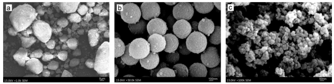
2. Experiment 2.1. MaterialsThe composition of all BG particles used in this study were same: 58% SiO2, 33% CaO, and 9% P2O5 (w/w) [15, 17, 18]. The following particle sizes were employed: microscale BG (m-BG) synthesized by acid-catalyzed sol-gel method: 2-20 μm (Fig. 1a) [17, 19]; mono-dispersed sm-BG synthesized by alkali-catalyzed sol-gel method: approximately 500 nm (Fig. 1b) [18, 20] and n-BG synthesized by acid-alkali-catalyzed sol-gel method: 20-30 nm (Fig. 1c) [15, 17]. Both the sm-BG and n-BG particles were regularly spherical, whereas the m-BG particles were irregularly agglomerate. Our early studies [17, 18] have reported the surface areas, total pore volumes and average mesopore sizes evaluated via N2 adsorption experiments, the data was shown in Table 1. All the BG particles used in this study were prepared by the National Engineering Research Center for Human Tissue Restoration and Reconstruction, South China University of Technology.

|
Download:
|
| Figure 1. Surface morphology of BG. Surface morphology of (a) m-BG, (b) sm-BG and (c) n-BG by FE-SEM analysis. | |
|
|
Table 1 The surface areas, total pore volumes and average pore diameters of BG. |

2.2. Dentin disc preparation
In total, 18 intact human third molars were collected with ethical approval and informed donor consent, and were stored in distilled water containing 0.9% NaCl at 4 8C. 1-mm-thick dentin discs were created using a hard histotome (SP1600, Leica, Germany) to discard the enamel and pulp and then the dentin surfaces were then wet ground using SiC paper (600 grit) so as to increase their smoothness. The dentin discs were subsequently wiped to remove the surrounding enamel and divided into four parts using diamond burrs. The dentin discs were immersed in 17% ethylenediaminetetraacetic acid (EDTA) (pH 7.4) for 1 wk, so that completely demineralized discs were obtained. The BG particles were mixed with deionized water to form BG suspensions with a liquid/powder ratio of 0.1 mL/0.05 g.
The dentin discs were randomly divided into four groups labeled m-BG, sm-BG, n-BG, and CTR (control). Each group contained 18 discs. The m-BG, sm-BG, and n-BG discs were separately embrocated with the m-BG, sm-BG, and n-BG suspensions, respectively, using a brush for 20 s. Next, they were washed with deionized water for 20 s and then placed into artificial saliva (AS). The CTR discs were placed into the AS directly. The AS was composition of 1.5 mmol/L of CaCl2, 50 mmol/L of KCl, 0.9 mmol/L of KH2PO4, and 20 mmol/L Tris, with a pH of 7.4 [21].
2.3. Evaluation of sealing abilityField-emission scanning electron microscopy (FE-SEM; S4800, JEOL, Tokyo, Japan) was used to observe the surface morphology of the dentin discs. After one week, six discs were collected randomly from each group and rinsed with deionized water for 20 s. The discs were then observed using FE-SEM for the cross sectional and longitudinal sectional views. Prior to the microscopic observation, the discs were coated with gold (5 min, 50 mTorr). The cross sectional surface elemental compositions of the 1-wk discs were evaluated using an energy-dispersive X-ray spectroscope (EDS) attached to the FE-SEM. FE-FEM and Image-J software (Version 1.48, National Institutes of Health, USA) were used together to assess the sealing ratios of the dentin tubules. The Image-J software was used to observe six 2000× magnified FE-SEM images of the dentin disc surfaces from each group. Hence, the overall open dentin tubule area (S), the average area of a single dentin tubule (s), and the number of dentin tubules (N) were measured in each case. The sealing ratios were calculated as

|
After two weeks, the remaining twelve discs were collected from each group and rinsed with deionized water for 20 s. Six discs were observed the morphological changes on the disc surfaces using FE-SEM.
2.4. Study of acid resistanceThe other six discs immersing in BG suspensions for two weeks were used to evaluate the roughness. A 3D profile measurement laser microscope (LM, VK-X200, Keyence, Japan) was used to measure the initial roughness (Ra) at two locations on each disk. The discs were then submerged in cola (pH 2.45) for 2 min and again rinsed with deionized water for 20 s. The roughness was measured for a second time (Ra'). After drying, the FE-SEM was used to observe the morphological changes on the disc surfaces.
2.5. Statistical analysisThe sealing ratio and Ra data were reported as mean±standard deviation (SD) values. Comparisons of the dentin tubule sealing ratios of the 1-wk groups were conducted with t-tests. The dentin surface Ra and Ra' values of the four groups before and after the cola immersion, and the corresponding roughness variance values, were also compared using t-tests. Differences were considered significant at P < 0.05.
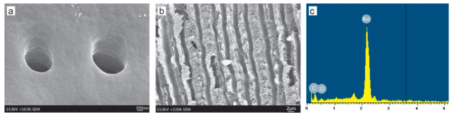
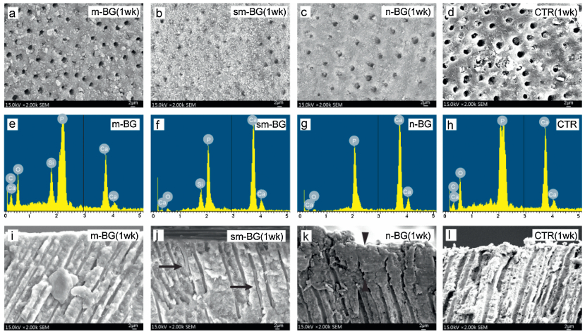
3. Results and discussion 3.1. Dentin-tubule sealingThe FE-SEM analysis showed that, following immersion in 17% EDTA for one week, the dentin tubules of the initial demineralized discs were completely open (Fig. 2a and b). Moreover, the EDS spectra indicated that the surface elements present on the demineralized dentin discs were carbon, oxygen and gold (Fig. 2c). Therefore, calcium and phosphorus were absent; this demonstrated that the discs were completely demineralized. As regards the 1-wk specimens treated with the three different BG suspensions, all the disc surfaces exhibited material and mineral deposition. In addition, the dentin tubules were partly sealed (Fig. 3a-c). Besides, the dentin surfaces and tubules of the CTR samples exhibited some deposition formation (Fig. 3d). The 1 wk sealing ratios of the m-BG, sm-BG, n-BG, and CTR groups were 38.63±7.5%, 48.01±6.85%, 68.02±4.22%, and 25.38±8.53% (n=6), respectively. Thus, the n-BG result was significantly higher than those of the other three groups (P < 0.05). Dentin tubule exposure is the physical pathogenesis of DH, and an in vivo study has shown that the majority of dentin tubules are open in dentin hypersensitive areas [22]. The m-BG, sm-BG, and n-BG specimens examined in this study all exhibited mineral formation promotion on the dentin surfaces, which would relieve DH.

|
Download:
|
| Figure 2. Characterization of demineralized dentin. FE-SEM images of (a) surface morphology and (b) exposed dental tubules. (c) Dentin-surface element composition. | |

|
Download:
|
| Figure 3. Representative surface FE-SEM images and element compositions. (a–d) Cross sectional FE-SEM images of the surface morphology after one week. (e–h) Surface element composition after one week. (i–l) Longitudinal sectional FE-SEM images of the dental tubules. Newly formed minerals bars in the sm-BG group tubules (j, indicated by the arrows). A mineral layer to a depth of 10 mm in the dental tubules for the n-BG group (k, between the arrowhead). | |
Previous studies have demonstrated that BG could promote mineral formation on dentin surfaces. Apatite formation occurs through a series of interdependent sequential reactions. Briefly, the BG dissolves through hydrolysis to release calcium ions (Ca2+) and phosphate ions (PO43-), the Si-O-Si bridges break to yield a SiO2-rich surface layer; this acts as a heterogenic nucleation site. Positively charged Ca2+ interacts with the negatively charged PO43- in the surrounding fluid to form an amorphous calcium phosphate. The amorphous calcium phosphate crystallizes to form apatite [23]. The CaO and SiO2 content of the BG and the particle size and volume surface area can influence the solubility, which in turn affects the mineral formation [24, 25]. Therefore, to prevent differences in the components from influencing the results, three BG particles with the same components but different particle sizes were used in this study. Thus, the effect of the BG size on the mineral formation on the dentin surface could be evaluated. Further, as residual minerals from partially demineralized dentin cannot be distinguished from newly formed minerals by FE-SEM, completely demineralized dentin was used.
The EDS spectra indicated that the surface elements for all the BG groups were carbon, phosphorus, and oxygen. In addition, the m-BG and sm-BG groups also contained silicon (Fig. 3e-g). Semiquantitative analyses indicated that molar composition for the dentin surface deposition of m-BG was 5.79% SiO2, 29.43% CaO, and 11.15% P2O5. The surface deposition molar composition of sm-BG was 6.71% SiO2, 49.92% CaO, and 27.05% P2O5, and that of the n-BG was 56.07% CaO and 26.97% P2O5. The CTR composition was 18.25% CaO and 8.42% P2O5 (Fig. 3h). The Ca/P ratios of the dentin surface depositions were higher than that of the natural hydroxyapatite (Ca/P=1.67), which might be explained by the residual BG on the dentin surfaces.
For the longitudinal section view, there seemed no obvious mineral deposited in the dental tubules for the m-BG and CTR groups (Fig. 3i and l), while some mineral bars can be seen in the sm-BG treated group (Fig. 3j, arrow showed). As regards the n-BG treated disc, about a 10 mm width newly formed mineral layer could be found in the dental tubules (Fig. 3k, between the arrowhead).
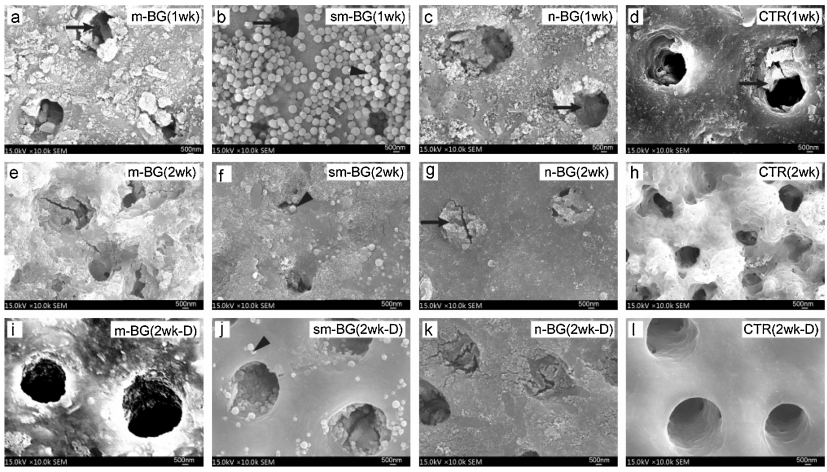
Based on the magnified images, it was apparent that some materials and small mineral deposition were present on the disc surface and in the tubules of the m-BG group (Fig. 4a, indicated by the arrow). Materials were present on the surface and in the tubules of the sm-BG group (Fig. 4b, indicated by the arrowhead), with newly formed minerals present in the tubules themselves (Fig. 4b, arrow). As regards the n-BG group, newly formed minerals were apparent in the tubules; they were attached closely to the tubular walls such that the dentin tubules were closed (Fig. 4c, arrow). Finally, only loose minerals were apparent on the CTR specimen disc surfaces (Fig. 4d).

|
Download:
|
| Figure 4. Representative surface FE-SEM images. (a–d) Magnified images of (Fig. 3a–d). Newly formed minerals were apparent in the BG groups tubules (indicated by the arrows). (e–h) FE-SEM images of the surface morphology after two weeks. Materials were obviously present on the sm-BG group dentin surface (arrowhead). Plug-like minerals were apparent in the tubules of the n-BG specimen (arrow). (i–l) FE-SEM images of the surface morphology after soaking in cola. Materials were still apparent on the sm-BG group dentin surface (arrowhead). | |
For the 2-wk discs, larger sealing areas compared to those of the 1-wk discs were observed for each group (Fig. 4e-h). Both the m-BG and sm-BG treated discs exhibited almost completely sealed tubules, with newly formed platelet-like minerals being apparent within the tubules (Fig. 4e and f). Some materials were also present on the sm-BG surface (Fig. 4f, arrowhead). For the n-BG treated discs, the tubules were completely sealed by plug-like minerals, which were adapted to the tubule outlines (Fig. 4g, arrow). Because of the smaller particle size, the n-BG particles can enter the tubules. The tubule structure then determines the Ca2+, PO43-, and OH- ion deposition and the subsequent apatite propagation [26]. Finally, the CTR group discs exhibited a large number of loose minerals on their surfaces and in the tubules and these tubules were partly sealed (Fig. 4h).
3.2. Acid resistanceAfter soaking in cola for 2 min, FE-SEM analysis showed that the m-BG group tubules were completely open (Fig. 4i). In the sm-BG case, the depositions collapsed and the dentin tubules were partly open; however, some materials remained on the surface (Fig. 4j, arrowhead). In contrast, the n-BG discs continued to exhibit completely closed tubules (Fig. 4k). Finally, the loose minerals on the CTR surface disappeared and the tubules were open (Fig. 4l).
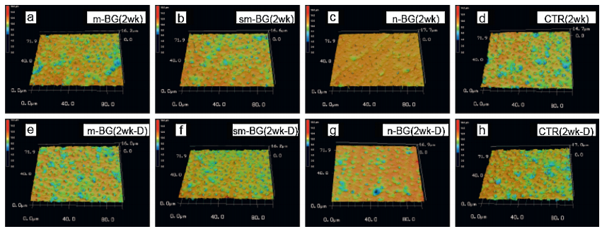
After immersion in AS for 2 wk, the 3-D profile displayed by LM showed smooth surface with a homogeneous jacinth spread for the n-BG treated discs (Fig. 5c), while the other three groups exhibited heterogeneous heights with color variation (Fig. 5a, b and d). The m-BG, sm-BG, n-BG, and CTR Ra values were 0.72±0.08, 0.60±0.07, 0.51±0.04, and 0.91±0.10 μm (n=12), respectively. Following the cola immersion, the n-BG treated discs retained a relatively smooth surface compared with the other three groups (Fig. 5e-h). The Ra' values were 1.03±0.13, 0.65±0.06, 0.52±0.04, and 1.27±0.12 mm (n=12), respectively. The BG group Ra and Ra' values were lower than those of the CTR specimens (P < 0.05), and the n-BG group values were the lowest overall (P < 0.001). The differences between Ra and Ra' were significant for both the m-BG and CTR groups (P < 0.001).

|
Download:
|
| Figure 5. Representative 3D profiles of dentin surfaces exhibited by LM. (a–d) 3D profiles of the surface morphology after two weeks. (e–h) 3D profiles of the surface morphology after soaking in cola. | |
Studies have shown that dentin surface roughness is related to mineralization, and sealing or blocking the tubules may yield a smoother surface [27]. Following treatment with the BG suspensions, the dentin surface roughness decreased as the BG group Ra values were significantly lower than those of the CTR, which indirectly confirms the occurrence of mineralization. After soaking in cola, the roughness variance for the BG groups was lower than that of the CTR, and the n-BG specimens retained their lower roughness. This suggested that the mineral formation was more acid-resistant in the n-BG case. This behavior may be explained by the fact that the plug-like apatite adapts to the tubule shapes, and the tubule angles and structures may limit the apatite displacement and contribute to mechanical retention, thus increasing the minerals stability. It should be noted that the surface roughness affects not only the dentin esthetics, but also the bacteria attachment and plague formation [28]. Through decreased surface roughness, the dentin obtains increased resistance to bacterial attachment, which prevents the occurrence of caries in demineralized dentin.
BG with smaller particle size has larger volume surface area and can, therefore, make greater contact with neighboring materials; thus, they exhibit increased dissolution rate [26]. Compared to the m-BG and sm-BG, it is more easily for the n-BG to promote mineral formation for dentin tubule sealing, which is effective in relieving DH.
4. ConclusionThe present study revealed that n-BG not only greatly promoted mineral formation on the dentin surfaces and tubules compared to m-BG and sm-BG, but also the minerals produced by n-BG exhibited greater stability and acid resistance. The n-BG could be considered as a potential material for the treatment of dentin hypersensitivity and the promotion of dentin mineralization. Future research should evaluate the bonding ability between the newly formed minerals and collagen, along with the mechanical properties of the mineralized dentin.
AcknowledgmentsThe authors are grateful for to the financial support from the National Natural Science Foundation of China (No. 51372005).
| [1] | Holland G.R., Narhi M.N., Addy M., Gangarosa L., Orchardson R.. Guidelines for the design and conduct of clinical trials on dentine hypersensitivity. J. Clin. Periodontol. 24 (1997) 808–813. DOI:10.1111/cpe.1997.24.issue-11 |
| [2] | Porto I.C.C.M., Andrade A.K.M., Montes M.A.J.R.. Diagnosis and treatment of dentinal hypersensitivity. J. Oral Sci. 51 (2009) 323–332. DOI:10.2334/josnusd.51.323 |
| [3] | Cummins D.. Dentin hypersensitivity:from diagnosis to a breakthrough therapy for everyday sensitivity relief. J. Clin. Dent. 20 (2009) 1–9. |
| [4] | Andrew D., Matthews B.. Displacement of the contents of dentinal tubules and sensory transductioninintradental nerves ofthecat. J. Physiol. 529 (2000) 791–802. DOI:10.1111/tjp.2000.529.issue-3 |
| [5] | Neuhaus K.W., Milleman J.L., Milleman K.R., et al. Effectiveness of a calcium sodium phosphosilicate containing prophylaxis paste in reducing dentine hypersensitivity immediately and 4 weeks after a single application:a double-blind randomized controlled trial. J. Clin. Periodontol. 40 (2013) 349–357. DOI:10.1111/jcpe.12057 |
| [6] | Chałas R., Wójcik-Chęciń ska I., Zamościń ska J., Bachanek T.. Assessment of pain intensity in patients with dentin hypersensitivity after application of prophylaxis paste based on calcium sodium phosphosilicate formula. Med. Sci. Monit. 21 (2015) 2950–2955. DOI:10.12659/MSM.894189 |
| [7] | Zhang Y., Santos J.D.. Crystallization and microstructure analysis of calcium phosphate-based glass ceramics for biomedical applications. J. Non-Cryst. Solids 272 (2000) 14–21. DOI:10.1016/S0022-3093(00)00115-0 |
| [8] | Mitchell J.C., Musanje L., Ferracane J.L.. Biomimetic dentin desensitizer based on nano-structured bioactive glass. Dent. Mater. 27 (2011) 386–393. DOI:10.1016/j.dental.2010.11.019 |
| [9] | Vollenweider M., Brunner T.J., Knecht S., et al. Remineralization of human dentin using ultrafine bioactive glass particles. Acta Biomater. 3 (2007) 936–943. DOI:10.1016/j.actbio.2007.04.003 |
| [10] | Hench L.L.. The story of Bioglass®. J. Mater. Sci.:Mater. Med. 17 (2006) 967–978. DOI:10.1007/s10856-006-0432-z |
| [11] | Saravanapavan P., Hench L.L.. Low-temperature synthesis, structure, and bioactivity of gel-derived glasses in the binary CaO-SiO2 system. J. Biomed. Mater. Res. 54 (2001) 608–618. DOI:10.1002/(ISSN)1097-4636 |
| [12] | Sepulveda P., Jones J.R., Hench L.L.. Characterization of melt-derived 45S5 and sol-gel-derived 58S bioactive glasses. J. Biomed. Mater. Res. 58 (2001) 734–740. DOI:10.1002/(ISSN)1097-4636 |
| [13] | Sun Y.S., Li A.L., Xu F.J., Qiu D.. A low-temperature sol-gel route for the synthesis of bioactive calcium silicates. Chin. Chem. Lett. 24 (2013) 170–172. DOI:10.1016/j.cclet.2013.01.009 |
| [14] | Zhong J.P., Greenspan D.C.. Processing and properties of sol-gel bioactive glasses. J. Biomed. Mater. Res. 53 (2000) 694–701. DOI:10.1002/(ISSN)1097-4636 |
| [15] | Lei B., Chen X.F., Wang Y.J., et al. Surface nanoscale patterning of bioactive glass to support cellular growth and differentiation. J. Biomed. Mater. Res. Part A 94A (2010) 1091–1099. |
| [16] | Hu Q., Li Y.L., Miao G.H., Zhao N.R., Chen X.F.. Size control and biological properties of monodispersed mesoporous bioactive glass sub-micron spheres. RSC Adv. 4 (2014) 22678–22687. DOI:10.1039/c4ra01276c |
| [17] | Wang S.N., Gao X.J., Gong W.Y., et al. Odontogenic differentiation and dentin formation of dental pulp cells under nanobioactive glass induction. Acta Biomater. 10 (2014) 2792–2803. DOI:10.1016/j.actbio.2014.02.013 |
| [18] | Liu S.Q., Gong W.Y., Dong Y.M., et al. The effect of submicron bioactive glass particles on in vitro osteogenesis. RSC Adv. 5 (2015) 38830–38836. DOI:10.1039/C5RA03786G |
| [19] | Lin C., Mao C., Zhang J.J., Li Y.L., Chen X.F.. Healing effect of bioactive glass ointment on full-thickness skin wounds. Biomed. Mater. 7 (2012) 045017. DOI:10.1088/1748-6041/7/4/045017 |
| [20] | Hu Q., Chen X.F., Zhao N.R., Li Y.L.. Facile synthesis and in vitro bioactivity of monodispersed mesoporous bioactive glass sub-micron spheres. Mater. Lett. 106 (2013) 452–455. DOI:10.1016/j.matlet.2013.04.075 |
| [21] | Wang Z.J., Sa Y., Sauro S., et al. Effect of desensitising toothpastes on dentinal tubule occlusion:a dentine permeability measurement and SEM in vitro study. J. Dent. 38 (2010) 400–410. DOI:10.1016/j.jdent.2010.01.007 |
| [22] | Joshi S., Shivananje Gowda A., Joshi C.. Comparative evaluation of NovaMin desensitizer and Gluma desensitizer on dentinal tubule occlusion:a scanning electron microscopic study. J. Periodontal Implant Sci. 43 (2013) 269–275. DOI:10.5051/jpis.2013.43.6.269 |
| [23] | Greenspan D.C.. NovaMin and tooth sensitivity-an overview. J. Clin. Dent. 21 (2010) 61–65. |
| [24] | Orsini G., Procaccini M., Manzoli L., et al. A double-blind randomized-controlled trial comparing the desensitizing efficacy of a new dentifrice containing carbonate/hydroxyapatite nanocrystals and a sodium fluoride/potassium nitrate dentifrice. J. Clin. Periodontol. 37 (2010) 510–517. DOI:10.1111/cpe.2010.37.issue-6 |
| [25] | Tirapelli C., Panzeri H., Lara E.H.G., et al. The effect of a novel crystallised bioactive glass-ceramic powder on dentine hypersensitivity:a long-term clinical study. J. Oral Rehabil. 38 (2011) 253–262. DOI:10.1111/jor.2011.38.issue-4 |
| [26] | Curtis A.R., West N.X., Su B.. Synthesis of nanobioglass and formation of apatite rods to occlude exposed dentine tubules and eliminate hypersensitivity. Acta Biomater. 6 (2010) 3740–3746. DOI:10.1016/j.actbio.2010.02.045 |
| [27] | Zurick K.M., Qin C.L., Bernards M.T.. Mineralization induction effects of osteopontin, bone sialoprotein, and dentin phosphoprotein on a biomimetic collagen substrate. J. Biomed. Mater. Res. Part A 101 (2013) 1571–1581. DOI:10.1002/jbm.b.32968 |
| [28] | Wang Z.J., Jiang T., Sauro S., et al. Dentine remineralization induced by two bioactive glasses developed for air abrasion purposes. J. Dent. 39 (2011) 746–756. DOI:10.1016/j.jdent.2011.08.006 |