 2016, Vol.27


Among the factors that cause healthy and environmental problems, hydrazine (N2H4) is the one that cannot be ignored. It is a highly toxic and unstable compound which can cause severe accident during production, transportation or application. It can act as a highly reactive base and reducing agent in the synthesis of many chemicals [1, 2, 3, 4]. It is also used as efficient fuels for various rockets and missiles [5, 6]. Although there is no hard evidence for endogenous hydrazine in living cells, it can easily get into the body through oral, breathing and skin contact. It has been reported that various diseases disorders related to the lungs, kidneys, liver and the nervous system are caused by hydrazine contamination [7, 8, 9]. Therefore, the development of convenient, efficient and inexpensive methods to detect trace amount of hydrazine in environmental and biological samples is of great importance.
During the past decades, many methods were utilized to detect hydrazine levels, such as chromatography-mass spectrometric, titrimetric and electrochemical methods [10, 11, 12]. These methods are unsuitable for measuring hydrazine levels in cellular systems. In recent years, fluorescent probes have been gradually utilized for the detection of hydrazine [13, 14, 15, 16, 17, 18, 19, 20, 21, 22, 23]. Some of these systems are limited by onerous synthetic procedures, low pH reaction system, imperfect limit of detection (~2 µmol/L) or narrow linear response range (0-10 µmol/L), which restrict further applications. Therefore, it is necessary to develop a fluorescent probe which has advantages including simple synthetic process and good biocompatibility.
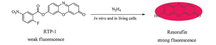
Herein, we designed and synthesized a novel resorufin based fluorescent “turn-on” probe for hydrazine detection in vitro and in living cells. Compared to other fluorophores, resorufin exhibits excellent physical and fluorescent properties including good water-solubility, high manoeuvrability and easy fluorescence quenching via 7-hydroxy substitution [24, 25, 26, 27, 28]. As illustrated in Scheme 1, the fluorophore resorufin is modified with 2-fluoro-5- nitrobenzoic acid, thus fluorescence of resorufin was quenched. Then, fluorescent probe RTP-1 is added to the detection system, incubated with hydrazine at ambient temperature. Hydrazine reacted with the ester group of fluorescent probe RTP-1 via nucleophilic addition-elimination reaction, accompanied by a distinct enhancement of the fluorescent intensity through the release of free resorufin.

|
Download:
|
| Scheme 1.Schematic illustration of the resorufin based fluorescent “turn-on” probe RTP-1 for detection of hydrazine levels. | |
All solvents and reagents were obtained from Alfa Aesar or Sigma-Aldrich, and the solvents were treated as required prior to use. Other chemicalswere commercially available and usedwithout further purification unless for special needs. HeLa cells were purchased from China Center for Type Culture Collection. UV/vis absorption spectra were collected on a Shimadzu UV-2550 spectrophotometer from 400 nm to 700 nm with 600 µL quartz cuvettes. Fluorescent emission spectra were collected from 570 nm to 650 nm on PerkinElmer LS 55 with an excitation wavelength of 550 nm, the excitation and emission slit widths were 15 nm and 3 nm, respectively. Quartz cuvettes with 2 mL volume were used for emission measurements. For determination of the fluorescence quantum yield (Yu), rhodamine B in water (Ys = 0.31) was used as a standard. Values were calculated according to the following equation.
where s means standard, umeans sample, A means absorbance at the excitation wavelength, F means integrate area under the fluorescence spectra on an energy scale. Unless otherwise specified, all spectra were taken at an ambient temperature. 1H and 13C NMR spectra were recorded on Varian Mercury 300 spectrometers, respectively. Data for 1H NMR spectra were recorded as follows: chemical shift (δ, ppm), multiplicity (s, singlet; d, doublet; t, triplet; q, quartet; m, multiplet), integration, coupling constant (Hz). HPLC data was collected with Lambo Model 2000. Mass spectrometry (MS) was recorded on Shimadzu LCMS-2010. High resolution mass spectrometry (HRMS) was recorded on Agilent 6224 Accurate-Mass Time-of-Flight (TOF) LC/MS.
Synthesis of probe RTP-1: Resorufin (213 mg, 1mmol), 2-fluoro- 5-nitrobenzoic acid (220 mg, 1.2 mmol) and 4-dimethylaminopyridine (DMAP, 12 mg, 0.1mmol) were dissolved in dry dichloromethane (20 mL) and stirred at 0 ℃. Then N, N-dicyclohexylcarbodiimide (DCC, 309 mg, 1.5 mmol) dissolved in dichloromethane (5 mL) were added to the solution dropwise and stirred at room temperature for 16 h. The precipitate of urea was removed by filtration and the filtratewas concentrated in high vacuumto give an oily residue. This residue was purified by silica gel column chromatography with eluent dichloromethane/ethyl acetate (v/v 20:1) to give RTP-1 (130 mg, 34%). 1H NMR (CDCl3, 300 MHz): δ 9.04 (d, 1H, J = 3.6 Hz), 8.56 (d, 1H, J = 9.6 Hz), 7.90 (d, 1H, J = 8.4 Hz), 7.48-7.42 (m, 2H), 7.33-7.26 (m, 2H), 6.91 (d, 1H, J = 9.9 Hz), 6.36 (s, 1H). 13C NMR (CDCl3, 75MHz): δ 186.4, 152.6, 149.2, 148.8, 135.4, 134.9, 131.7, 131.4, 130.8, 130.7, 128.7, 119.1, 118.8, 109.8, 107.5. HRMS calcd. for C19H9FN2O6 [M+H]+: 381.05174; found: 381.05177.
2.2. Cell cultureHeLa cells (CCTCC, China) were cultured in DMEM medium (Hyclone, Thermo Fisher Scientific Inc., MA) supplemented with 10% FBS (Hyclone, Thermo Fisher Scientific Inc., MA), 1% penicillin and streptomycin. For all cell experiments, cells were trypsinized and seeded in a 96-well culture plates, and for confocal experience, cells were seeded in 35 mmglass-bottomed dishes, then incubated in a humidified 37 ℃ incubator supplied with 5.0% CO2.
2.3. MTT assayHeLa cells were digested by trypsin and washed with PBS buffer (10 mmol/L, pH 7.4). The cells were seeded into a 96-well plate and incubated in a 5% CO2-containing incubator at 37 ℃ overnight for attachment. Then the cellswere treated with RTP-1 (1-500 µmol/L) for 48 h. Cell viability was evaluated via MTT assay. At first, freshly prepared MTT solution (10 µL, 5 mg/mL in PBS) was added to each well. After incubated for 4 h, the medium was removed and DMSO (100 µL) was added to each well. The optical density values were detected at 492 nm and the cytotoxicity data was expressed as relative cell viability.
2.4. Confocal imagingWhen the cells grew to the 50% confluence, RTP-1 (2 µmol/L, final concentration) was added to the complete culture medium from a 10 mmol/L stock in DMSO. After incubating with RTP-1 for 30 min, cells were washed 3 times in PBS followed by incubation with hydrazine (100 µmol/L) for another 30 min. Confocal fluorescence microscopy images of RTP-1-labeled cells were obtained by Laser Scanning Confocal microscope (Nikon, TE2000, Japan) with an oil objective lens (×60). The fluorescence emission at 560-610 nm was recorded using an excitation wavelength of 532 nm.
3. Results and discussion 3.1. Synthesis and characterizationIn our initial study, resorufin was used as the starting material for the synthesis of the fluorescent probe RTP-1 (Scheme 2). Resorufin reacted with 2-fluoro-5-nitrobenzoic acid using 4- dimethylaminopyridine (DMAP) as catalyst and N, N-dicyclohexyl- carbodiimide (DCC) as dehydrating agent. After one-step synthesis, the fluorescent probe RTP-1 was obtained and characterized by NMR and HRMS (Figs. S9-S11 in Supporting information).

|
Download:
|
| Scheme 2.Synthesis of the fluorescent “turn-on” probe RTP-1. | |
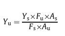
Firstly, the time-dependent fluorescent property of the probe RTP-1 toward hydrazine was evaluated. Since the pH value in human physiological environment has been reported to lie between pH 7 and 7.4, a phosphate-buffered saline (PBS, 10 mmol/L, pH 7.4) was chosen as simulated physiological conditions. In our experiments, fluorescent probe RTP-1 (5 µmol/L) in PBS (30% CH3CN, v/v) was incubated in the presence of hydrazine (100 µmol/L) within 1.5 h at 25 ℃. The emission intensity changes of the fluorescent probe RTP-1 at 589 nm was monitored continuously. As shown in Fig. 1, the fluorescent intensity of probe RTP-1 increased remarkably at the initial 2000 s, and then the rate of fluorescence enhancement tended to slowdown. Eventually, the fluorescence of the solution reached to a maximum after 5000 s. Since the reaction could be nearly completed within 30 min, we set 30 min as reaction time for further experiments.

|
Download:
|
| Fig. 1.Time-dependent fluorescent intensity changes of RTP-1 (5 µmol/L) in the presence of hydrazine (100 µmol/L) within 5000 s. The reactions were carried out at 25 ℃ in PBS buffer (10 mmol/L, pH 7.4, 30% CH3CN, v/v). All samples were excited at 550 nm and the data represent the fluorescent intensity at 589 nm. | |
Next, the pH dependency of RTP-1 was investigated (Fig. S1 in Supporting information). The results demonstrate that RTP-1 is stable when pH ranging from 7.0 to 8.0, which indicate RTP-1 is suitable to detect hydrazine under physiological conditions.
Afterwards, the absorption and fluorescent properties of the RTP-1 (5 µmol/L) solution containing different concentrations of hydrazine (changing from 0 to 100 µmol/L) were evaluated. The intensity of RTP-1 absorption band at 456 nm gradually decreased along with the increase of hydrazine concentration, while the intensity of a new absorption band at 576 nm gradually increased simultaneously. The color of the solution clearly changed from colorless to pink, indicate that RTP-1 could be utilized for hydrazine detection by naked eye (Fig. 2).

|
Download:
|
| Fig. 2.Absorbance changes of RTP-1 (5 µmol/L) with the addition of hydrazine at different concentrations (0, 5, 10, 20, 30, 40, 50, 60, 70, 80, 90, 100 µmol/L). The reactions were carried out for 30 min at 25 ℃ in PBS buffer (10 mmol/L, pH 7.4, 30% CH3CN, v/v). | |
In the fluorescence spectra (Fig. 3), we observed that the probe RTP-1 exhibited little fluorescence in the absence of hydrazine, while the peak emission intensity at 589 nm was highly enhanced along with the increasing concentration of hydrazine and ultimately reached to a value that was approximately 10 times higher compared to the initial emission intensity. The fluorescence quantum yield of RTP-1 (5 µmol/L), before and after the addition of hydrazine (100 µmol/L), was found to be 0.048 and 0.333, respectively. Hence, these results indicated the dose-dependent change in emission spectra of RTP-1 upon the addition of hydrazine. The fluorescent enhancement displayed a decent linear relationship (R2 = 0.9975) with hydrazine concentration ranging from 0 to 50 µmol/L, with a detection limit of 0.84 µmol/L (LOD = 3σ/S), thus indicating that RTP-1 is suitable for detecting hydrazine at micromolar concentrations.

|
Download:
|
| Fig. 3.(Left) Fluorescent emission changes of RTP-1 (5 µmol/L) with the addition of hydrazine at different concentrations (0, 5, 10, 20, 30, 40, 50, 60, 70, 80, 90, 100 µmol/L).The reactions were carried out for 30 min at 25 ℃ in PBS buffer (10 mmol/L, pH 7.4, 30% CH3CN, v/v). (Right) Plot of the fluorescent intensity at 589 nm against hydrazine concentration (0–50 µmol/L). | |
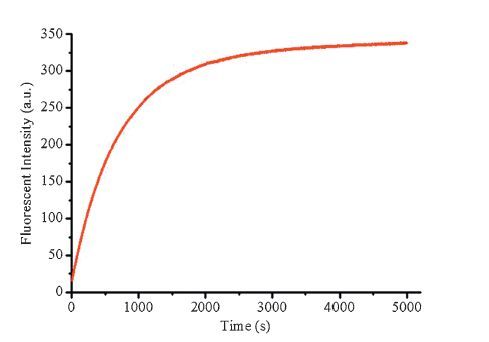
As the probe RTP-1 can sensitively detect hydrazine, we went on to evaluate the specificity of RTP-1 for hydrazine to various species that may interfere the detection of hydrazine. We investigated the effect of a number of metal ions, anions and amines on detection process, such as Cu2+ , Mg2+, Fe3+, Cr3+, Mn2+, F-, Cl-, Br-, SO3 2-, SO4 2-, S2O3 2-, HSO4 -, NH4+, H2PO4 -, NO2 -, CH3COO-, N3 -, n-butylamine, ethylenediamine (Fig. 4, Fig. S2 in Supporting information). The reaction between fluorescent probe RTP-1 and hydrazine exhibited a dramatic enhancement of the emission intensity, while the solutions of other interferent showed little changes in fluorescent intensity. Moreover, the antiinterference experiments also showed that the fluorescent intensity of RTP-1 in the presence of single hydrazine was similar to that in the presence of hydrazine and other interferent. Taken together, these observations suggested that all the interferent above would not interfere with hydrazine detection in the living cells.

|
Download:
|
| Fig. 4.Fluorescent intensity of RTP-1 (5 µmol/L) at 589 nm. Blue bars represent the addition of a single interferent. Red bars represent the fluorescence response after addition of hydrazine to the mixture.(1)Blank;(2)Cu2+;(3)Mg2+;(4)Fe3+;(5)Cr3+;(6)Mn2+;(7)F-;(8)Cl-;(9)Br-;(10)SO3 2-;(11)SO4 2-;(12)S2O3 2-;(13)HSO4 -;(14)NH4 +;(15)H2PO4 -;(16)NO2 -;(17)CH3COO-;(18)N3 -;(19)n-butylamine;(20)ethylenediamine. All solutions were incubated for 30 min at 25 ℃ in PBS buffer (10 mmol/L, pH 7.4, 30% CH3CN, v/v). | |
Based on the spectral changes, we propose that a nucleophilic addition-elimination reaction induced by hydrazine was occurred in the detection process. In order to verify the proposed mechanism, RTP-1 and the reaction product of RTP-1 and hydrazine were analysed by HPLC (Fig. S3 in Supporting information). RTP-1 displayed an absorption peak with a retention time of 19.0 min. When the mixture of RTP-1 (excess) and hydrazine was injected, a new peak at 8.8 min appeared which was consistent with the retention time of resorufin. When the mixture of RTP-1 and excess hydrazine was injected, the peak at 19.0 min completely disappeared leaving only a single peak at 8.8 min. Next, the product separated by HPLC was analysed by ESI MS (Fig. S4 in Supporting information). A peak at [M-H]- m/z 212.10, corresponding to resorufin was observed. Moreover, the density functional theory (DFT) with the B3LYP/6-31+G method basis set using the Gaussian 09 program was utilized to verify the hypothesis. The energy minimized form and charge surface diagram of RTP-1 are in favour of our hypothesis that the nucleophilic attack of hydrazine possibly takes place at the positively charged C-atom of ester group (Figs. S5, S6 in Supporting information).
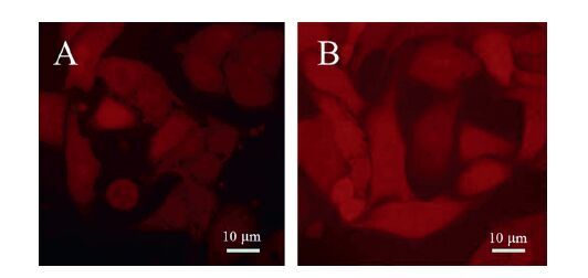
3.4. Cell imagingEncouraged by these results, we then assessed the feasibility of RTP-1 as a fluorescent probe for hydrazine detection in living cells. At first, we investigated the fluorescence response of RTP-1 toward hydrazine under a condition that suitable for living cells (Fig. S7 in Supporting information). RTP-1 showed a good enhancement of fluorescent intensity at 585 nm when excited at 550 nm. This result encouraged us to utilize RTP-1 for cell imaging. Hela cells were chosen for imaging of hydrazine by confocal fluorescence microscopy. The confocal fluorescence image of the HeLa cells labelled with probe RTP-1 (2 µmol/L) showed a relatively lower background emission. However, the fluorescence was clearly observed when the cells were further incubated with hydrazine (100 µmol/L) for 30 min (Fig. 5). The MTT assay confirmed cell viability during the imaging experiments (Fig. S8 in Supporting information). Herein, these results supported our hypothesis that RTP-1 can be used as a fluorescent “turn-on” probe for sensing hydrazine in living cells.

|
Download:
|
| Fig. 5.Confocal fluorescence images of HeLa cells: cells incubated with RTP-1 (2 µmol/L) for 30 min (A). Images of HeLa cells pre-incubated with probe RTP-1 followed by hydrazine (100 µmol/L) incubation for 30 min (B). Live Cells were used for imaging. Bar = 10 µm. | |
In summary, we have developed a novel resorufin based fluorescent “turn-on” probe RTP-1 for the detection of hydrazine levels. The synthesis of this probe is very facile, which only needs one step. RTP-1 could detect hydrazine levels sensitively and selectively within 30 min, along with obvious color changes from colorless to pink and enhancement of fluorescent intensity. We also used HPLC, ESI MS and DFT to verify that hydrazine was reacted with the ester group of fluorescent probe RTP-1 via nucleophilic addition-elimination reaction. Moreover, the probe was utilized to visualize hydrazine levels in living cells by confocal fluorescence microscopy. We expect that this fluorescent probe RTP-1 will be further useful to identify the influence of hydrazine in cellular systems.
AcknowledgmentsThis work was supported by the National Basic Research Program of China (973 Program, Nos. 2012CB720600, 2012CB720603), the National Science Foundation of China (Nos. 91413109, 21202126) and East Lake High-tech Zone 3551 Talents Scheme.
Appendix A. Supplementary dataSupplementary data associated with this article can be found, in the online version, at http://dx.doi.org/10.1016/j.cclet.2016.01.024.
| [1] | S.S. Narayanan, F. Scholz, A comparative study of the electrocatalytic activities of some metal hexacyanoferrates for the oxidation of hydrazine, Electroanalysis 11(1999) 465-469. |
| [2] | U. Ragnarsson, Synthetic methodology for alkyl substituted hydrazines, Chem. Soc. Rev. 30(2001) 205-213. |
| [3] | K. Yamada, K. Yasuda, N. Fujiwara, et al., Potential application of anion-exchange membrane for hydrazine fuel cell electrolyte, Electrochem. Commun. 5(2003) 892-896. |
| [4] | S. Garrod, M.E. Bollard, A.W. Nicholls, et al., Integrated metabonomic analysis of the multiorgan effects of hydrazine toxicity in the rat, Chem. Res. Toxicol. 18(2005) 115-122. |
| [5] | H.W. Schiessl, Hydrazine and its derivatives, in:Kirk-Othmer (Ed.), Encyclopedia of Chemical Technology, John Wiley & Sons, New York, 2000, pp. 562-607. |
| [6] | J.W. Mo, B. Ogorevc, X.J. Zhang, B. Pihlar, Cobalt and copper hexacyanoferrate modified carbon fiber microelectrode as an all-solid potentiometric microsensor for hydrazine, Electroanalysis 12(2000) 48-54. |
| [7] | E.H. Vernot, J.D. MacEwen, R.H. Bruner, et al., Long-term inhalation toxicity of hydrazine, Fundam. Appl. Toxicol. 5(1985) 1050-1064. |
| [8] | S.M. Sanins, J.A. Timbrell, C. Elcombe, J.K. Nicholson, Proton NMR spectroscopic studies on the metabolism and biochemical effects of hydrazine in vivo, Arch. Toxicol. 66(1992) 489-495. |
| [9] | C.A. Reilly, S.D. Aust, Peroxidase substrates stimulate the oxidation of hydralazine to metabolites which cause single-strand breaks in DNA, Chem. Res. Toxicol. 10(1997) 328-334. |
| [10] | J.A. Oh, J.H. Park, H.S. Shin, Sensitive determination of hydrazine in water by gas chromatography-mass spectrometry after derivatization with ortho-phthalaldehyde, Anal. Chim. Acta 769(2013) 79-83. |
| [11] | Z.K. He, B. Fuhrmann, U. Spohn, Coulometric microflow titrations with chemiluminescent andamperometricequivalencepoint detection:bromimetric titrationof low concentrations of hydrazine and ammonium, Anal. Chim. Acta 409(2000) 83-91. |
| [12] | A. Benvidi, P. Kakoolaki, H.R. Zare, R. Vafazadeh, Electrocatalytic oxidation of hydrazine at a Co(Ⅱ) complex multi-wall carbon nanotube modified carbon paste electrode, Electrochim. Acta 56(2011) 2045-2050. |
| [13] | X.X. Zhao, J.F. Zhang, W. Liu, et al., A unique dansyl-based chromogenic chemosensor for rapid and ultrasensitive hydrazine detection, J. Mater. Chem. B 2(2014) 7344-7350. |
| [14] | S. Goswami, S. Das, K. Aich, et al., A chemodosimeter for the ratiometric detection of hydrazine based on return of ESIPT and its application in live-cell imaging, Org. Lett. 15(2013) 5412-5415. |
| [15] | B. Chen, X. Sun, X. Li, H. Ågren, Y.S. Xie, TICT based fluorescence "turn-on" hydrazine probes, Sens. Actuators B 199(2014) 93-100. |
| [16] | Y. Sun, D. Zhao, S.W. Fan, L. Duan, A 4-hydroxynaphthalimide-derived ratiometric fluorescent probe for hydrazine and its in vivo applications, Sens. Actuators B 208(2015) 512-517. |
| [17] | Y.Q. Tan, J.C. Yu, J.K. Gao, et al., A new fluorescent and colorimetric probe for trace hydrazine with a wide detection range in aqueous solution, Dyes Pigments 99(2013) 966-971. |
| [18] | S. Goswami, S. Das, K. Aich, D. Sarkar, T.K. Mondal, A coumarin based chemodosimetric probe for ratiometric detection of hydrazine, Tetrahedron Lett. 55(2014) 2695-2699. |
| [19] | J. Zhao, Y.Q. Xu, H.J. Li, A.P. Lu, S.Q. Sun, A facile intracellular fluorescent probe for detection of hydrazine and its application, New J. Chem. 37(2013) 3849-3852. |
| [20] | L.L. Xiao, J. Tu, S.Q. Sun, et al., A fluorescent probe for hydrazine and its in vivo applications, RSC Adv. 4(2014) 41807-41811. |
| [21] | M.H. Lee, B. Yoon, J.S. Kim, J.L. Sessler, Naphthalimide trifluoroacetyl acetonate:a hydrazine-selective chemodosimetric sensor, Chem. Sci. 4(2013) 4121-4126. |
| [22] | Y. Qian, J. Lin, L.J. Han, L. Lin, H.L. Zhu, A resorufin-based colorimetric and fluorescent probe for live-cell monitoring of hydrazine, Biosens. Bioelectron. 58(2014) 282-286. |
| [23] | S. Goswami, K. Aich, S. Das, et al., A reaction based colorimetric as well as fluorescence 'turn on' probe for the rapid detection of hydrazine, RSC Adv. 4(2014) 14210-14214. |
| [24] | M. Sun, D.H. Shangguan, H.M. Ma, et al., Simple PbⅡ fluorescent probe based on PbⅡ-catalyzed hydrolysis of phosphodiester, Biopolymers 72(2003) 413-420. |
| [25] | W.Z. Gao, B.G. Xing, R.Y. Tsien, J.H. Rao, Novel fluorogenic substrates for imaging b-lactamase gene expression, J. Am. Chem. Soc. 125(2003) 11146-11147. |
| [26] | S.Y. Kim, J.I. Hong, Chromogenic and fluorescent chemodosimeter for detection of fluoride in aqueous solution, Org. Lett. 9(2007) 3109-3212. |
| [27] | M.G. Choi, S.Y. Cha, J.E. Park, et al., Selective perborate signaling by deprotection of fluorescein and resorufin acetates, Org. Lett. 12(2010) 1468-1471. |
| [28] | M.G. Choi, J. Hwang, S. Eor, S.K. Chang, Chromogenic and fluorogenic signaling of sulfite by selective deprotection of resorufin levulinate, Org. Lett. 12(2010) 5624-5627. |