 2016, Vol.27


b Laboratoire d'Electrochimie Physique et Analytique, Ecole Polytechnique Fédérale de Lausanne, Switzerland
Microfludic system has been recently reported as an interface between liquid chromatography and mass spectrometry (MS) hyphenation for online protein digestion [1] and enrichment [2]. It also provides an attractive concept for the online coupling of electrophoresis and MS by incorporating the miniaturized microchips into commercial electrospray ionization (ESI)-MS instruments [3]. Alternatively,the microchip electrophoresis can be hyphenated offline with matrix-assisted laser desorption/ionization (MALDI)-MS by collecting the fractionated samples in a rotating ball inlet [4] of MALDI source,or introducing the separated samples to MALDI chamber through on-chip "monitoring windows" [5]. Furthermore,electrophoresis has been reported to be performed in completely open channels [6],closed-open-closed channels [7],or pseudo-closed channels with removable covers [8] for subsequent offline MALDI-MS detection.
Recently,a new ambient ionization strategy,namely electrostatic spray ionization (ESTASI),has been presented for the in situ generation of molecular ions from samples deposited on an insulating surface [9]. It features the screening of dried samples under ambient conditions,showing great flexibility for MS-based chemical and biochemical analysis. As an attractive alternative to MALDI for the offline coupling of MS with electrophoresis,ESTASI has been reported for the ionization of isoelectric focusing electrophoretic bands in polyacrylamide gels [10]. Herein,ESTASI is further proposed for in situ ionization and detection of dried electrophoretic bands obtained in open channel.
2. ExperimentalInformation about the chemicals and materials,as well as the microchip fabrication procedure,is included in Supporting information. Open channel based microchip electrophoresis was performed for two fluorescent dyes as illustrated in Fig. 1a to optimize experimental conditions. After the electrophoretic separation and solvent evaporation,dried fractions formed in the open channel were in situ ionized by ESTASI,as schematically shown in Fig. 1b. Electrophoretic separation and MS detection of antibiotics was afterwards carried out with the same protocol for further proof-of-concept. More details about the experimental procedure can be found in Supporting information.

|
Download:
|
| Fig. 1.Schematic representation of (a) open channel-based microchip electrophoresis and (b) ESTASI-MS scanning of the open channel. EOF: electroosmotic flow; HV: high voltage. | |
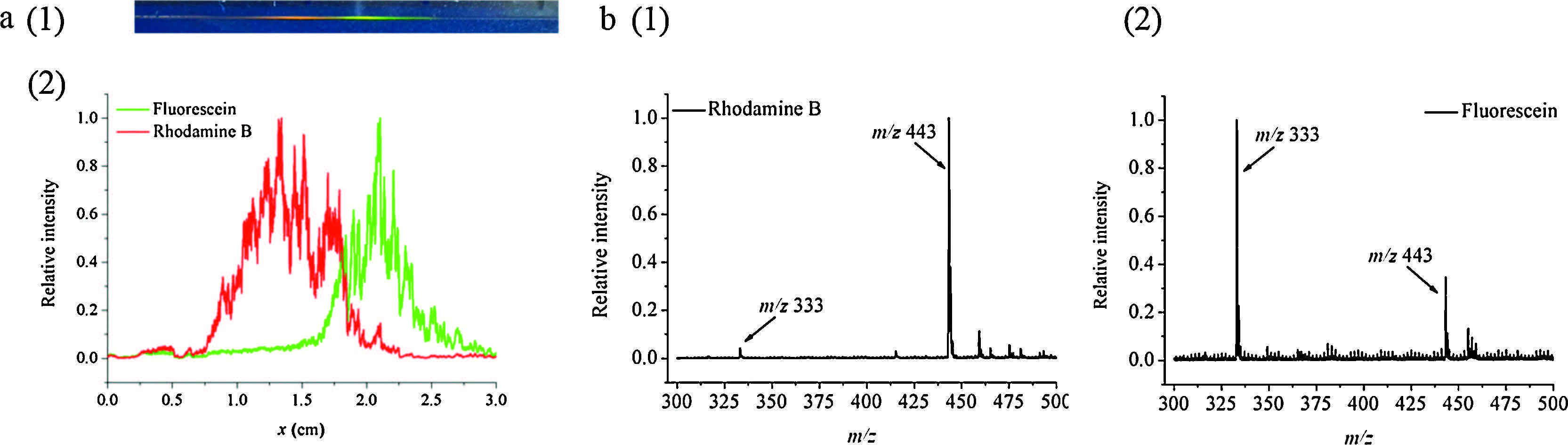
The optimization of conditions for the microchip electrophoresis and ESTASI-MS detection is in the Supporting information. Glycerol was employed to reduce the longitudinal diffusion of electrophoretic bands during solvent evaporation in the open channel. The electrophoretic bands of fluorescent dyes obtained under optimized separation conditions were imaged by a digital camera (Fig. 2a (1)),and analyzed by in situ ESTASI-MS to generate the electropherogram (Fig. 2a (2)). The MS peak intensities for rhodamine B (RhB) and fluorescein were normalized,respectively,and plotted as a function of position in the microchannel. The wide electrophoretic bands in both detection modes were attributed to the sample overloading in order to make the dyes easily visible under UV excitation.

|
Download:
|
| Fig. 2.(a) Bands of fluorescent dyes (0.5 mg/mL fluorescein,and 0.1 mg/mL RhB) by open channel-based microchip electrophoretic separation with digital camera imaging under UV excitation at 254 nm (1) and electropherogram from in situ scanning ESTASI-MS (2). (b) Mass spectra of RhB obtained at x = 1.32 cm (1) and fluorescein at x = 2.09 cm (2). | |
Compared with those in Fig. 2a (1),the peak widths in the extracted ion electropherogram were slightly broadened,possibly resulted from the increased mobility of glycerol under the high temperature of the ionization source. A less important factor was contributed to the delayed detection in offline hyphenation. However,its influence on the band broadening is almost negligible because of the very high viscosity of glycerol under room temperature. The channel with fractionated sample in glycerol can be stored under 4 ℃ for long term.
Fig. 2b presents the mass spectra extracted from the electropherogram. At x = 1.32 cm,the migration distance of RhB,the mass spectrum demonstrates a very strong peak of RhB with a tiny peak of fluorescein. In contrast,at x = 2.09 cm,the peak of fluorescein is predominant,while RhB still produces a peak with noticeable signal-to-noise ratio (S/N) due to its higher ionization efficiency. All in all,open channel microchip electrophoresis and ESTASI-MS experiments performed for fluorescent dyes demonstrate the in situ detection of electrophoretic bands by MS,which was further proven by the proof-of-concept results obtained with antibiotic mixture as the test sample (Fig. S2 in Supporting information).
4. ConclusionA novel interface between open channel-based microchip electrophoresis and MS is developed via ESTASI. In contrast with the prevalent offline coupling of CE to MALDI-MS based on CE effluent collection,the main characteristic of the proposed interface is the in situ ionization and detection of electrophoretic bands. Remaining challenges lie in the loss of the separation resolution and detection sensitivity,which could be further improved by applying narrow channel or highly efficient electrophoretic mode. Nevertheless,the proposed new interface can be potentially applied for two-dimension scanning of microarray-based high throughput electrophoresis.
AcknowledgmentYan Deng thanks the Chinese Scholarship Council for financial support.
Appendix A. Supplementary dataSupplementary data associated with this article can be found,in the online version,at http://dx.doi.org/10.1016/j.cclet.2015.09.017.
| [1] | J. Ji, L. Nie, L. Qiao, et al., Proteolysis in microfluidic droplets:an approach to interface protein separation and peptide mass spectrometry, Lab Chip 12(2012) 2625-2629. |
| [2] | J. Ji, L. Nie, Y.X. Li, P.Y. Yang, B.H. Liu, Simultaneous online enrichment and identification of trace species based on microfluidic droplets, Anal. Chem. 85(2013) 9617-9622. |
| [3] | A.G. Chambers, J.S. Mellors, W.H. Henley, J.M. Ramsey, Monolithic integration of two-dimensional liquid chromatography-capillary electrophoresis and electrospray ionization on a microfluidic device, Anal. Chem. 83(2011) 842-849. |
| [4] | H.K. Musyimi, J. Guy, D.A. Narcisse, S.A. Soper, K.K. Murray, Direct coupling of polymer-based microchip electrophoresis to online MALDI-MS using a rotating ball inlet, Electrophoresis 26(2005) 4703-4710. |
| [5] | M. Brivio, N.R. Tas, M.H. Goedbloed, H.J. Gardeniers, W. Verboom, A. van den Berg, D.N. Reinhoudt, A MALDI-chip integrated system with a monitoring window, Lab Chip 5(2005) 378-381. |
| [6] | J. Liu, K. Tseng, B. Garcia, et al., Electrophoresis separation in open microchannels. A method for coupling electrophoresis with MALDI-MS, Anal. Chem. 73(2001) 2147-2151. |
| [7] | J. Jacksen, T. Frisk, T. Redeby, et al., Off-line integration of CE and MALDI-MS using a closed-open-closed microchannel system, Electrophoresis 28(2007) 2458-2465. |
| [8] | M.L.S. Mok, L. Hua, J.B.C.Phua, M.K.T. Wee, N.S.K. Sze, Capillary isoelectric focusing in pseudo-closed channel coupled to matrix assisted laser desorption/ionization mass spectrometry for protein analysis, Analyst 129(2004) 109-110. |
| [9] | L. Qiao, R. Sartor, N. Gasilova, et al., Electrostatic-spray ionization mass spectrometry, Anal. Chem. 84(2012) 7422-7430. |
| [10] | L. Qiao, E. Tobolkina, B. Liu, H.H. Girault, Coupling isoelectric focusing gel electrophoresis to mass spectrometry by electrostatic spray ionization, Anal. Chem. 85(2013) 4745-4752. |