 2015, Vol.26


In recent years,a variety of inorganic-organic hybrid nanomaterials have emerged as new platforms for controlled release applications [1, 2, 3, 4]. In these systems,inorganic nanomaterials offer a kind of robust framework,while the incorporated organic building blocks give functionalities. Among the existing inorganic nanomaterials,mesoporous silica nanoparticles (MSNs) can serve as ideal containers for drug controlled release systems due to their simple,controllable and cost-effective fabrication,extremely high surface areas,uniformly sized pores,and good biocompatibility [5, 6]. Organic nanomaterials acted as the “gate” of MSNs [7, 8, 9, 10],which can allow the releasing of entrapped molecules just in specific environments in response to external stimuli [11, 12, 13, 14, 15]. Of the various applied stimuli,pH-sensitive system has received particular attention,because it has been shown that the pH values of interstitial or extracellular environment in tumors were lower than that of normal tissues and bloodstream,and lysosomes exhibited even lower pH value [16]. Therefore,pH-sensitive controlled release containers are highly desired in practical applications.
Smart polymers can undergo conformational and chemical changes in response to external stimuli,and therefore are often prepared for controlled release application. It has been reported that the biocompatible polymer coatings can not only reduce the toxicity of MSNs but also provide colloidal stability,handle conjugation of targeting moieties and improve blood circulation lifetimes [17]. Hence,many stimuli-responsive polymers were grafted onto the external surface of MSNs for pH-sensitive controlled release application. For instance,Pan et al. used poly[2-(diethylamino)ethyl methacrylate] (PDEAEMA) as the stimuli-responsive polymer to construct a MSN-based pH-controlled release system. For PDEAEMA was a hydrophobic collapsed state at high pH and was a hydrophilic swollen state as a weak cationic polyelectrolyte at low pH [18]. However,in polymer capped MSN system,the leakage at neutral and alkaline conditions limited its usage in practical applications,so minimal release of drugs at these conditions is meaningful.
Herein,we integrated the advantages of MSNs and organic polymers to develop a method to adjust the release behavior of pH-sensitive hybrid nanoparticles. Four kinds of poly(4-vinylphenybronic acid-co-2-(dimethylamino)ethyl acrylate) [P(VPBADMAEA)] capped MSNs were synthesized,which have different mole ratio of two kinds of sensor moieties,including [2-(dimethylamino)ethyl acrylate] (DMAEA) (pKa ≈ 7.5) and 4-vinylphenybronic (VPBA) (pKa ≈ 8.9) [19]. At high pH,the polymer collapsed onto the mesopores of the silica nanoparticles,and gated the model molecules within the MSNs. By contrast,low pH condition lead to positive charge and solubilization of the polymer,resulting in the release of the cargo from the MSNs (Fig. 1). Under the same pH condition,the release behavior of the hybrid nanoparticles could be adjusted. At neutral and alkaline conditions,the leakage of model molecules from the hybrid nanoparticles could be decreased by increasing the mole ratio of VPBA.

|
Download:
|
| Fig. 1.Schematic illustration for the “off” and “on” release of Ru(bipy)32+ fromRu(bipy)32+/MSN@polymer at different pH. | |
Tetraethylorthosilicate (TEOS) was obtained from Xilong Reagent Company (Guangdong,China). N-Cetyltrimethylammonium bromide (CTAB) was purchased from Alfa Aesar. 4- Vinylphenylboronic acid (VPBA),2-(dimethylamino)ethyl acrylate (DMAEA),3-(methacryloxy)porpyltrimethoxysilane (MPS),2,2'-azobis(2-methylpropionamidine) dihydrochloride (AAPH),[Ru(bipy)3]Cl2 (bipy = 2,2'-bipyridine) were bought from J & K Chemical Technology. All of the chemicals were of analytical grade and used without further purification. All solutions were prepared with ultrapure Milli-Q water (Resistance > 18.2 MΩ cm).
2.2. CharacterizationThe morphology images of the nanoparticles were studied using a JEOL 3010 Transmission electron microscopy (TEM). Smallangle powder X-ray diffraction patterns (XRD) of the MSN-based nanoparticles were obtained from a Scintag XDS-2000 powder diffractometer,using Cu Kα irradiation (λ = 0.154 nm). Fourier transform infrared (FTIR) spectra were obtained from a TENSOR 27 spectrometer,Bruker Instruments Inc.,Germany. Zeta potential experiments were performed at 25°C using a Malvern Zeta Sizer Nano instrument. N2-adsorption-desorption isotherms were obtained on a Micromeritics ASAP 2010 sorptometer. Brunauer- Emmett-Teller (BET) surface area was calculated from the linear part of the BET plot according to IUPAC recommendations. Pore size distribution was estimated from the adsorption branch of the isotherm by the Barrett-Joyner-Halenda (BJH) method. All fluorescence spectra were recorded on a Hitachi F-7000 spectrophotometer in PBS buffer.
2.3. Synthesis of Ru(bipy)32+/MSN@polymer nanoparticlesFirstly,3-(methacryloxy)porpyltrimethoxysilane modified mesoporous silica nanoparticles (MSN-MPS) were prepared following the literature procedure [20]. The experiment details were provided in S-1 of Supporting information. Secondly,MSNMPS (50 mg) was added to 50 mL of 1 mmol/L Ru(bipy)32+ solution,and the mixture was stirred at room temperature for 24 h. Thirdly,Ru(bipy)32+/MSN@polymer nanoparticles were synthesized according to literature procedure [18] (Fig. S1b,see S-2 of Supporting information for further details). The total mole of the sensor moieties was 0.396 mmol. Ru(bipy)32+/MSN@polymer nanoparticles gated with DMAEA and VPBA were coded as Ru(bipy)32+/MSN@p1,Ru(bipy)32+/MSN@p2,Ru(bipy)32+/ MSN@p3,and Ru(bipy)32+/MSN@p4,and the mole ratios of DMAEA and VPBA were 5:0,3:2,2:3 and 1:4,respectively. The MSN@polymer nanoparticles used in cytotoxicity experiment were synthesized by the same method,but without the loading of Ru(bipy)32+.
2.4. Ru(bipy)32+ releaseRu(bipy)32+ release experiments were performed in phosphate buffer solution (PBS) at pH values of 2.0,4.0,5.0,6.0,7.0,8.0,and 10.0,respectively. 1 mg Ru(bipy)32+/MSN@polymer nanoparticles were shaken in 1 mL buffer solution at 37°C at 300 rpm. After a predetermined time interval,10 μL of supernatant buffer solution was taken out after centrifugation,and 10 μL fresh buffer solution was refilled. Subsequently,the release profiles of Ru(bipy)32+ from the pores to aqueous solution were monitored via the fluorescence intensity of the dye centered at 595 nm. Time-dependent release of Ru(bipy)32+ from the Ru(bipy)32+/MSN@polymer nanoparticles was studied for 330 min.
2.5. Cell culture and cytotoxicity assayThe cytotoxicity of the particles was evaluated using HeLa cells by MTT assay. HeLa cells were purchased from the Cancer Institute & Hospital (Chinese Academy of Medical Sciences). The culture and the viability measurement of cells were performed as described previously (see S-3 of Supporting information for further details) [21]. The concentrations of MSN@polymer nanoparticles incubated with cells ranging from 0 to 400 μg/mL for 24 h.
3. Results and discussion 3.1. Synthesis and characterization of polymer-coated nanoparticlesFor the design of the hybrid nanoparticles with pH-sensitive valve,MSN-41-type MSNs were selected as framework and P(VPBA-DMAEA) as the pH-sensitive nanovalve. Ru(bipy)32+/ MSN@polymer nanoparticles were synthesized,and the size and pore structure of MSN-MPS and Ru(bipy)32+/MSN@polymer nanoparticles were characterized by TEM and XRD. The TEM image in Fig. 2(a) showed that the MSN-MPS had a diameter of 100 nm and a typical hexagonal channel-like pore. After loading Ru(bipy)32+ and capping polymers,the surface of the hybrid nanoparticles (Fig. 2(b)-(e)) were not as smooth as MSN-MPS. The XRD in the 2° < 2θ < 7° range both exhibited three low-angle reflections typical of hexagonal array [21],which could be indexed as (1 0 0),(1 1 0),and (2 0 0) (Fig. S2,Supporting information). For this material,reflections (1 1 0) and (2 0 0) were mostly lost due to a reduction in contrast related to the functional process and the filling of mesopores with Ru(bipy)32+. Nonetheless,the intensity of the (1 0 0) peak in this pattern strongly indicates that the loading process with the dye and the additional functionalization with polymer did not modify the mesoporous MCM-41 scaffold. Furthermore,the N2-adsorption-desorption isotherm of MSN-MPS nanoparticles showed a typical type IV curve with a surface area of 1140.63 m2/g. Barrett-Joyner-Halenda (BJH) pore-size distribution exhibited a single peak and implied the formation of uniform pores of size 2.5 nm (Fig. S3,Supporting information). Moreover,in contrast to MSNs with zeta potential of 27.4 mV,the zeta potential of MSN-MPS before and after removed template changed from 13.4 mV to -22.2 mV indicating the successful modification of MPS and the remove of templates. Comparative analysis of FTIR spectra of MSNs (without template),MSN-MPS,and MSN@polymer nanoparticles provided evidence for the grafting of polymers onto MSNs (Fig. S4,Supporting information). A new band attributed to -CH3 from MPS and DMAEA appeared at 2930 cm-1,and the peak at 1426 cm-1 was the characteristic absorbance band of -B-O-,which proved evidences of the grafting successfully. In addition,the variation zeta potential of MSN@polymer nanoparticles was from about 30 mV to -30 mV with change in the range of pH 3.0-10.0 (Fig. S5,Supporting information),indicating realized pH controlled release.

|
Download:
|
| Fig. 2.TEM images of MSN-MPS and Ru(bipy)32+/MSN@polymer nanoparticles. (a) MSN-MPS; (b) Ru(bipy)32+/MSN@p1; (c) Ru(bipy)32+/MSN@p2; (d) Ru(bipy)32+/MSN@p3;(e) Ru(bipy)32+/MSN@p4. All scale bars are 50 nm. | |
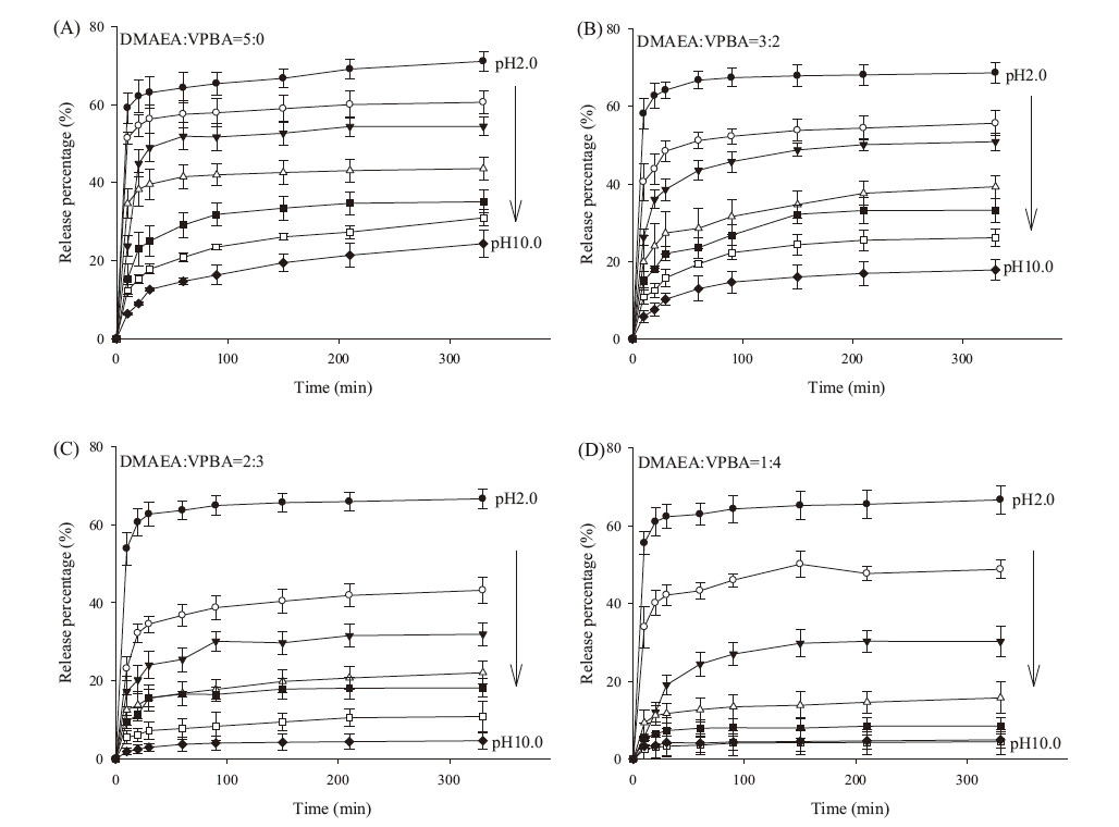
To investigate the pH effect upon Ru(bipy)32+ release behavior,Ru(bipy)32+/MSN@polymer nanoparticles were treated at pH 2.0,4.0,5.0,6.0,7.0,8.0 and 10.0,then fluorescence intensity of the released Ru(bipy)32+ in the supernatant was measured. As shown in Fig. 3,the release behavior of Ru(bipy)32+/MSN@polymer nanoparticles demonstrated that the release of Ru(bipy)32+ was pH-dependent,i.e.,all these Ru(bipy)32+/MSN@polymer nanoparticles can release Ru(bipy)32+ at low pH. When pH value of the solution was increased,the amount of released model molecules was reduced. It can be explained that at low pH environment the polymers were protonated and swollen,then the preloaded molecules could release from the nanopores.

|
Download:
|
| Fig. 3.The release of Ru(bipy)32+ from Ru(bipy)32+/MSN@polymer nanoparticles at different pH values (2.0, 4.0, 5.0, 6.0, 7.0, 8.0, 10.0). (A) Ru(bipy)32+/MSN@p1; (B)Ru(bipy)32+/MSN@p2; (C) Ru(bipy)32+/MSN@p3; (D) Ru(bipy)32+/MSN@p4. | |
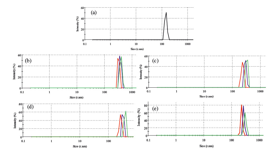
To prove this,TEM images of the Ru(bipy)32+/MSN@polymer nanoparticles after incubation in pH 2.0 were given in Fig. S6 in Supporting information. As shown in the images,there is almost no difference between the nanoparticles in Fig. 2 and Fig. S6,since the nanoparticles should be dried when TEM samples were prepared. Dynamic light scattering of MSN-MPS and Ru(bipy)32+/MSN@polymer nanoparticles were given in Fig. 4. The results showed that the size of nanoparticles increased when the pH value decreased.

|
Download:
|
| Fig. 4.Size distribution by intensity of MSN-MPS and Ru(bipy)32+ /MSN@polymer nanoparticles. (a) MSN-MPS; (b) Ru(bipy)32+ /MSN@p1; (c) Ru(bipy)32+ /MSN@p2; (d)Ru(bipy)32+ /MSN@p3; (e) Ru(bipy)32+ /MSN@p4. Black represent water, red, duck purple and green represent PBS (pH 7.0, 4.0 and 2.0). | |
It should be noted that the release behavior of Ru(bipy)32+/ MSN@polymer nanoparticles could be adjusted by changing the mole ratio of DMAEA and VPBA. For Ru(bipy)32+/MSN@p1,the release percentage in high pH solutions were quite large (pH 8.0 and pH 10.0 was 31.0% and 24.3%); for Ru(bipy)32+/MSN@p4,the release percentage in high pH solutions were decreased obviously (pH 8.0 and pH 10.0 was 4.4% and 4.9%) (Fig. 3). In other words,addition of VPBA can lower the release percentage of the model molecules at neutral and alkaline conditions. The possible reason may be the generation of a strong intramolecular Bδ-...Nδ+ bond between DMAEA and VPBA [19],the shrinking of the polymers were different with the mole ratio of sensor moieties changing. Considering the release behaviors of these hybrid nanoparticles,we believe that this method have promise for designing controlled release system in practical applications.
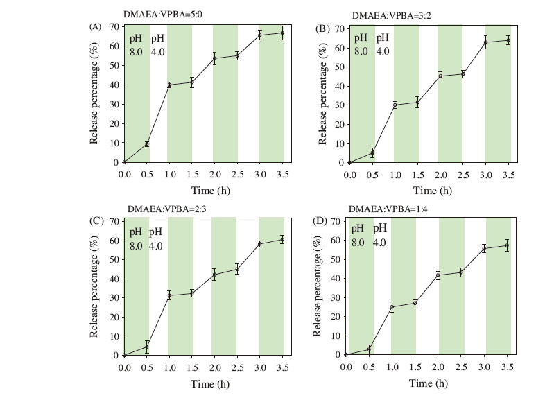
In the design of controlled release system,not only was it important to achieve drug delivery as required,but also to be reversible for some applications [22]. To investigate whether the release of model molecules from this hybrid nanoparticles could be switched "on" and "off",the release of Ru(bipy)32+ from Ru(bipy)32+/MSN@polymer nanoparticles were carried out at pH 4.0 and 8.0. Fig. 5 shown that the valves of the hybrid nanoparticles were closed at pH 8.0,while were open at pH 4.0. The results also indicated that the release of the model molecules can be open and closed reversibly.

|
Download:
|
| Fig. 5.The “on” and “off” property of Ru(bipy)32+ release from Ru(bipy)32+/MSN@polymer nanoparticles, while pH vary between 4.0 and 8.0. (A) Ru(bipy)32+/MSN@p1; (B)Ru(bipy)32+/MSN@p2; (C) Ru(bipy)32+/MSN@p3; (D) Ru(bipy)32+/MSN@p4. | |
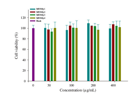
Biocompatibility of MSNs should be investigated since it is one of the primary concerns of nanocontainers for drug delivery application. Cells without the treatment of nanoparticles were taken as the control group and the viability was set as 100%. The final report data were expressed as a percentage of the control group. The cytotoxicity of MSN@polymer nanoparticles wasmeasured by MTT assay in HeLa cell,which was widely used to measure the mitochondria activity to quantify the cell growth. The HeLa cells were treated with MSN@polymer nanoparticles samples at various concentrations (50,100,200 and 400 μg/mL) for 24 h (Fig. 6). The toxicity was not observed even the concentration was up to 400 μg/mL. Therefore,we confirmed that using MSN@polymer nanoparticles as nanocontainers had good biocompatibility,which revealed the potential use for developing a smart drug delivery system in vivo.

|
Download:
|
| Fig. 6.Cell viability of HeLa cell after incubation with MSN@polymer nanoparticles for 24 h. | |
In summary,we have reported a simple method to decrease the leakage of model molecules from pH-sensitive MSNs at neutral and alkaline conditions. In this study,we synthesized four kinds of pH regulated polymer-gated MSNs with different mole ratio of DMAEA and VPBA. The addition of VPBA decreased the release percentage of the model molecules from MSNs in neutral and alkaline media solutions. Moreover,the polymer-gated nanocontainers could release model molecules reversibly and showed good biocompatibility. Therefore,they would be potential candidates for application in biomedical fields.
AcknowledgmentsThis work was supported by the National Natural Science Foundation of China (Nos. 21190040,21175035,21375034),National Basic Research Program of China (No. 2011CB911002) and International Science & Technology Cooperation Program of China (No. 2010DFB30300).
Appendix A. Supplementary dataSupplementary data associated with this article can be found,in the online version,at http://dx.doi.org/10.1016/j.cclet.2015.08.005.
| [1] | Y.M. Yang, F. Liu, X.G. Liu, B.G. Xing, NIR light controlled photorelease of siRNA and its targeted intracellular delivery based on upconversion nanoparticles, Nanoscale 5(2013) 231-238. |
| [2] | T. Chen, N.W. Yang, F.J. Fu, Controlled release of cargo molecules from hollow mesoporous silica nanoparticles based on acid and base dual-responsive cucurbit[7] uril pseudorotaxanes, Chem. Commun. 49(2013) 6555-6557. |
| [3] | A. Al-Nahain, S.Y. Lee, I. In, K.D. Lee, S.Y. Park, Triggered pH/redox responsive release of doxorubicin from prepared highly stable graphene with thiol grafted Pluronic, Int. J. Pharm. 450(2013) 208-217. |
| [4] | Y. Zhou, H. Li, Y.W. Yang, Controlled drug delivery systems based on calixarenes, Chin. Chem. Lett.26(2015)825-828. |
| [5] | X.X. Hu, Y. Wang, B. Peng, Chitosan-capped mesoporous silica nanoparticles as pHresponsive nanocarriers for controlled drug release, Chem. Asian J. 9(2014) 319-327. |
| [6] | Y. Xiao, T. Wang, Y. Cao, et al., Enzyme and voltage stimuli-responsive controlled release system based on β-cyclodextrin-capped mesoporous silica nanoparticles, Dalton Trans. 44(2015) 4355-4361. |
| [7] | S.S. Wu, X. Huang, X.Z. Du, Glucose- and pH-responsive controlled release of cargo from protein-gated carbohydrate-functionalized mesoporous silica nanocontainers, Angew. Chem. Int. Ed. 125(2013) 5690-5694. |
| [8] | M.H. Yu, S. Jambhrunkar, P. Thorn, et al., Hyaluronic acid modified mesoporous silica nanoparticles for targeted drug delivery to CD44-overexpressing cancer cells, Nanoscale 5(2013) 178-183. |
| [9] | H. Yan, C. Teh, S. Sreejith, et al., Functional mesoporous silica nanoparticles for photothermal-controlled drug delivery in vivo, Angew. Chem. Int. Ed. 51(2012) 8373-8377. |
| [10] | C. Giménez, C. de la Torre, M. Gorbe, et al., Gatedmesoporous silica nanoparticles for the controlled delivery of drugs in cancer cells, Langmuir 31(2015) 3753-3762. |
| [11] | W. Feng, X.J. Zhou, C.L. He, et al., Polyelectrolyte multilayer functionalized mesoporous silica nanoparticles for pH-responsive drug delivery:layer thickness-dependent release profiles and biocompatibility, J. Mater. Chem. B 1(2013) 5886-5898. |
| [12] | Y.F. Jiao, Y.F. Sun, B.S. Chang, D. Lu, W.L. Yang, Redox- and temperature-controlled drug release from hollow mesoporous silica nanoparticles, Chem., Eur. J. 19(2013) 15410-15420. |
| [13] | N.Ž. Knězević, V.S.-Y. Lin, A magnetic mesoporous silica nanoparticle-based drug delivery system for photosensitive cooperative treatment of cancer with a mesopore-capping agent and mesopore-loaded drug, Nanoscale 5(2013) 1544-1551. |
| [14] | A. Popat, B.P. Ross, J. Liu, et al., Enzyme-responsive controlled release of covalently bound prodrug from functional mesoporous silica nanospheres, Angew. Chem. Int. Ed. 51(2012) 12486-12489. |
| [15] | L. Sun, X.G. Zhang, Z.M. Wu, C. Zheng, C.X. Li, Oral glucose- and pH-sensitive nanocarriers for simulating insulin release in vivo, Polym. Chem. 5(2014) 1999-2009. |
| [16] | J. Zheng, X.J. Tian, Y.F. Sun, D. Lu, W.L. Yang, pH-sensitive poly (glutamic acid) grafted mesoporous silica nanoparticles for drug delivery, Int. J. Pharm. 450(2013) 296-303. |
| [17] | M. Chen, X.X. He, K.M. Wang, et al., A pH-responsive polymer/mesoporous silica nano-container linked through an acid cleavable linker for intracellular controlled release and tumor therapy in vivo, J. Mater. Chem. B 2(2014) 428-436. |
| [18] | J.T. Sun, C.Y. Hong, C.Y. Pan, Fabrication of PDEAEMA-coated mesoporous silica nanoparticles and pH-responsive controlled release, J. Phys. Chem. C 114(2010) 12481-12486. |
| [19] | W.T. Wu, N. Mitra, E.C.Y. Yan, S.Q. Zhou, Multifunctional hybrid nanogel for integration of optical glucose sensing and self-regulated insulin release at physiological pH, ACS Nano 4(2010) 4831-4839. |
| [20] | Y. Tian, A. Glogowska, W. Zhong, T. Klonisch, M. Xing, Polymeric mesoporous silica nanoparticles as a pH-responsive switch to control doxorubicin intracellular delivery, J. Mater. Chem. B 1(2013) 5264-5272. |
| [21] | Z. Zou, D.G. He, X.X. He, et al., Natural gelatin capped mesoporous silica nanoparticles for intracellular acid-triggered drug delivery, Langmuir 29(2013) 12804-12810. |
| [22] | D.G. He, X.X. He, K.M. Wang, et al., Intracellular acid-triggered drug delivery system using mesoporous silica nanoparticles capped with T-Hg2+-T base pairs mediated duplex DNA, J. Mater. Chem. B 1(2013) 1552-1560. |