


 , Yan Yanga, Jie Weia, Jun Wanga, Ming-Wei Wangb,c,d , Hong Yanga, Ying-Jian Zhangb,c,d, Shi-Ping Yanga
b Department of Nuclear Medicine, Fudan University Shanghai Cancer Center, Shanghai 200032, China;
c Center for Biomedical Imaging, Fudan Univerisity, Shanghai 200032, China;
d Department of Oncology, Shanghai Medical College, Fudan University, Shanghai 200032, China
In recent years,two-dimensional (2D) nanomaterials have attracted great research efforts because of their fascinating electronic and optical properties. Graphene with excellent electronic, optical,thermal,and mechanical properties has been investigated for a long time [1, 2, 3]. Beyond graphene,its analogs of layered transition metal dichalcogenides (MX2) such as MoS2 [4], MoSe2 [5],WS2 [6],and WSe2 [7] have become a hot research field. Although all of these materials combine each layer with the strong covalent bond,they have weak van der Waals forces between different MX2 sheets. Therefore,it provides a possibility to prepare the nanosheets by overcoming the van der Waals forces between sheets.With regard to their applications,MX2 nanosheets have been already extensively investigated in the fields of integrated circuits [8],scanning probe microscopy [9, 10],catalysts [11, 12],field-effect transistors [13, 14],and thermoelectric devices [15, 16]. Very few reports have been reported for the theranostic application.
Photothermal therapy (PTT),which depends on a photosensitizer to absorb the near-infrared (NIR) light and generate heat from the optical energy,leads to the thermal ablation of cancer cells. During the past decade,a number of nanomaterials with a relatively high photothermal conversion efficiency have been widely explored by many research groups for PTT ablation of cancer cells,such as metal nanomaterials (Au,Pd) [17, 18, 19],carbon nanomaterials (carbon nanotube and graphene) [20, 21],metal sulfide and oxide nanomaterials [22, 23] as well as organic nanoparticles [24]. Recently,we have developed WO3-x nanorods for X-ray computed tomography (CT) guided photothermal therapy [25],and Fe@Fe3O4 nanoparticles for magnetic targeted photothermal therapy [26]. X-ray CT based on different substance with different density is an important molecular imaging technology with the better spatial and density resolution than other imaging modalities. Up to date,the clinical iodine-based CT contrast agents have severe limitations including the relatively short blood circulation time due to the rapid renal clearance, nonspecific biodistribution,renal toxicity,and vascular permeation [27]. Therefore,novel nanoparticulated CT contrast agents have been recently investigated,such as Au [28, 29],FePt [30],Bi2S3 [31],TaOx [32],and ytterbium-based nanomaterials [33].
In this paper,we demonstrated the use of WS2 nanosheets for CT imaging and NIR photothermal ablation of cancer cells. The photothermal therapy effect of PEGylated WS2 nanosheets was confirmed in vitro under the irradiation of an 808 nm laser (1 W/cm2). Furthermore,the in vivo lymph vessel CT imaging was preformed after the injection in the right rear footpad of a mouse.
2. Experimental 2.1. MaterialsWS2 powder was purchased from Alfa Aesar. N-Butyl lithium (2.5 mol/L in hexane) was purchased from Energy Chemical. Thiolmodified polyethylene glycol (PEG-SH,MW= 5000 Da) was purchased from Shanghai Yare Biotech. All other reagents were used without further purification. Water used in all experiments was purified using a Milli-Q Plus 185 water purification system (Millipore,Bedford,MA) with resistivity higher than 18 MV cm.
2.2. CharacterizationThe phase and crystallography of the products were characterized with a Rigaku DMAX 2000 diffractometer equipped with Cu/ Kα radiation at a scanning rate of 4°/min in the 2θ range of 10-80° (λ = 0.15405 nm,40 kV,40 mA). Transmission electron microscopy (TEM) images were obtained on a JEOL JEM-2010 transmission electron microscope operating at 200 kV. The concentration of tungsten was determined by inductively coupled plasma atomic emission spectroscopy (ICP-AES) (Vistampxicp Varian,America). The absorption spectra and zeta potential were obtained on the spectrometer of Beckman Coulter DU730 Life Science and Malvern Zetasizer Nano ZS90,respectively. An 808 nm laser was bought from Shanghai Xilong Optoelectronics Technology Co.,Ltd. The temperature was monitored by a thermal imaging system (FLIR A300,USA). The optical absorption of formazan at 490 nm was measured on an enzyme-linked immunosorbent assay reader (Multiskan MK3,USA). CT information was performed on a GE Light Speed VCT 64-detector CT (GE Amersham Healthcare System, Milwaukee,WI). Cell morphology was observed on an inverted optical microscope (Olympus,IX71,Japan) with a magnification of 200×. Laser scanning confocal microscopic imaging was performed on a Leica TCS SP5-II inverted microscope (Germany). The in vivo CT imaging was obtained on a Siemens Biograph mCT scanner.
2.3. Synthesis of WS2 nanosheetsWS2 powder (0.4 g) was reacted with n-butyl lithium in hexane (2.5 mol/L,5 mL) in a Teflon-lined autoclave (10 mL) at 120 ℃ for 7 h. After the autoclave was cooled to room temperature,the upper solution was removed. The obtained precipitation was washed with dry hexane to remove the remaining n-butyl lithium twice. For the exfoliation,100 mL water was added under the condition of ultrasound with the power of 500W for 4 h. The formed opaque suspension was centrifuged at 10,000 rpm for 5 min to remove the unexfoliated WS2. The final product was collected by centrifugation with 12,000 rpm for 8 min,purified with ethanol for three times,and dialyzed against water using dialysis bag of MW 3500 for 4 days.
2.4. Synthesis of PEG-conjugated nanosheets (WS2-PEG)PEG-SH (25 mg) was dissolved in the opaque suspension of WS2 nanosheets (15 mL,10 μg/mL). The mixture solution was stirred at room temperature for 24 h,then separated by centrifugation with 17,000 rpm for 15 min and washed with ethanol for three times.
2.5. Photothermal experiments of WS2-PEG nanosheetsAn aqueous suspension containing WS2-PEG nanosheets with different concentrations (0,10,25,50,75 and 100 μg/mL, respectively) was put in a quartz cuvette with an optical path length of 1 cm. The cuvette was illuminated by an 808 nm laser with a power density of 1 W/cm2 for 600 s. The diameter of the laser spot was 1 cm. The temperature was monitored by a thermal imaging system.
2.6. Photothermal conversion efficiencyThe photothermal conversion efficiency (η) was calculated using the following equation [34]:

where h is heat transfer coefficient,S is the surface area of the container,Tmax is the equilibrium temperature,Tsurr is the beginning temperature. Qdis is the loss of heat which is absorbed by the quartz sample cell. I is the power of laser. A808 is the absorbance of the aqueous solution of WS2-PEG nanosheets at 808 nm. 2.7. CT Imaging and HU measurements
WS2-PEG nanosheets or the clinically used CT contrast agent (Iohexol) with different concentrations were prepared in 2 mL Eppendorf tubes and placed in a self-designed scanning holder. The imaging parameters were shown as follows: slice thickness, 0.625 mm; pitch,0.984:1; voltage,80 kV; current,500 mA; field of view,512 × 512; gantry rotation time,0.4 s; table speed, 40 mm/rotation; view,84 × 84.
2.8. Photothermal ablation of HeLa cells in vitroPBS (500 mL) or the solution of WS2-PEG nanosheets with different concentrations (0,10,25,50,75 and 100 μg/mL, respectively) were added to a 12-well cell culture plate containing HeLa cells. Then,HeLa cells were incubated for 4 h at 37 ℃. The adherent cell solution was exposed to an 808 nm laser for 10 min (0.4 W/cm2). After the laser irradiation,HeLa cells were cultured for another 1 h for MTT assay. All measurements were done in triplicate.
2.9. Trypan blue stainingPBS (100 μL) or the solution of WS2-PEG nanosheets (75 μg/mL in PBS buffer solution with 10% FBS) were added to a 96-well cell culture plate containing HeLa cells,then HeLa cells were incubated for 4 h. The adherent cell solution was exposed to an 808 nm laser for 10 min (0.4 W/cm2),then HeLa cells were cultured for another 1 h. After that,HeLa cells were stained with 0.4% trypan blue solution for 3 min. After removal of the medium,the adherent cells were washed with PBS for three times. Cell morphology of the adherent cells in PBS (100 μL) was observed. Cells stained by trypan blue were counted as dead cells. Each experiment was carried out in triplicate.
2.10. Laser scanning confocal microscopyFor Calcein-AM/PI (3,8-diamino-5-[3-(diethylmethylammonio) propyl]-6-phenylphenanthridinium diiodide) assay,500 μL PBS or the solution of WS2-PEG nanosheets (75 μg/mL in PBS buffer solution with 10% FBS) was added to a 12-well cell culture plate containing HeLa cells. HeLa cells were incubated for 4 h. The adherent cell solution was exposed to an 808 nm laser for 10 min (0.4 W/cm2),then HeLa cells were cultured for another 1 h. After that,a mixture solution (0.8mL) containing Calcein-AM(2 μmol/L) and PI (4 μmol/L) was added to the cells,then the cells were incubated for additional 15 min. Calcein-AM and PI was excited by the 488 nm and 543 nm lasers,respectively. A 63 × oil-immersion objective lens was used.
2.11. CT imaging in vivoKunming mice (~20 g body weight) were purchased from Shanghai SLAC Laboratory Animal Co.,Ltd. The mice were anesthetized by intraperitoneal injection of chloral hydrate solution (10 wt%),then WS2-PEG nanosheets in the physiological saline solution (200 μL) with a dose of 20 mg/kg body weight were subcutaneously injected into the right rear footpad. After the injection,the imaging parameters were shown as follows: voltage, 60 kV; current,500 μA; field of view,512 × 512; gantry rotation time,0.5 s.
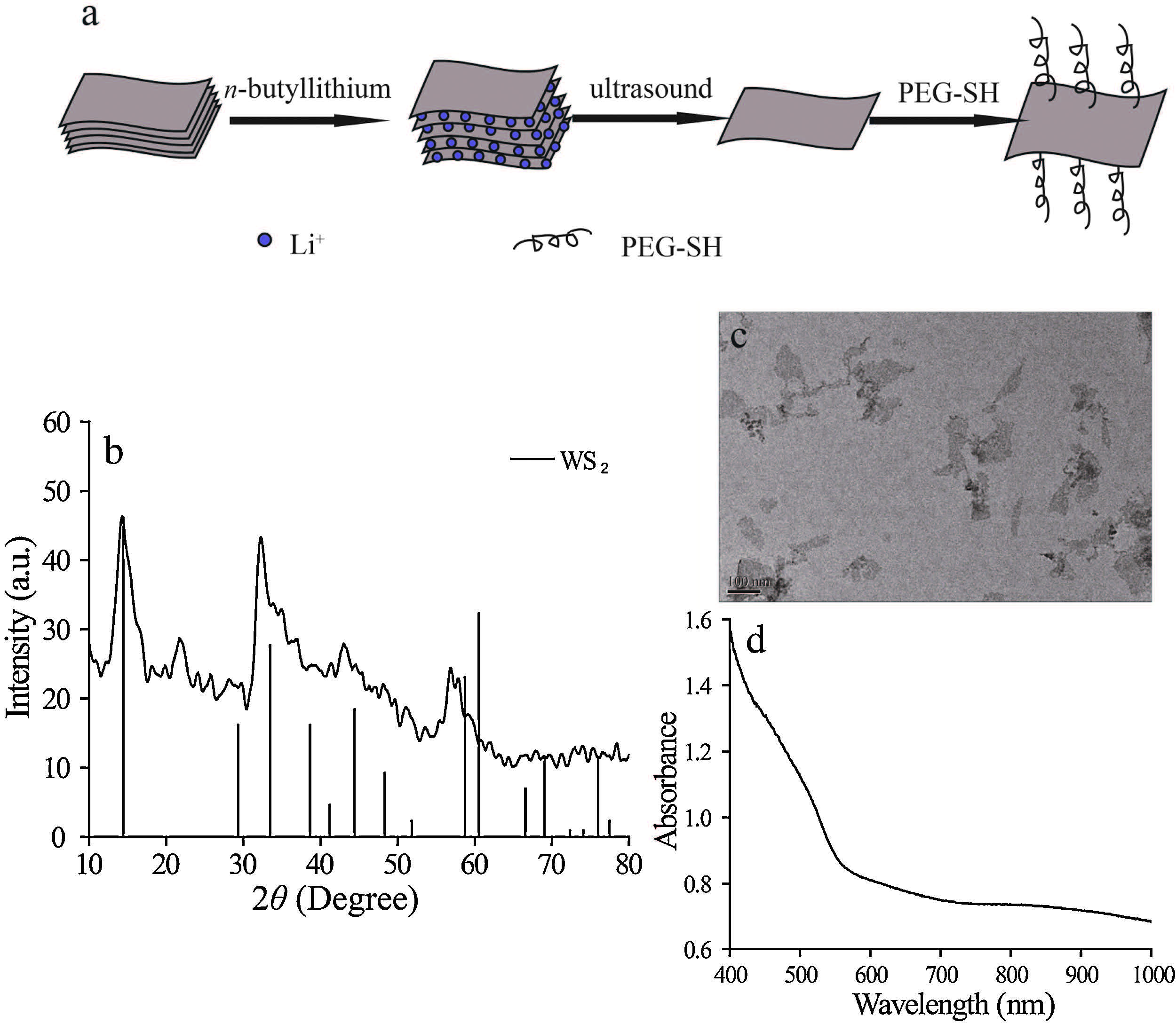
3. Results and discussion 3.1. Synthesis and characterization of PEGylated WS2 nanosheetsWS2 nanosheets were synthesized with two steps [35]. Firstly, the precursor of LixWS2 was prepared by the solvent-thermal method in the hexane solution of n-butyl lithium (2.5 mol/L) at 120 ℃ for 7 h. During this process,Li+ can be sufficiently inserted between the layers of WS2. Secondly,LixWS2 could react with the distilled water under the condition of ultrasound,which results in the exfoliation of LixWS2 to nanosheets. To improve their biocompatibility,WS2 nanosheets were conjugated with thiolmodified polyethylene glycol (PEG-SH) as shown in Fig. 1a. The phase and crystallography of the product were characterized by XRD. As shown in Fig. 1b,the XRD pattern of the as-prepared WS2 was well matched with the standard WS2 phase (PDF#08-0237). Due to the randomness of the exfoliation process,the formed WS2 nanosheets had not a good crystalline structure. The morphology of sheet-like structure was identified by TEM (Fig. 1c).

|
Download:
|
| Fig. 1.(a) The synthetic route of WS2 nanosheets. (b) Powder X-ray diffraction patterns of WS2 nanosheets referred by the standard WS2 phase (JCPDS: 08-0237). (c) A typical TEM image of WS2 nanosheets. (d) The absorption spectrum of WS2-PEG nanosheets in the aqueous solution (100 μg/mL). | |
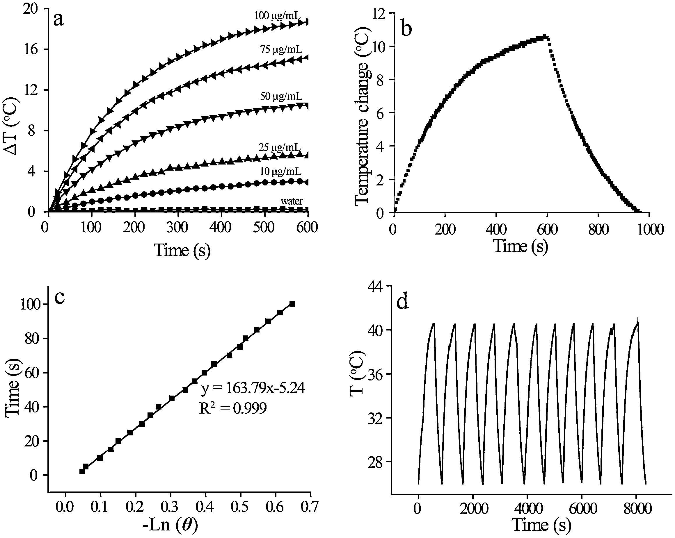
The aqueous solution of WS2-PEG nanosheets (100 μg/mL) showed a broad NIR absorption band from 700 nm to 1000 nm as presented in Fig. 1d,which was attributed to the strong localized surface plasmon resonances (LSPR) of the nanosheets [36]. The strong NIR absorption of WS2 nanosheets encouraged us to investigate their application as photothermal agents. In order to show the photothermal properties of WS2-PEG nanosheets,we investigated the temperature increase of the aqueous solution with the concentration of WS2-PEG nanosheets from 10 μg/mL to 100 μg/mL. The higher concentration of WS2-PEG nanosheets is, the more prominent temperature of WS2-PEG nanosheets under the 808 nm laser irradiation (1 W/cm2) will be (Fig. 2a). At the concentration of 100 μg/mL,the temperature increased 18 ℃ within 10 min. In strong contrast,the pure water solution showed no obvious temperature increase under the similar experimental condition. To calculate the photothermal conversion efficiency, the aqueous solution of WS2-PEG nanosheets (50 μg/mL) was irradiated by an 808 nm laser with the power density of 1 W/cm2 until the temperature remained stable. Subsequently,an 808 nm laser was shut off. The heat was transferred from the solution to the environment and the temperature of the aqueous solution decreased rapidly. To measure the photothermal conversion efficiency,we control the surface area of the container is 1 cm2. According to Fig. 2b (Tmax - Tsurr) was 10.6 ℃,the time constant for heat transferring from the system was determined to be 164.8 s of τs by applying the linear time data from the cooling period versus negative natural logarithm of driving force temperature (Fig. 2c). According to the equation shown in the experimental section,the photothermal conversion efficiency was calculated to be ~35%. To assess the NIR photostability of WS2-PEG nanosheets,Laser on/off were performed by irradiating the aqueous solution of WS2-PEG nanosheets via an 808 nm laser (1 W/cm2) for 10 min (Laser on), followed by cooling down to room temperature without the NIR laser irradiation (Laser off). As shown in Fig. 2d,the temperature increase of 14.6 ℃ was able to be achieved after the first laser on for the WS2-PEG nanosheets concentration of 120 μg/mL. No significant change in the temperature increase was observed after ten Laser ON/OFF cycles.

|
Download:
|
| Fig. 2.(a) The changes of the temperature as a function of the concentration of WS2-PEG nanosheets under the 808 nm laser irradiation for 10 min (1 W/cm2). (b) Photothermal effect of the irradiation of the aqueous dispersion of WS2-PEG nanosheets (50 μg/mL) with an 808 nmlaser (1 W/cm2) for 10 min,then the laser was shut off. (c) The linear time data from the cooling period versus negative natural logarithm of driving force temperature. (d) Temperature changes of the aqueous solution of WS2-PEG nanosheets (120 μg/mL) over ten Laser ON/OFF cycles (1 W/cm2). | |
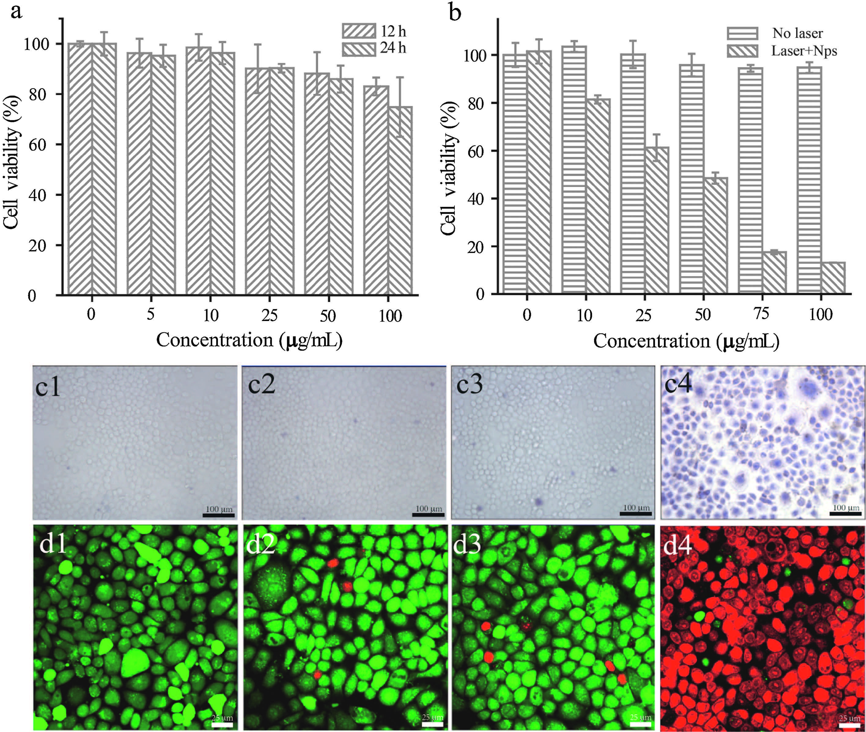
Considering the high photothermal conversion efficiency of WS2-PEG nanosheets,it is feasibility that the WS2-PEG nanosheets can be used as a photothermal agent for cancer therapy. For the application of WS2-PEG nanosheets in biomedicine,cytotoxicity was firstly necessary to be evaluated. HeLa cells as a model cell line was used to investigate the cytotoxicity ofWS2-PEG nanosheets via MTT assay. It is clear that the viability of HeLa cells incubated with WS2-PEG nanosheets is greater than 80% even at the high concentrations up to 100 μg/mL (Fig. 3a). The data suggested that WS2-PEG nanosheets exhibited the low cytotoxicity under our experimental condition. We firstly evaluated the capability of WS2-PEG nanosheets for the ablation of cancer cells in vitro by trypan blue staining which was used to distinguish dead cells from live ones under the laser irradiation. As shown in the optical microscopic images in Fig. 3c,negligible cell ablation was observed only with the 808 nm laser irradiation for 10 min at 0.4 W/cm2 (Fig. 3c2) and with WS2-PEG nanosheets of 75 μg/mL alone (Fig. 3c3). In contrast,HeLa cells incubated with WS2-PEG nanosheets (75 μg/mL) were stained blue after the 808 nm laser irradiation at 0.4 W/cm2 for 10 min (Fig. 3c4),indicating that HeLa cells incubated with WS2-PEG nanosheets were effectively ablated by the 808 nm laser irradiation (0.4 W/cm2). The photothermal ablation of HeLa cells was also demonstrated by laser confocal fluorescence microscopy. After exposed to the NIR laser at 0.4 W/cm2 for 10 min,HeLa cells were co-stained with Calceine AM and Propidium Iodide (PI) to differentiate live (green) and dead cells (red),respectively. The majority of HeLa cells were able to be ablated after the incubation with WS2-PEG nanosheets (75 μg/mL) under the 808 nm laser irradiation (0.4 W/cm2),suggesting that WS2-PEG nanosheets are able to kill HeLa cells via the photothermal destruction (Fig. 3d4). However,HeLa cells were not affected for the above control groups (Fig. 3d2 and d3). These results were matched with the above optical microscopic imaging data.

|
Download:
|
| Fig. 3.Cell viability determined by MTT assay under different concentration of WS2-PEG nanosheets (a) and under different concentration of WS2-PEG nanosheets (b) with or without the 808 nm laser irradiation for 10 min (0.4 W/cm2). (c) Optical images of HeLa cells stained by trypan blue (c) and laser confocal fluorescence images stained by Calcein AM/PI (d). (c1) and (d1),control HeLa cells. (c2) and (d2),HeLa cells treated with the 808 nm laser irradiation for 10 min(0.4 W/cm2). (c3) and (d3),HeLa cells incubated with WS2-PEG nanosheets. (c4) and (d4),HeLa cells incubated with WS2-PEG nanosheets under the 808 nmlaser irradiation for 10 min. The power density is 0.4 W/ cm2. The incubation concentration of WS2 nanosheets is 75 μg/mL. | |
The MTT assay was carried out to further quantitatively investigate the photothermal ablation of HeLa cells,which was shown in Fig. 3b. HeLa cells incubated with the different concentration of WS2-PEG nanosheets for 4 h were irradiated under the 808 nm laser at 0.4 W/cm2 for 10 min. It was determined that with the increase of concentration of WS2-PEG nanosheets, more cells incubated with WS2-PEG nanosheets were killed after the NIR laser irradiation. With the incubation concentration of WS2-PEG nanosheets from 10 to 100 μg/mL,the cell viability decreased from (81.4 ± 1.7)% to (13.1 ± 0.1)%. The median lethal dose induced by an 808 nmlaser at 0.4 W/cm2 was~50 μg/mL. These experimental results demonstrate that the combination of WS2-PEG nanosheets and NIR laser irradiation is able to ablate cancer cells in vitro. Therefore,WS2-PEG nanosheets have a great potential to be used for in vivo photothermal tumor therapy.
3.4. X-Ray attenuation propertyThe high atomic number and X-ray absorption coefficient of tungsten makes it possible that WS2-PEG nanosheets can be used as a CT contrast agent,the X-ray attenuation coefficient of the W atom at 100 keV is 4.438 cm2/kg,which is higher than that of iodine (1.94 cm2/kg at 100 keV). Iohexol,a commercial iodine-based CT contrast agent,was used in the clinic. The CT values of different concentrations of WS2-PEG nanosheets or Iohexol in water were measured in Hounsfield units (HU), respectively. The Hounsfield value was increased linearly as a function of the concentration of W and I,respectively. However, the increasing trend of the WS2-PEG nanosheets (the slope of the CT value for WS2-PEG nanosheets was ~15.65) was much higher than that of Iohexol with a slope of CT value of ~6.07 (Fig. 4b). At equal concentrations of W or I element,the CT contrast enhancement of WS2-PEG nanosheets was much higher than that of Iohexol,which is primarily due to the higher X-ray attenuation coefficient of W than that of I. The CT images were presented in Fig. 4a.

|
Download:
|
| Fig. 4.(a) In vitro CT images of WS2-PEG nanosheets (upper panel) and Iohexol (lower panel) with the different concentration of tungsten or iodine. (b) CT value (HU) of WS2-PEG nanosheets and Iohexol as a function of the concentration of tungsten (circle) and iodine (square),respectively. | |
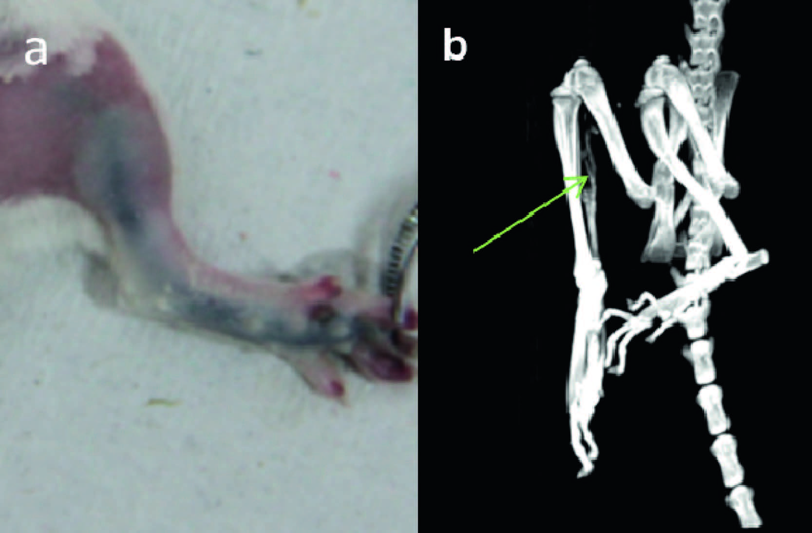
As a proof of concept,we studied the feasibility of WS2-PEG nanosheets as a CT contrast agent for in vivo application. WS2-PEG nanosheets dispersed in the physiological saline were injected into the right rear footpad of a mouse with a dose doe of 20 mg/kg of body weight. As shown in Fig. 5b,the left lymph vessel has an obvious signal. However,no obvious signal was observed from its right foot as a control.

|
Download:
|
| Fig. 5.The photograph (a) and 3-D renderings of in vivo CT images (b) after the injection of WS2-PEG nanosheets into the left rear footpad. The dose of WS2-PEG nanosheets was 20 mg/kg of body weight. | |
In summary,WS2-PEG nanosheets have been synthesized by the intercalation of n-butyllithium through the solvent-thermal method,the exfoliation with the ultrasound,and the conjugation with PEG-SH onto the surface of WS2 to improve their biocompatibility. The obtained nanosheets have the high photothermal conversion efficiency and show a good X-ray attenuation property. These properties enable the application WS2-PEG nanosheets to be extended into photothermal therapy and CT imaging. We anticipate that WS2-PEG nanosheets conjugated with the targeted ligand could be applied to the targeted theranostics.
AcknowledgmentsThis work was partially supported by National Natural Science Foundation of China (Nos. 21271130,21371122,and 11275050), Program for Changjiang Scholars and Innovative Research Team in University (No. IRT1269),Shanghai Science and Technology Development Fund (Nos. 12ZR1421800 and 13520502800), Shanghai Pujiang Program (No. 13PJ1406600),Shanghai Municipal Education Commission (No. 13ZZ110),Shanghai Normal University (Nos. DXL122 and SK201339),and International Joint Laboratory on Resource Chemistry (IJLRC).
| [1] | A.H. Castro Neto, N.M.R. Peres, K.S. Novoselov, A.K. Geim, The electronic properties of graphene, Rev. Mod. Phys. 81 (2009) 109-162. |
| [2] | X. Huang, X. Qi, F. Boey, et al., Graphene-based composites, Chem. Soc. Rev. 41 (2012) 666-686. |
| [3] | Y. Liu, X. Dong, P. Chen, Biological and chemical sensors based on graphene materials, Chem. Soc. Rev. 41 (2012) 2283-2307. |
| [4] | K.S. Novoselov, D. Jiang, F. Schedin, et al., Two-dimensional atomic crystals, Proc. Natl. Acad. Sci. U. S. A. 102 (2005) 10451-10453. |
| [5] | B. Chamlagain, Q. Li, N.J. Ghimire, et al., Mobility improvement and temperature dependence in MoSe2 field-effect transistors on parylene-C substrate, ACS Nano 8 (2014) 5079-5088. |
| [6] | H.S. Matte, A. Gomathi, A.K. Manna, et al., MoS2 and WS2 analogues of graphene, Angew. Chem. Int. Ed. 49 (2010) 4059-4062. |
| [7] | H.-J. Chuang, X. Tan, N.J. Ghimire, et al., High mobility WSe2 p-and n-type fieldeffect transistors contacted by highly doped graphene for low-resistance contacts, Nano Lett. 14 (2014) 3594-3601. |
| [8] | M. Chhowalla, H.S. Shin, G. Eda, et al., The chemistry of two-dimensional layered transition metal dichalcogenide nanosheets, Nat. Chem. 5 (2013) 263-275. |
| [9] | J. Chen, S.-L. Li, Q. Xu, et al., Synthesis of open-ended MoS2 nanotubes and the application as the catalyst of methanation, Chem. Commun. (2002) 1722-1723. |
| [10] | M. Viršek, A. Jesih, I. Milošević, et al., Raman scattering of the MoS2 and WS2 single nanotubes, Surf. Sci. 601 (2007) 2868-2872. |
| [11] | X. Zong, H. Yan, G. Wu, et al., Enhancement of photocatalytic H2 evolution on CdS by loading MoS2 as cocatalyst under visible light irradiation, J. Am. Chem. Soc. 130 (2008) 7176-7177. |
| [12] | N. Harada, S. Sato, N. Yokoyama, Computational study on electrical properties of transition metal dichalcogenide field-effect transistors with strained channel, J. Appl. Phys. 115 (2014) 034505. |
| [13] | RadisavljevicB, RadenovicA, BrivioJ, et al., Single-layer MoS2 transistors, Nat. Nanotechnol. 6 (2011) 147-150. |
| [14] | Q.H. Wang, K. Kalantar-Zadeh, A. Kis, et al., Electronics and optoelectronics of two-dimensional transition metal dichalcogenides, Nat. Nanotechnol. 7 (2012) 699-712. |
| [15] | X. Liu, G. Zhang, Q.-X. Pei, et al., Phonon thermal conductivity of monolayer MoS2 sheet and nanoribbons, Appl. Phys. Lett. 103 (2013) 133113. |
| [16] | N. Perea-López, A.L. Elías, A. Berkdemir, et al., Photosensor device based on few-layered WS2Films, Adv. Funct. Mater. 23 (2013) 5511-5517. |
| [17] | G. von Maltzahn, J.H. Park, A. Agrawal, et al., Computationally guided photothermal tumor therapy using long-circulating gold nanorod antennas, Cancer Res. 69 (2009) 3892-3900. |
| [18] | J. Shao, R.J. Griffin, E.I. Galanzha, et al., Photothermal nanodrugs: potential of TNF-gold nanospheres for cancer theranostics, Sci. Rep. 3 (2013) 1293. |
| [19] | S.R. Asemi, A. Farajpour, M. Borghei, et al., Thermal effects on the stability of circular graphene sheets via nonlocal continuum mechanics, Lat. Am. J. Solids Struct. 11 (2014) 704-724. |
| [20] | M.B.A. Kunze, D.W. Wright, N.D. Werbeck, et al., Loop interactions and dynamics tune the enzymatic activity of the human histone deacetylase 8, J. Am. Chem. Soc. 135 (2013) 17862-17868. |
| [21] | K. Yang, S. Zhang, G. Zhang, et al., Graphene in mice: ultrahigh in vivo tumor uptake and efficient photothermal therapy, Nano Lett. 10 (2010) 3318-3323. |
| [22] | Q. Tian, M. Tang, Y. Sun, et al., Hydrophilic flower-like CuS superstructures as an efficient 980 nm laser-driven photothermal agent for ablation of cancer cells, Adv. Mater. 23 (2011) 3542-3547. |
| [23] | Z. Chen, Q. Wang, H. Wang, et al., Ultrathin PEGylated W18O49 nanowires as a new 980 nm-laser-driven photothermal agent for efficient ablation of cancer cells in vivo, Adv. Mater. 25 (2013) 2095-2100. |
| [24] | K. Yang, H. Xu, L. Cheng, et al., In vitro and in vivo near-infrared photothermal therapy of cancer using polypyrrole organic nanoparticles, Adv. Mater. 24 (2012) 5586-5592. |
| [25] | Z. Zhou, B. Kong, C. Yu, et al., Tungsten oxide nanorods: an efficient nanoplatform for tumor CT imaging and photothermal therapy, Sci. Rep. 4 (2014) 3653. |
| [26] | Z. Zhou, Y. Sun, J. Shen, et al., Iron/iron oxide core/shell nanoparticles for magnetic targeting MRI and near-infrared photothermal therapy, Biomaterials 35 (2014) 7470-7478. |
| [27] | J. Li, F. Jiang, B. Yang, et al., Topological insulator bismuth selenide as a theranostic platform for simultaneous cancer imaging and therapy, Sci. Rep. 3 (2013) 1998. |
| [28] | Y. Wang, K.C.L. Black, H. Luehmann, et al., Comparison study of gold nanohexapods, nanorods, and nanocages for photothermal cancer treatment, ACS Nano 7 (2013) 2068-2077. |
| [29] | D. Kim, Y.Y. Jeong, S. Jon, A drug-loaded aptamer-gold nanoparticle bioconjugate for combined CT imaging and therapy of prostate cancer, ACS Nano 4 (2010) 3689-3696. |
| [30] | S.-W. Chou, Y.-H. Shau, P.-C. Wu, et al., In vitro and in vivo studies of FePt nanoparticles for dual modal CT/MRI molecular imaging, J. Am. Chem. Soc. 132 (2010) 13270-13278. |
| [31] | O. Rabin, J. Manuel Perez, J. Grimm, et al., An X-ray computed tomography imaging agent based on long-circulating bismuth sulphide nanoparticles, Nat. Mater. 5 (2006) 118-122. |
| [32] | Q. Xiao, W. Bu, Q. Ren, et al., Radiopaque fluorescence-transparent TaOx decorated upconversion nanophosphors for in vivo CT/MR/UCL trimodal imaging, Biomaterials 33 (2012) 7530-7539. |
| [33] | Y. Liu, K. Ai, J. Liu, et al., A high-performance ytterbium-based nanoparticulate contrast agent for in vivo X-ray computed tomography imaging, Angew. Chem. Int. Ed. 51 (2012) 1437-1442. |
| [34] | Q. Tian, J. Hu, Y. Zhu, et al., Sub-10 nm Fe3O4@Cu(2-x)S core-shell nanoparticles for dual-modal imaging and photothermal therapy, J. Am. Chem. Soc. 135 (2013) 8571-8577. |
| [35] | S.S. Chou, B. Kaehr, J. Kim, et al., Chemically exfoliated MoS2 as near-infrared photothermal agents, Angew. Chem. Int. Ed. 52 (2013) 4160-4164. |
| [36] | J.A. Faucheaux, A.L.D. Stanton, P.K. Jain, Plasmon resonances of semiconductor nanocrystals: physical principles and new opportunities, J. Phys. Chem. Lett. 5 (2014) 976-985. |