


 , Umer Rashida, Muhammad Anis-ur-Rehmanb, Wajid Rehmana, Khalid Mahmooda
b Department of Physics, COMSATS Institute of Information Technology, Islamabad 44000, Pakistan
1. Introduction
Lead is a one of the metals that have many industrial applications. However,its presence in ionic form in industrial wastewater is extremely toxic to both biota and abiota. From the industrial effluents,its concentration has been reported in the range of 40-100 mg/L [1, 2, 3]. In the last few decades the pollution of water resources due to indiscriminate disposal of lead has become a worldwide threat. Lead is non-biodegradable and can be readily absorbed into the human body after accumulated in living tissues. The presence of lead in drinking water may cause diseases such as anemia,encephalopathy,hepatitis and nephritic syndrome [4, 5]. Different physiochemical methods such as chemical precipitation, filtration,reverse osmosis,electrochemical treatment membrane technologies and ion exchange have been widely applied for the removal of toxic metals. However,sometimes these processes are ineffective and expensive [6]. Adsorption is considered to be one of the most effective physiochemical methods for the removal of heavy metal ions from the aqueous solutions. It is preferred over the other removal techniques because of its ease of operation and low cost [7, 8].
The sol-gel technique has several advantages over other methods for the preparation of various adsorbents [9]. It can be used to control the shape,morphology and textual properties of composite materials,in which the mixing of two or more metal oxide phases can be controlled even at nanometer scale level [10]. In literature,the sol-gel method has been widely used for coating silica over a large number of metal ions [11]. SiO2 has diverse applications in numerous processes such as manufacture of glass, electronic substrates,thin film substrates,electrical insulators,and thermal insulators and humidity sensors. It has interesting physical properties that have been found to have wide applications in various antireflective and protective coating [12]. In the present work,NiO coated silica and silica coated NiO are given preference because the surface groups contributed by the parent materials collectively provide the active sites for the removal of toxic metal ions. In literature limited data are available regarding the physical properties and Pb2+ adsorption studies toward NiO coated silica or silica coated NiO. In this study,we will focus on the synthesis and physical characteristics of silica coated NiO and NiO coated silica by the sol-gel method. To explore the physical properties of the materials,various techniques like surface area measurements,PZC, SEM/EDX and XRD were employed. The solid was finally tested for the removal of Pb2+ ions from the aqueous solutions. 2. Experimental
The chemicals used in the present work were of analytical grade and were used as received. Sodium silicate was purchased from Scharlau,HCl and NaOH were provided by Riedel-de Haen while sodium nitrate and lead nitrate were supplied by Merck chemicals. All the solutions were prepared in doubly distilled water. The glassware was first washed with detergent,then 10% nitric acid and finally with doubly distilled water. SiO2,NiO coated silica and silica coated NiO were prepared by the sol-gel method,while NiO was provided by BDH chemicals. 2.1. Synthesis of SiO2 gel and coated materials
SiO2 gel has been synthesized by the method described elsewhere [13]. For the synthesis of silica coated NiO,a 4 mol/L Na2SiO3 solution and an 8 mol/L HCl solution were added drop wise to the reaction vessel that contained 3 g of NiO powders. The suspension was magnetically mixed for 1 h and the gel thus formed was aged for 20 h. Finally it was washed three times and dried in an oven at 100 ℃. For the preparation of Ni coated silica,a 2 mol/L solution of Na2SiO3 (4.45 mL) was prepared in 25 mL of deionized water,which was then mixed in drop wise fashion with a 4 mol/L solution of HCI (9.8 mL),prepared in 25 mL of doubly distilled water. Then 3 g of NiO powder was dispersed to this solution with continuous stirring for 1 h and was aged for 20 h. The coated material thus obtained was well washed and stored in polythene bottle after drying at 100 ℃. 2.2. Characterization of coated and uncoated materials
The powder samples of NiO,SiO2,silica coated NiO and NiO coated silica were analyzed by various analytical techniques such as surface area analysis,point of zero charge (PZC) measurements, X-ray diffraction (XRD),scanning electron microscopy (SEM) and energy dispersive X-ray (EDX) spectroscopy. The instruments and analytical conditions at which these physicochemical tests were performed are mentioned below.
The PZC values of the solid were determined by the salt addition method with a Mettler Toledo MP 220 pH meter (Germany). The specific surface areas (SBET),average pore volume (Vp) and average pore density (Dv) for the coated and their parent materials were determined by the nitrogen adsorption method at 77 K using surface area analyzer model Quantachrome Nova 1200e. Before measuring the surface area and pore size parameters,all the samples were degassed at 105 ℃ for 1 h. The phenomenon of diffraction occurs when penetrating radiation,such as X-ray, enters a crystalline substance and are scattered. The direction and intensity of the scattered (diffracted) beams depend on the orientation of the crystal lattice with respect to the incident beam. To find the crystallinity,X-ray diffraction patterns of the air-dried finely powdered pure and coated materials were recorded using a JEOL X-ray diffractometer,model JDX-7E with Mn filtered Cu Ka radiations. The diffractograms were recorded from 10° to 70° (2θ). The acquisition time was 0.5 s per step,and the step was 0.018 (2θ). The morphology of the samples was determined by SEM. The micrographs of the samples were obtained by an SEM model JSM 5910 (JEOL Co.,Japan) at 20 keV. The elemental percentages present in the solid are determined in INCA 200 micro analyzer (UK). Before analysis,the samples were sputter-coated with gold in SPI-MODULE sputter coater (USA) for 90 s at 30 mA. 3. Results and discussion
3.1. Point of zero charge for coated and uncoated materials
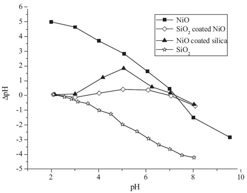
The curves for the PZC values of the coated and uncoated materials are shown in the Fig. 1. The isoelectric point (IEP) for silica was observed at pH 2.2,whereas for the NiO particles,pH vs. △pH curve crosses at pH 7.2. For SiO2 coated NiO and NiO coated SiO2,two PZC values were observed. The PZC value of SiO2 coated NiO was found to be at the pH 3.1 and 7,whereas the values for NiO coated SiO2 are 2.8 and 7.2. For NiO coated SiO2 the surface of the solid is mainly positively charged between the PZC values. Further, it was observed that the second PZC value for SiO2 coated NiO (7) was lower than the second PZC value of NiO coated SiO2 (7.2). This decrease in the PZC value may be due to the dissolution of silica from the surface of the coated material,which results in the lowering of surface charge due to the subsequent adsorption of SiOH groups onto NiO. Similar results were reported in literature where,it was found that the PZC of iron oxide/silica mixed system decreases upon the adsorption of the silicate anions [14, 15].

|
Download:
|
| Fig. 1. PZC of silica, NiO, SiO2 coated NiO and NiO coated SiO2. | |
The surface morphology of the solids was investigated by SEM and EDX analyses and the micrographs are displayed in Fig. 2. The SEM micrograph of SiO2 shows that the particles are regular in shape with definite boundaries and with very little aggregation. The size of the silica particles was found to be in the range of 100 nm to 250 nm. The SEM image of NiO reveals that most of the particles are discrete while several are assembled like a compact sack of small sized particles. The size of particles varies from 1.5 μm to 20μm. From the SEM image of SiO2 coated NiO,a uniform coating can be seen on the surface of NiO particles, whereas in case of NiO coated SiO2,a honeycomb like appearance was observed with multiple pores in the structure. These particles can be useful in many applications especially for adsorption of toxic metals,catalysis and drug delivery. The percent weight of the elements in the coated and uncoated samples is shown in Table 1, whereas Table 2 lists the BET surface area,pore size and pore volume of these materials.

|
Download:
|
| Fig. 2. SEM images of (A) SiO2, (B) NiO, (C) SiO2 coated NiO and (D) NiO coated SiO2. | |
| Table 1 EDX analysis of coated and uncoated materials showing % weight. |

| Table 2 Surface areas, pore size and pore volume of coated and uncoated materials. |

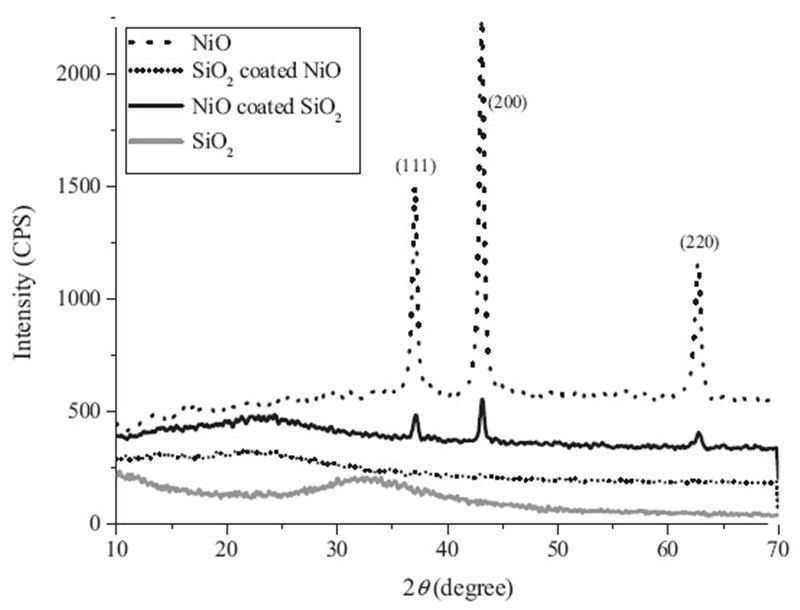
The XRD patterns of the silica coated NiO and NiO coated silica along with the parent materials are shown in Fig. 3. The silica sample was totally amorphous where a broad hump ranging from 10° to 30° is considered to be a characteristic of amorphous silica [16]. The sharp peaks appeared at 2θ values of 368,438 and 638 correspond to the (1 1 1),(2 0 0) and (2 2 0) planes respectively. These diffraction peaks are indexed to pure NiO phase according to the JCPDS card No. 47-1049,which are considered to be the characteristic pattern of the NiO particles. Similar characteristic peaks for NiO were reported elsewhere [17, 18]. In NiO coated silica,the intensity of diffractions peaks drastically decreases after reacting the NiO with the amorphous silica. However,the 2u values for these peaks remained at the same position as those in pure NiO. On the other hand,in silica coated NiO,the diffraction peaks completely vanish showing the disappearance of the crystallinity in this composite material as was observed for silica.

|
Download:
|
| Fig. 3. XRD patterns of coated and uncoated materials.. | |
Fig. 4 displays the adsorption isotherm for coated and their parent materials with respect to the pH of the aqueous solution. The uptake of Pb2+ ions were found to increase with rise in the pH value of cationic solution. The sorption trend for Pb2+ ions was in the order of NiO coated SiO2 > SiO2 coated NiO > NiO > SiO2. This trend confirms that the coated materials and specially NiO coated silica have an excellent adsorption capacity toward Pb2+ ions. It was observed that the percent removal of Pb2+ ions at pH values of 5 and 6 was about 81% and 97% respectively. While at pH near PZC, the metal removal was about 98%. This shows that at neutral point the removal capacity of Pb2+ ions was enhanced,however,at pH 8 and above aqueous metal hydroxide formation may become a significant mechanism in the lead removal process. The high adsorption capacity of NiO coated silica may be due to its porous nature as well as the high surface area of adsorbent. In future,the coated materials would be used for the detailed adsorption studies of Pb2+ ions from aqueous solutions at different values of pH and temperature. The metal uptake study will be modeled by applying various adsorption isotherms. Further,the adsorption mechanism will also be explored after doing the comparative sorption study toward the adsorbents synthesized in the present work.

|
Download:
|
| Fig. 4. Percent uptake of Pb2+ removal by coated and their parent materials.. | |
Two PZC values were observed for SiO2 coated NiO and NiO coated SiO2. In NiO coated SiO2 the surface was mainly positively charged between the PZC values. The second PZC value for SiO2 coated NiO was found to be lower than the second PZC value of NiO coated SiO2,which may be due to the dissolution of silica from the surface resulting in the lowering of surface charge due to the subsequent adsorption of SiOH groups onto NiO. From the SEM image of SiO2 coated NiO,a uniform coating can be seen on the surface of NiO particles,whereas in case of NiO coated SiO2,a honeycomb like appearance was observed with porous structures. The XRD analysis showed that silica was amorphous with a broad hump ranging from 10° to 30°. In NiO coated silica,the intensity of diffractions peaks decreases whereas in silica coated NiO,these peaks completely disappear,which however,results in the phase shifting from crystalline to amorphous. From the Pb2+ removal studies,it was concluded that NiO coated silica has a better adsorption capacity than the rest of the materials synthesized in the present study do. The high metal uptake may be due to the porous nature as well as the high surface area of the adsorbent.
| [1] | J. Perrone, B. Fourest, E. Giffaut, Sorption of nickel on carbonate fluoroapatites, J.Colloid Interf. Sci. 239 (2001) 303–313. |
| [2] | A. Clearfield, Inorganic Ion Exchange Materials, CRC Press, Boca Raton, FL, 1982. |
| [3] | B.B. Sahu, H.K. Mishra, K. Parida, Cation exchange and sorption properties ofTIN(IV) phosphate, J. Colloid Interf. Sci. 225 (2000) 511–519. |
| [4] | K.K. Wong, C.K. Lee, K.S. Low, M.J. Haron, Removal of Cu and Pb by tartaric acidmodified rice husk from aqueous solutions, Chemosphere 50 (2003) 23–28. |
| [5] | W. Lo, H. Chua, K.H. Lam, S.H. Bi, A comparative investigation on the biosorption oflead by filamentous fungal biomass, Chemosphere 39 (1999) 2723–2736. |
| [6] | N. Li, R.B. Bai, Copper adsorption on chitosan–cellulose hydrogel beads: behaviorsand mechanisms, Sep. Purif. Technol. 42 (2005) 237–247. |
| [7] | J.C.Y. Ng, W.H. Cheung, G. Mckay, Equilibrium studies of the sorption of Cu(II) ionsonto chitosan, J. Colloid Interf. Sci. 255 (2002) 64–74. |
| [8] | S. Babel, T.A. Kurniawan, Low-cost adsorbents for heavy metals uptake fromcontaminated water: a review, J. Hazard. Mater. 97 (2003) 219–243. |
| [9] | C.J. Brinker, G.W. Scherer, Sol–Gel Science, The Physics and Chemistry of Sol–GelProcessing, Academic Press, Boston, 1990. |
| [10] | J.D. Wright, N.A.J.M. Sommerdijk, Sol–Gel Materials, Chemistry and Applications,Gordon and Breach Science, Amsterdam,2001. |
| [11] | C. Mansuy, R. Mahiou, J.M. Nedelec, A new sol–gel route to Lu2SiO5 (LSO)scintillator, powders and thin films, Chem. Mater. 15 (2003) 3242–3244. |
| [12] | O. De Sanctis, L. Gomez, N. Pellegri, et al., Protective glass coatings on metallicsubstrates, J. Non-Cryst. Solid 121 (1990) 338–343. |
| [13] | M. Waseem, S. Mustafa, A. Naeem, K.H. Shah, I. Shah, Mechanism of Cd (II)sorption on silica synthesized by sol–gel method, Chem. Eng. J. 169 (2011)78–83. |
| [14] | U. Schwertmann, H. Fechter, The point of zero charge of natural and syntheticferrihydrites and its relation to adsorbed silicate, Clay Miner. 17 (1982) 471–476. |
| [15] | X.G. Meng, R.D. Letterman, Effect of component oxide interaction on the adsorptionproperties of mixed oxides, Environ. Sci. Technol. 27 (1993) 970–975. |
| [16] | A. Autef, E. Joussein, G. Gasgnier, S. Rossignol, Role of the silica source on thegeopolymerization rate, J. Non-Cryst. Solid 358 (2012) 2886–2893. |
| [17] | D.D. Han, P.C. Xu, X.Y. Jing, et al., Trisodium citrate assisted synthesis of hierarchicalNiO nanospheres with improved supercapacitor performance, J. PowerSources 235 (2013) 45–53. |
| [18] | R.R. Wang, Q.R. Li, H.Y. Xiao, H.X. Lu, Synthesis of NiO using pine as template andadsorption performance of Pb(II) from aqueous solution, Appl. Surf. Sci. 279(2013) 129–136. |