


 , Lan-Ping Zhangb, Tong Yaoa,b, Cheng Chenb, Lian-Gang Maoa,b, Yin Wangb, Jie Wuc
b JARI Pharmaceutical Co., Ltd, Lianyungang 222006, China;
c Department of Pharmaceutical Engineering, Wuhan Bioengineering Institute, Wuhan 430415, China
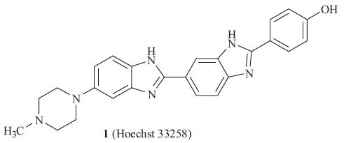
Bis-benzimidazole derivatives have been proven to be potent antitumor agents via DNA minor groove binding,such as Hoechst 33258 (Scheme 1). Hoechst 33258 [3],a fluorescent compound with a head-to-tail bis-benzimidazole structure,was initially found to be active against L1210 murine leukemia. During phase I clinical trials in humans,positive responses were observed in pancreatic cancer. However,a subsequent phase II clinical trial failed to show any objective responses [4].
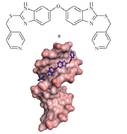
Due to the high DNA minor groove binding ability of Hoechst 33258,several groups have synthesized bis-benzimidazole derivatives derived from Hoechst 33258 [5, 6, 7, 8]. To the best of our knowledge,most derivatives ofHoechst33258 are planarmolecules, and there is little literature related to adding a linker between two benzimidazoles. Molecular modeling studies (shown in Fig. 1) suggest that the novel benzimidazole (9) could effectively bind into the DNA minor groove. Therefore we combined two benzimidazole derivatives into one moleculewith an oxygen atom. The interaction of compound 9 with CT (calf thymus)-DNA has been investigated using UV-vis,fluorescence spectroscopy. These results showed that compound 9 interacted with the DNA by binding into the minor groove. All of these compounds were screened for anti-tumor activity in vitro. Among them,compound 9 had good activity against three tumor cell lines (HeLa,HL60 and U937).

|
Download:
|
| Scheme 1.The synthetic routes to the NDAZO and PANDAZO. | |
An AutoDock 3.05 was used to perform theoretical docking calculations [9]. The molecular structure of the A-tract DNA dodecamer d(CGCAAATTTGCG) (PDB code: 2DND) was as receptor. The graphical front-end,AutoDock Tools,was used to add polar hydrogens and partial charges for proteins using the Kollman United Atom charges. Atomic solvation parameters were assigned using the add solubility function in the AutoDock package. Affinity grid fields were generated using the auxiliary program Auto- Grid3.0. The Lamarckian genetic algorithm (LGA) was used to find the appropriate binding positions,orientations,and conformations of the ligands. The optimized AutoDocking parameters are as follows: The maximum number of energy evaluations was increased to 25,000,000 per run; the iterations of Solis & Wets local search was 3000; the number of individuals in the population was 300. All other parameters were maintained as default. Cluster analysis with AutoDock results was performed to determine if different binding sites have been produced from multiple runs. 2.2. Chemical and instruments
All commercially available reagents and solvents were used without further purification unless otherwise specified. Solvents were dried and re-distilled prior to use according to standard methods. Melting points were determined on a Bu¨ chi Melting Point B-540 apparatus (Bu¨ chi Labortechnik,Flawil,Switzerland) and are uncorrected. 1H NMR spectra were measured in DMSO-d6 on a Bruker ARX 300 spectrometer (Bruker,Rheinstetten,Germany). Chemical shifts are reported in parts per million (ppm) using tetramethylsilane (TMS) as the internal standard if not specifically mentioned (J in Hz). Mass spectra were measured on a Waters Micromass Quattro Micro API mass spectrometer (Waters Corporation, Milford,United States). The UV-vis spectral measurements were recorded on a Cary Varian double beam spectrophotometer (Cary BIO 100,Australia). The sample cuvette used was a pair quartz cells of 1.00 cm path length. All scanning parameters were optimized to obtain the best spectra and in general the parameters were scanned in a wavelength range of 230-300 nm with a step of 0.5 and all measurements were carried out at room temperature. Fluorescence measurements were performed using a Spectrofluorimeter model FS920 of Edinburgh Instruments,U.K. equipped with a xenon arc lamp. The temperature of the sample holder was regulated with a peltier cooled thermostat. Quartz cuvettes of 3 mL capacity and path length 1 cm were used for all measurements. 2.3. General procedure for the synthesis of substituted compounds 8- 15
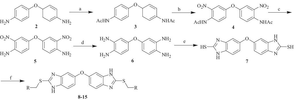
The key intermediate compounds 3-6 were prepared according to described procedures using 4,4'-diaminodiphenyl ether as a starting material [10]. To a stirred acetone (50 mL) solution of 4,4'-diaminodiphenyl ether (2) (5.0 g,19.6 mmol) at 0 °C,acetic anhydride (10.0 mL,105.8 mmol) was added dropwise and reacted for 3 h. The reaction was monitored by TLC. To stop the reaction, triethylamine (25 mL,179.4 mmol) was added dropwise,neutralizing the solution and producing a white solid,which was filtrated, washed with acetone,and dried to obtain the compound 3. Then, obtained compound 3 (6.2 g,16.6 mmol) was dissolved with acetic acid (50mL) and stirred at 0 °C for 10 min. Fuming nitric acid (8mL, 171.7mmol) was added dropwise to the above solution over 2 h. After that,the ice-bath was removed and the reaction mixture was further stirred at roomtemperature for 2 h. Themixturewas poured into ice (100 mL) and the resulting yellow solid (4) was filtered, washed with water,dried in vacuum,dissolved in a mixed water (10mL) and ethanol (30mL) solution of potassiumhydroxide (7.3 g, 106.7mmol),and then refluxed for 4 h. After stopped the reaction, the above solution was poured into ice (100mL). The resulting red solid was filtrated,washed with water,and dried to give 5.2 g (17.9 mmol) of compound 5. Then,it was dissolved in methanol (50mL) with Pd-C (0.3 g,10%) in it,enabling hydrogen gas passing through continuously at a flowrate of 10mL/min for 3 h. After that,it was filtrated to collect the filtrate and evaporated under vacuum to give 3.7 g of compound 6. Then,compound 6 with potassium ethylxanthate gave 2,2'-dithiol-5,5'-bis-1H,10H-benzimidazole ether (7),which reacted with various substituted chloromethylpyridine analogs in ethanol at reflux to synthesize the desired compounds 8- 15 [11]. 2.4. Biological assays
The anti-proliferational effects of the compounds on tumor cells were tested by the same methods. Tumor cells in RPMI1640 medium with 10% fetal bovine serum were plated in 96-well microtiter plates (4.0 × 10⁴ cells per well) and allowed to adhere at 37 8C with 5% CO2 for 4 h. The test compound was then added,and the cells were incubated at 37 °C with 5% CO2 for 72 h. The cell viability was assessed using a standard MTT assay [12]. 3. Results and discussion
Molecular modeling studies (shown in Fig. 1) suggest that the novel symmetric head-to-head benzimidazole (9) could effectively bind into the DNA minor groove. The predicted binding free energy was -14.35 kcal/mol. From the modeling,it was shown that the compound should adopt a concave shape,exactly fitting the convex minor groove,allowing for deeper penetration into the DNA minor groove.

|
Download:
|
| Fig. 1.Close-up view of compound 9 binding in the minor groove. | |
The designed compounds were successfully synthesized as described in Scheme 2. Protection of the amino group of commercially available 4,4'-diaminodiphenyl ether with acetic anhydride at 0 °C gave 4,4'-diacetamido-diphenyl (3). Nitration of compound 3 with nitric acid in acetic acid formed compound 4. Treatment of compound 4 with a solution of potassium hydroxide in water and ethanol gave 3,30-dinitro-4,4'-diaminodiphenyl ether (5),followed by reduction of the nitro group to afford 3,30,4,4'- tetraaminodiphenyl ether (6). Treatment of compound 6 with Potassium ethylxanthate gave 2,2'-dithiol-5,5'-bis-1H,10H-benzimidazole ether (7),which reacted with sodium hydroxide and various substituted chloromethylpyridine analogs in ethanol at reflux to synthesize the desired compounds 8-15.

|
Download:
|
| Scheme 2.Reagents and conditions: (a) (CH3CO)2O/NEt3,acetone,0 °C,2 h,98%; (b) conc. HNO3,acetic acid,0-50 °C,5 h,98%; (c) 42% KOH aq,MeOH,reflux,2 h,97%; (d) NH2NH2 H2O,MeOH,r.t.,6 h,89%; (e) KOH,CS2,ethanol,reflux,r.t.,6 h,82%; (f) various hy-CH2Cl (heterocyclic),NaOH,MeOH,reflux,3 h,54-93%. | |
In vitro antitumor activities of all the above final products were screened against three selected tumor cell lines. The biological assay results are summarized in Table 1. It was found that all compounds showed noticeable antitumor activities. Worthy of noting,compounds 8-10without substituents onthe pyridineringshowedmore potent antitumor activities than compounds 11-15,which contain electron-withdrawing halogen substituents (Cl,F or Br) or electrondonating substituents (hydroxyl or methoxyl) on the pyridine ring. Compounds 8-10 showed low cytotoxicity at a concentration of 15 mmol/L,while compounds 11-15 were less potent with IC50 values more than 20 mmol/L. Among them,compound 9 was most potent with IC50 values of 5.95 mmol/L for the U937 tumor cell line and 5.58 mmol/L for the HeLa tumor cell line.
| Table 1 Structure and anti-tumor activity of bis-benzimidazoles.a |

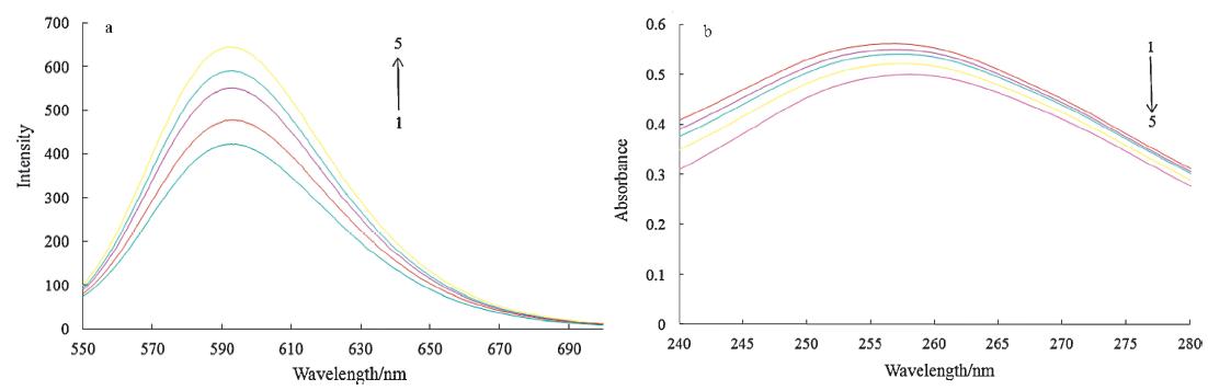
UV-vis spectroscopy on CT-DNA in the presence of the small molecules provided useful information related to the nature of the interaction between the two. Fig. 2a shows the absorbance of CT-DNA at 257 nm progressively increasing when the concentration of compound 9 solution was increased from 0 to 5 mmol/L. Not only was there an increase in the absorbance of CT-DNA upon the addition of compound 9,the appearance of a distinct blue shift upon the formation of the DNA-compound 9 complex in the 257 nm region was also observed. These results indeed indicate that compound 9 may bind in the minor groove of CT-DNA.

|
Download:
|
| Fig. 2.(a) The absorption spectra of calf thymus DNA (1,7.8 × 10-5 mol/L) in Tris-HCl buffer upon addition of compound 9 (2-5,1-4 × 10⁻⁶ mol/L,respectively); (b) fluorescence emission spectra (excited at 520 nm) of EB,EB-DNA complexes in the absence (1) and presence (2-5) of increasing concentrations of the compound 9 (2 mmol/L, 1 L per scan). | |
Fluorescence quenching measurements are effective to monitor the binding nature of the small molecules to DNA. The molecular fluorophore EB (ethidium bromide) has a conjugate planar structure,and its fluorescence intensity is very weak,but it emits intense fluorescence at about 600 nm in the presence of DNA due to its strong intercalation between the adjacent DNA base pairs. Many DNA minor groove agents could quench the intense fluorescence [13]. Similar quenching was observed in compound 9 (Fig. 2b). Therefore,it is concluded that compound 9 could bind into the minor groove of DNA.
The DNA-binding ability of compound 9 was also evaluated by the ultrafiltration method (Table 2) [14, 15]. It can be seen that the DNA-binding ability for compound 9 was 65.8 and 69.9 for Hoechst 33258. The binding constant (Ka) for compound 9 was 6.5 × 10³ L/mol for Hoechst 33258. The similar DNA-binding ability and similar binding constant (Ka) indicate that compound 9 may bind in the minor groove of CT-DNA.
| Table 2 DNA-binding abilities of compounds 9 and Hoechst 33258 determined by ultrafiltration assay using calf thymus DNA. |

In summary,we have successfully designed and synthesized a new series of bis-benzimidazole derivatives based upon molecular modeling experiments of docking within the minor grooves of DNA. These compounds exhibited desirable anti-tumor activity and have better DNA minor groove binding ability. Such compounds are of interest in the context of the future development of novel anti-tumor agents. Acknowledgment
This study was supported by Wuhan City Department of Education (No. 2009K106).
| [1] | H.H. Jardosh, C.B. Sangani, M.P. Patel, R.G. Patel, One step synthesis of pyrido[1,2-a]benzimidazole derivatives of aryloxypyrazole and their antimicrobial evaluation, Chin. Chem. Lett. 24 (2013) 123.126. |
| [2] | X.J. Wang, M.Y. Xi, J.H. Fu, et al., Synthesis, biological evaluation and SAR studies of benzimidazole derivatives as H1-antihistamine agents, Chin. Chem. Lett. 23 (2012) 707.710. |
| [3] | B.S. Reddy, S.M. Sondhi, J.W. Lown, Synthetic DNA minor groove-binding drugs, Pharmacol. Ther. 84 (1999) 1.111. |
| [4] | P.G. Baraldi, A. Bovero, F. Fruttarolo, et al., DNA minor groove binders as potential antitumor and antimicrobial agents, Med. Res. Rev. 24 (2004) 475.528. |
| [5] | J. Mann, A. Baron, Y. Opoku-Boahen, et al., A new class of symmetric bisbenzimidazole- based DNA minor groove-binding agents showing antitumor activity, J. Med. Chem. 44 (2001) 138.144. |
| [6] | S. Neidle, DNA minor-groove recognition by small molecules, Nat. Prod. Rep. 18 (2001) 291.309. |
| [7] | M. Singh, V. Tandon, Synthesis and biological activity of novel inhibitors of topoisomerase I: 2-aryl-substituted 2-bis-1H-benzimidazoles, Eur. J. Med. Chem.46 (2011) 659.669. |
| [8] | J. Valdez, R. Cedillo, A. Hernández-Campos, et al., Synthesis and antiparasitic activity of 1H-benzimidazole derivatives, Bioorg. Med. Chem. Lett. 12 (2002) 2221.2224. |
| [9] | A.J. Olson, D.S. Goodsell, Automated docking and the search for HⅣ protease inhibitors, SAR QSAR Environ. Res. 8 (1998) 273.285. |
| [10] | F. Dawans, C.S. Marvel, Polymers from ortho aromatic tetraamines and aromatic dianhydrides, J. Polym. Sci. A: Gen. Pap. 3 (1965) 3549.3571. |
| [11] | Analytical data for compounds 7.15:; Compound 7: yield: 54.8%, mp 211.6 213.8 8C; 1HNMR(300MHz,DMSO-d6): d 6.84 (d, 1H, J = 8.4, Bz-7-H), 7.30 (d, 1H, J = 1.8), 7.52 (dd, 1H, J = 8.4,1.8,Bz-6-H), 12.61 (br, s, 2H), 13.56 (br, s, 2H); 13CNMR(100MHz,DMSO-d6): d 105.6, 112.6, 115.2, 133.8, 138.1, 151.7, 168.0; ESI-HRMS: m/z 314.0288. (Calcd. for C26H20N6OS2: 314.0296). Compound 8: yield (last step): 92.6%, mp 120.3 122.2 8C; 1H NMR (300MHz, DMSO-d6): d 4.65 (s, 4H), 6.80.7.20 (m, 4H), 7.27 (m, 2H), 7.36 (d, 2H, J = 7.8 Hz), 7.51 (m, 2H), 7.73 (d, 2H, J = 7.8), 8.51 (m, 2H), 12.65 (br s, 2H); 13C NMR (100 MHz, DMSO-d6): d 41.2, 106.7, 114.2, 115.1, 124.5, 125.3, 134.2, 136.7, 139.4, 148.6, 149.3, 151.9, 159.6; ESI-HRMS: m/z 496.1152. (Calcd. for C26H20N6OS2: 496.1140). Compound 9: yield (last step): 88.7%, mp 118.9 122.2 8C; 1H NMR (300MHz, DMSO-d6): d 4.55(s, 4H), 6.85 (d, 2H, J = 8.6 Hz), 7.00 (s, 2H), 7.33 (s, 2H), 7.44 (d, 2H, J = 8.6 Hz), 7.86 (d, 2H, J = 7.7 Hz), 8.47 (d, 2H, J = 7.7 Hz), 8.54 (s, 2H), 12.59 (br s, 2H); 13C NMR (100 MHz, DMSO-d6): d 39.6, 106.7, 114.2, 115.1, 125.3, 134.2, 139.4, 146.7, 148.4, 148.6, 151.9; ESI-HRMS: m/z 496.1146. (Calcd. for C26H20N6OS2: 496.1140). Compound 10: yield (last step): 89.3%, mp 134.1 134.9 8C; 1H NMR (300 MHz, DMSO-d6): d 4.53 (s, 4H), 6.83 (dd, 2H, J = 8.7, 2.1 Hz), 6.98 (s, 2H), 7.43 (d, 4H, J = 8.7 Hz), 7.44 (s, 2H), 8.48 (d, 4H, J = 2.1 Hz), 12.60 (br s, 2H); 13C NMR (100MHz, DMSO-d6): d 38.3, 106.7, 114.2, 115.1, 125.9, 133.4, 134.2, 134.8, 139.4, 148.6, 151.9, 152.6; ESI-HRMS: m/z 496.1149. (Calcd. for C26H20N6OS2: 496.1140). Compound 11: yield (last step): 90.7%, mp 94.9 96.1 8C; 1H NMR (300 MHz, DMSO-d6): d 3.79 (s, 12H), 3.88 (s, 6H), 4.64 (s, 4H), 6.80.7.20 (m, 4H), 7.07 (d, 2H, J = 5.7 Hz), 8.15 (d, 2H, J = 5.7 Hz), 12.60 (br s, 2H); 13C NMR (100MHz, DMSO-d6): d 19.5, 20.6, 42.5, 57.3, 106.7, 114.2, 115.1, 118.4, 132.0, 139.4, 134.2, 143.1, 148.2, 148.6, 150.8, 151.9; ESI-HRMS: m/z 612.1981. (Calcd. for C32H32N6O3S2: 612.1977). Compound 12: yield (last step): 90.7%,mp 94.9 96.1 8C; 1H NMR (300 MHz, DMSOd6): d 3.79 (s, 6H), 3.88 (s, 6H), 4.64 (s, 4H), 6.80.7.20 (m, 4H), 7.07 (d, 2H, J = 5.7 Hz), 7.42 (m, 2H), 8.15 (d, 2H, J = 5.7 Hz), 12.60 (br s, 2H); 13C NMR (100 MHz, DMSO-d6): d 35.1, 56.2, 56.9, 106.2, 106.8, 114.2, 115.1, 134.2, 142.7, 139.4, 142.6, 148.2, 148.6, 151.9, 157.9; ESI-HRMS: m/z 616.1567. (Calcd. for C26H28N6O5S2: 616.1563). Compound 13: yield (last step): 87.2%, mp 123.7 126.5 8C; 1H NMR (300MHz, DMSO-d6): d 2.25 (s, 6H), 4.71 (s, 4H), 4.91 (dd, 4H, J = 8.7, 5.4 Hz), 6.80.7.00 (m, 4H), 7.09 (d, 2H, J = 5.4 Hz), 7.45 (s, 2H), 8.31 (d, 2H, J = 8.7 Hz), 12.60 (br s, 2H); 13C NMR (100MHz, DMSO-d6): d 16.7, 39.0, 83.9, 105.8, 106.7, 112.3, 114.2, 115.1, 124.6, 134.2, 139.4, 148.9, 148.6, 151.9, 161.2, 168.7; ESI-HRMS: m/z 720.1428. (Calcd. for C32H26F6N6O3S2: 720.1412). Compound 14: yield (last step): 76.2%, mp 121.8 124.1 8C; 1H NMR (300MHz, DMSO-d6): d 3.96 (6H, s), 4.79 (s, 4H), 6.88 (dd, 2H, J = 8.6, 2.1 Hz), 7.04 (s, 2H), 7.47 (d, 2H, J = 8.6 Hz), 7.57 (d, 2H, J = 2.1 Hz), 8.27 (m, 2H), 12.61 (br s, 2H); 13C NMR (100 MHz, DMSO-d6): d 35.6, 56.3, 106.7, 114.2, 115.1, 122.8, 124.0, 134.2, 139.4, 143.9, 148.6, 149.2, 151.9, 156.2; ESI-HRMS: m/z 624.0560. (Calcd. for C28H22Cl2N6O3S2: 624.0572). Compound 15: yield (last step): 53.7%, mp 111.2 113.1 8C; 1H NMR (300MHz, DMSO-d6): d 4.54 (s, 2H), 6.80.7.30 (m, 2H), 7.43 (m, 1H), 7.46 (d, 1H, J = 8.1 Hz), 7.93 (dd, 1H, J = 8.1, 2.4 Hz), |
| [12] | L.M. Green, J.L. Reade, C.F. Ware, Rapid colormetric assay for cell viability: application to the quantitation of cytotoxic and growth inhibitory lymphokines, J. Immunol. Methods 70 (1984) 257.268. |
| [13] | F. Barceló,M. Ortiz-Lombard.a, M. Martorell, et al., DNA binding characteristics of mithramycin and chromomycin analogues obtained by combinatorial biosynthesis, Biochemistry 49 (2010) 10543.10552. |
| [14] | K. Bielawski, A. Bielawska, T. Anchim, S. Wo.czyn丩 ski, Synthesis, DNA binding, topoisomerase inhibition and cytotoxic properties of 2-chloroethylnitrosourea derivatives of Hoechst 33258, Biol. Pharm. Bull. 28 (2005) 1004.1009. |
| [15] | M. Shichita, R. Shimazawa, O. Nakajima, et al., Non-intercalative and sequence- selective interaction of nitropyrene/acridine-skeleton with nucleotides: application of the dextran-coupling method, Biol. Pharm. Bull. 18 (1995) 637.639. |