


 , Hua Yuana, Yi-Ying Yanga, Hai-Lin Conga,b , Tian-Zi Haoa, Xiao-Dan Xua, Xiu-Lan Zhanga, Shu-Jing Yanga, Li-Xin Zhanga
b Collaborative Innovation Center for Marine Biomass Fibers, Materials and Textiles of Shandong Province, Qingdao University, Qingdao 266071, China
Dopamine (DA) is a natural catecholamine formed by the decarboxylation of 3,4-dihydroxyphenylalanine and belongs to the family of excitatory chemical neurotransmitters [1]. DA is one of the important neurotransmitters and is widely distributed in the mammalian central nervous system for signaling. It is mainly responsible for the reward sensation,and transmits the information of excitement and fun,but at the same time,is related to the addiction [2]. It is of great clinical importance to measure the DA level in extracellular fluids to monitor neurotransmission processes and diagnose Parkinson’s disease [3]. Therefore,it is of great importance to sensitively and selectively monitor DA not only for biomedical chemistry and neurochemistry research but also for diagnostic and pathological purposes. To date,several impressive techniques have demonstrated the feasibility for the assay of DA, including electrochemical [4, 5, 6, 7, 8, 9],colorimetric [10, 11, 12, 13],fluorescence spectrometry [14],liquid chromatography/electrospray tandem mass spectrometry [15],and high performance liquid chromatographymethods [16].Among these,electrochemical approaches have attracted most attention because of their simplicity,low instrumental cost,and capability in real-time and even in vivo measurements. Although the electrochemical responses have been continuously improved,the development of more reliable and efficient sensors for sensitive analysis of DA still remains very challenging. Therefore,the choice of material is essential and important to construct sensors with excellent performance. In recent years,various self-assembly methodologies have achieved great diversity [17, 18, 19]. Diazoresin (DR),as a photosensitive polyelectrolyte,has been massively utilized in self-assembly systems to improve the stability of the self-assembled multilayer films via the photocrosslinking technique [20, 21, 22, 23, 24]. For example,the unstable ionic linkages involved in the multilayers will convert to stable covalentbondswithexposure of theDRcontainingfilms toUV light. In this article,we applied the self-assembly method combined with the photocrosslinking technique to the preparation of DR/ SWNTmodified Au electrodes,and investigated the electrochemical behavior of DA on the electrodes by the methods of cyclic voltammeter (CV) and differential pulse voltammeter (DPV). 2. Experimental 2.1. Materials
Dopamine (DA),uric acid (UA) and ascorbic acid (AA) were purchased from Sigma. Single-walled carbon nanotubes (SWNTs,95% purity,diameter 10-20 nm,length 1-5 mm) were purchased from Nanoport Company (Shenzhen,China). Dopamine hydrochloride injection (DHI) was purchased from Shandong provincial hospital (Jinan,China). All other chemicals were of analytical grade and used as obtained without further purification. Phosphate buffer solutions (PBS,100 mmol/L) of pH 5-8 were used as a supporting electrolyte. The preparation of aqueous solution was done with deionized water. Solutions were deoxygenated by purging with prepurified nitrogen gas. DR was synthesized according to the method described elsewhere [20]. Carboxylic acid modified SWNT (COOH-SWNT) was prepared according to a reported method [22] 2.2. Apparatus
Electrochemical measurements were carried out on a CHI-832C electrochemical analyzer (CH Instruments,China). A threeelectrode cell was used with a Ag/AgCl electrode (KCl electrolyte concentration: CKCl = 3.0 mol/L) as a reference electrode,a Pt wire as a counter electrode and a bare gold electrode with a diameter of 3 mm (modified and unmodified) as a working electrode, respectively. All the electrodes were purchased from CH Instruments. The pH values of solutions were measured with a PB-10 pH meter (Renhe Instruments,China). All the measurements were performed at room temperature. 2.3. Fabrication of DR/SWNT modified electrodes
The schematic fabrication process was illustrated in Fig. 1. Prior to the self-assembly process,the bare gold electrode was polished to a mirror finish using aqueous alumina (particle size 0.05 mm) slurry,and rinsed with deionized water. The treated gold electrode was immersed in an aqueous solution of 0.1 mol/L thioglycollic acid (TGA) at room temperature for 3 h. And then,the electrode was rinsed by anhydrous alcohol and deionized water to remove non-specifically adsorbed TGA. After N2 drying,the electrode was alternately immersed in an aqueous polycation DR solution (0.05 mol/L) and an aqueous COOH-SWNT dispersion (0.05 mol/L) for 5min and 10min,respectively. The electrode was then rinsed thoroughlywith deionized water and dried with N2 to complete the assembly process. Finally,themodified electrodewas exposed toUV light from a medium-pressure mercury lamp with an intensity of 12mW/cm2 at 365 nm for 30 s to finish the whole fabrication process.

|
Download:
|
| Fig. 1.Illustration of self-assembly process of DR/SWNT modified electrodes. | |
The electrocatalytic activity of the DR/SWNT modified electrodes toward the oxidation of DA is characterized by cyclic voltammetry (CV) ranging from -0.1 V to 0.6 V. The CV responses of 2 mmol/L DA in 0.1 mol/L PBS (pH 6.0) at the bare and DR/SWNT modified electrodes are shown in Fig. 2. Compared to bare Au electrodes,the DR/SWNT modified electrodes show well-defined and resolved voltammetric responses for the direct oxidation of DA. The oxidation peak current at the modified electrode is much higher than that of the bare electrode,which indicates that the DR/ SWNT modified electrode is an effective electrocatalyst for the oxidation of DA.

|
Download:
|
| Fig. 2.CV responses at the bare Au (a) and DR/SWNT modified electrodes (b) in 0.1 mol/L PBS (pH 6.0) containing 2 mmol/L DA. Scan rate: 50 mV/s. | |
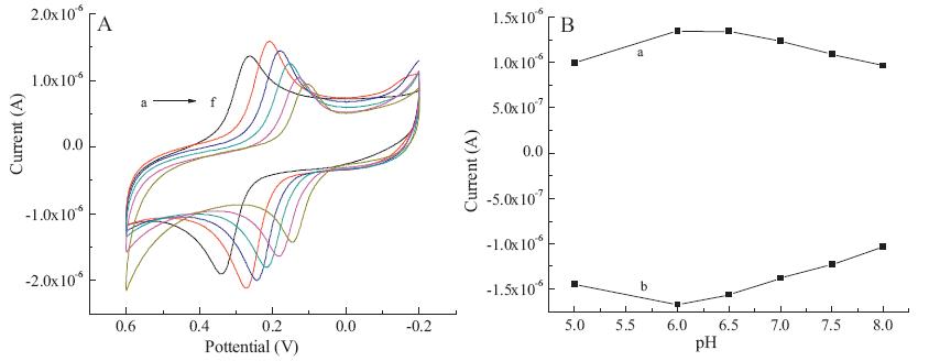
Proton is always involved in the electrochemical reactions of organic compounds and exerts significant impact on the reaction rate. Therefore,the effect of pH is investigated by the CV method in 0.1 mol/L PBS over the range from pH 5.0 to 8.0. Fig. 3A shows the CV results in 0.1 mol/L PBS containing 20 mmol/L of DA at different pH values. We can see that the electro-oxidation behavior of DA is dependent on the pH value of the solution. The peak potential for oxidation of DA shifts to more positive potentials with the changes of the pH of the solutions from alkaline to acidic values. Plots of peak current against pH value are shown in Fig. 3B. It is apparent that the highest peak currents are obtained at pH 6.0. Therefore,pH 6.0 is chosen for the subsequent analytical experiments.

|
Download:
|
| Fig. 3.Effect of pH on the electro-oxidation of DA: (A) CV responses of 20 mmol/L DA on DR/SWNT modified electrodes in 0.1 mol/L PBS at different pH values (a–f: 5.0, 6.0, 6.5, 7.0, 7.5, and 8.0, respectively); (B) relationship between the peak currents and different pH values (a: anodic peak potentials, b: cathodic peak potentials). Scan rate: 50 mV/s. | |
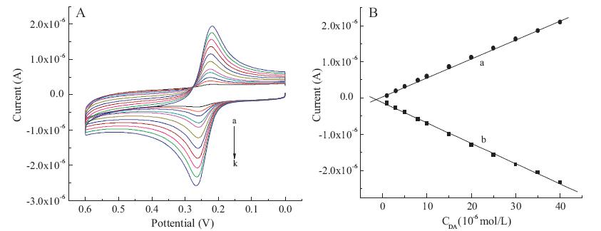
The experiment is performed with the CV method in 0.1 mol/L PBS (pH 6.0). As shown in Fig. 4,with the concentration of DA increased from 1 mmol/L to 40 mmol/L,the anodic peak grew proportionally. According to Fig. 4B,the anodic peak and the cathodic peak exhibit linear relationshipwith the concentration,and the linear regression equations are: Ipa = -5.61763 × 10⁻⁸ CDA - 1.2585 × 10⁻⁷ (R = 0.99905) and Ipc = 5.1885 × 10⁻⁸ CDA + 5.80066 × 10⁻⁸ (R = 0.99929),respectively. Where Ipa is the anodic peak current,Ipc is the cathodic peak current,CDA is the DA concentration,and R is the correlation coefficient.

|
Download:
|
| Fig. 4.Effect of concentrations on the electro-oxidation of DA: (A) CV responses at DR/SWNT modified electrodes in pH 6.0 PBS of different DA concentrations (a–f: 1, 3, 5, 8, 10, 15, 20, 25, 30, 35, and 40 mmol/L, respectively). (B) Relationship between the peak currents and the concentrations of DA (a: reduction peak current versus the concentrations of DA, b: oxidation peak current versus the concentrations of DA). Scan rate: 50 mV/s. | |
Fig. 5 presents the effect of scan rate on the electrochemical behaviors of 20 mmol/L DA in 0.1 mol/L PBS (pH 6.0) at the DR/ SWNT modified electrodes. From Fig. 5A,we can see that the oxidation peak currents of DA increase with increasing scan rates. According to Fig. 5B,the anodic peak and the cathodic peak exhibit linear relationship with the scan rates,and the linear regression equations are: Ipa = -7.16654 × 10⁻⁹ υ - 2.91766 × 10⁻⁷ (R = -0.99835) and Ipc = 3.57398 × 10⁻⁹ υ + 2.89475 × 10⁻⁷ (R = 0.99048),respectively,where n is the scan rate. It is indicated that the electrochemical redox behavior of DA at the modified electrodes surface is an adsorption-controlled process.

|
Download:
|
| Fig. 5.Effect of scan rate on the electro-oxidation of DA: (A) CV responses of 20 mmol/L DA on DR/SWNT modified electrodes in pH 6.0 PBS at various scan rates (a–n: 10.0, 30.0, 50.0, 70.0, 90.0, 110.0, 130.0, 150.0, 170.0, 190.0, 210.0, 230.0, 260.0, 280.0, and 300.0 mV/s, respectively). (B) Relationship between the peak currents and the scan rates (a: anodic peak potentials versus the scan rates, b: cathodic peak potentials versus the scan rates). | |
The DPV method is used for the determination of DA in the presence of 10 mmol/L UA. The voltammograms of solutions with different concentrations of DA are shown in Fig. 6A. The peak currents are proportional to DA concentrations in the range of 2-80 mmol/L with a detection limit of 2.1 × 10⁻⁹ mol/L (S/N = 3). According to Fig. 6B,the relationship between oxidation peak current and DA concentration can be divided into two sections: at DA concentrations from 2 to 50 mmol/L,the linear regression equation is described as: IDA = -0.18132 - 0.03248 CDA (R = 0.99916); while at DA concentrations from 60 to 120 mmol/L,the linear regression equation is described as: IDA = -1.19304 - 0.0142 CDA (R = 0.99715),where IDA is the peak current of DA.

|
Download:
|
| Fig. 6.Selective detection of DA in the presence of UA. (A) DPV responses at DR/SWNT modified electrodes in 0.1 mol/L PBS (pH 6.0) containing 10 mmol/L of UA and different concentrations of DA from 2 mmol/L to 120 mmol/L (a–q: 2, 6, 12, 16, 22, 26, 32, 36, 42, 46, 52, 60, 70, 80, 90, 100, 110 and 120 mmol/L, respectively). Scan rate: 50 mV/s. (B) Relationship between the oxidation peaks current versus concentration of DA. | |
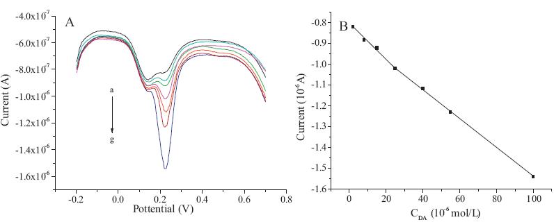
The DPV method is used for the determination of DA in the presence of 10 mmol/L AA. The voltammograms of solutions with different concentrations of DA are shown in Fig. 7A. The peak currents are proportional to DA concentrations in the range of 2-100 mmol/L with a detection limit of 2.5 × 10⁻⁹ mol/L (S/N = 3). According to Fig. 7B,the relationship between oxidation peak current and DA concentration can be divided into two sections: at DA concentrations from 2 mmol/L to 25 mmol/L,the linear regression equation is described as: IDA =-0.80593-0.00843 CDA (R = 0.99427); while at DA concentrations from 25 mmol/L to 100 mmol/L,the linear regression equation is described as: IDA = -0.84454 - 0.00695 CDA (R = 0.99988).

|
Download:
|
| Fig. 7.Selective detection of DA in the presence of UA. (A) DPV responses at DR/SWNT modified electrodes in 0.1 mol/L PBS (pH 6.0) containing 10 mmol/L of UA and different concentrations of DA from 2 mmol/L to 120 mmol/L (a–q: 2, 6, 12, 16, 22, 26, 32, 36, 42, 46, 52, 60, 70, 80, 90, 100, 110 and 120 mmol/L, respectively). Scan rate: 50 mV/s. (B) Relationship between the oxidation peaks current versus concentration of DA. | |
The stability of the DR/SWNT modified electrodes is evaluated. After one-week storage at room temperature,98.2% of the initial current signal was retained,and after two-week storage,97.2% signal remained,indicating that the prepared electrodes have excellent long-term stability. Meanwhile,the reproducibility of these modified electrodes is investigated. When five modified electrodes from the same fabrication procedure are prepared and used for the DA determination,the RSD is 3.4%. These results indicate that the modified electrodes have good detecting and fabrication reproducibility. 3.8. Dopamine determinations in real sample
Our recent experiments demonstrate that the modified electrodes not only can determine DA in the presence of main interferences such as UA and AA,but also can determine DA.HCl in the real samples of dopamine hydrochloride injection. As shown in Fig. 8,the peak currents are proportional to DA.HCl concentration in the range of 0.01-100 mmol/L. Therefore,the DR/SWNT modified electrode showed high sensitivity and selectivity for detection of DA.HCl in the real sample of DHI. The recovery test performed in Table 1 indicated that the DR/SWNT modified electrodes had very good recovery rates for the direct electrochemical determination of DA.HCl in the real samples.

|
Download:
|
| Fig. 8.Selective detection of DA.HCl in real sample of dopamine hydrochloride injection. (A) DPV responses at DR/SWNT modified electrodes in 0.1 mol/L PBS (pH 6.0) containing different concentrations of DA.HCl from 0.01 mmol/L to 100 mmol/L (a–g: 0.01, 2, 4, 10, 20, 40 and 100 mmol/L, respectively). Scan rate: 50 mV/s. (B) Relationship between the oxidation peaks current versus concentration of DA.HCl. | |
| Table 1 Recovery test of the DR/SWNT modified electrode to dopamine hydrochloride. |

In this paper,the self-assembly method combined with the photocrosslinking technique was applied to the preparation of DR/ SWNT modified Au electrodes successfully. The electrochemical behavior of DA on the modified electrodes was investigated by the CV and DPV methods. The modified electrodes exhibited good stability and reproducibility for the electrochemical determination of DA. Moreover,the modified electrodes showed very high electrocatalytic activity toward DA oxidation with a significant enhancement in peak current. Under the optimal conditions,a linear CV response to DA concentrations from 1 mmol/L to 40 mmol/L was observed,and the detection limit of DA was 2.1×10⁻³ mmol/L via the DPV method in the presence of 10 mmol/L of UA or 2.5 × 10⁻³ mmol/L via the DPV method in the presence of 10 mmol/L of AA. Additionally,the DR/SWNT modified electrodes showed high sensitivity and selectivity for detection ofDA.HCl inthe real samples of dopamine hydrochloride injection. The advantage of thismodified electrode is that photosensitiveDRas a polyelectrolyte is applied to the layer-by-layer self-assembly modification process, and afterUV photocrosslinking,the unstable ionic linkages involved in the self-assembled multilayers convert to stable covalent bonds, which makes the modified electrodes have better stability and fabrication reproducibility. Acknowledgments
This work is financially supported by the National Key Basic Research Development Program of China (973 Special Preliminary Study Plan,No. 2012CB722705),the Natural Science Foundation of China (Nos. 21375069 and 21344005),the Fok Ying Tong Education Foundation (No. 131045),the Scientific Research Foundation for the Returned Overseas Chinese Scholars of State Education Ministry (No. 20111568),and the Science and Technology Program of Qingdao (No. 1314159jch).
| [1] | R.M. Wightman, L.J. May, A.C. Michael, Detection of dopamine dynamics in the brain, Anal. Chem. 60 (1988) 769A-779A. |
| [2] | D. Han, T. Han, C. Shan, A. Ivaska, L.Niu, Simultaneous determination of ascorbic acid, dopamine and uric acid with chitosan-graphene modified electrode, Electroanalysis 22 (2010) 2001-2008. |
| [3] | P. Damier, E.C. Hirsch, Y. Aqid, A.M. Graybiel, The substantia nigra of the human brain. Ⅱ. Patterns of loss of dopamine-containing neurons in Parkinson's disease, Brain 122 (1999) 1437-1448. |
| [4] | U. Chandra, B.E.K. Swamy, O. Gilbert, et al., Poly(amaranth) film based sensor for resolution of dopamine in the presence of uric acid: a voltammetric study, Chin. Chem. Lett. 21 (2010) 1490-1492. |
| [5] | W. Song, Y. Chen, J. Xu, D.B. Tian, A selective voltammetric detection for dopamine usingpoly(gallic acid) filmmodified electrode, Chin.Chem. Lett.21 (2010) 349-352. |
| [6] | A.E. Poliakov, A.V. Dumshakova, S.V. Muginova, T.N. Shekhovtsova, A peroxidasebased method for the determination of dopamine, adrenaline, and a-methyldopa in the presence of thyroid hormones in pharmaceutical forms, Talanta 84 (2011) 710-716. |
| [7] | S. Liu, J. Yan, G. He, et al., Layer-by-layer assembled multilayer films of reduced graphene oxide/gold nanoparticles for the electrochemical detection of dopamine, J. Electroanal. Chem. 672 (2012) 40-44. |
| [8] | Z.H. Sheng, X.Q. Zheng, J.Y. Xu, et al., Electrochemical sensor based on nitrogen doped graphene: simultaneous determination of ascorbic acid, dopamine and uric acid, Biosens. Bioelectron. 34 (2012) 125-131. |
| [9] | E. Farjami, R. Campos, J.S. Nielsen, et al., RNA aptamer-based electrochemical biosensor for selective and label-free analysis of dopamine, Anal. Chem. 85 (2013) 121-128. |
| [10] | B. Kong, A. Zhu, Y. Luo, et al., Sensitive and selective colorimetric visualization of cerebral dopamine based on double molecular recognition, Angew. Chem. Int. Ed. 50 (2011) 1837-1840. |
| [11] | H. Su, B. Sun, L. Chen, Z. Xu, S. Ai, Colorimetric sensing of dopamine based on the aggregation of gold nanoparticles induced by copper ions, Anal. Methods 4 (2012) 3981-3986. |
| [12] | J.M. Liu, X.X. Wang, M.L. Cui, et al., A promising non-aggregation colorimetric sensor of AuNRs-Ag+ for determination of dopamine, Sens. Actuators B 176 (2013) 97-102. |
| [13] | J.J. Feng, H. Guo, Y.F. Li, et al., Single molecular functionalized gold nanoparticles for hydrogen-bonding recognition and colorimetric detection of dopamine with high sensitivity and selectivity, ACS Appl. Mater. Interfaces 5 (2013) 1226-1231. |
| [14] | S.S. Li, H.L. Wu, Y.J. Liu, H.W. Gu, R.Q. Yu, Simultaneous determination of tyrosine and dopamine in urine samples using excitation-emission matrix fluorescence coupled with second-order calibration, Chin. Chem. Lett. 24 (2013) 239-242. |
| [15] | A. El-Beqqali, A. Kussak, M. Abdel-Rehim, Determination of dopamine and serotonine in human urine samples utilizing microextraction online with liquid chromatography/electrospray tandem mass spectrometry, J. Sep. Sci. 30 (2007) 421-424. |
| [16] | P.S. Rao, N. Rujikarn, J.M. Luber Jr., D.H. Tyras, A specific sensitive HPLC method for determination of plasma dopamine, Chromatographia 28 (1989) 307-310. |
| [17] | J. Cho, K. Char, J.D. Hong, K.B. Lee, Fabrication of highly ordered multilayer films using a spin self-assembly method, Adv. Mater. 13 (2001) 1076-1078. |
| [18] | M. Grzelczak, J. Vermant, E.M. Furst, L.M. Liz-Marzan, Directed self-assembly of nanoparticles, ACS Nano 4 (2010) 3591-3605. |
| [19] | R. Deng, S. Liu, J. Li, et al., Mesoporous block copolymer nanoparticles with tailored structures by hydrogen-bonding-assisted self-assembly, Adv. Mater. 24 (2012) 1889-1983. |
| [20] | H. Cong, J. Chen, W. Cao, Covalently attached sandwich structure from colloidal particles and diazoresin, J. Colloid Interface Sci. 263 (2003) 665-668. |
| [21] | B. Yu, H.L. Cong, H.W. Liu, et al., Fabrication and characterization of stable ultrathin film micropatterns containing DNA and photosensitive polymer diazoresin, Anal. Bioanal. Chem. 384 (2006) 385-390. |
| [22] | F. Pompeo, D.E. Resasco, Water solubilization of single-walled carbon nanotubes by functionalization with glucosamine, Nano Lett. 2 (2002) 369-373. |
| [23] | B. Yu, W. Cui, H. Cong, et al., A novel diazoresin/polyethylene glycol covalent capillary coating for analysis of proteins by capillary electrophoresis, RSC Adv. 3 (2013) 20010-20015. |
| [24] | B. Yu, X.M. Liu, H.L. Cong, Z.H. Wang, J.G. Tang, Fabrication of stable ultrathin transparent conductive graphene micropatterns using layer by layer self-assembly, Sci. Adv. Mater. 5 (2013) 1533-1538. |