 2019, Vol. 35


2. Institute of Materials Science and Department of Civil and Environmental Engineering, The George Washington University, Washington, DC 20052
2. Institute of Materials Science and Department of Civil and Environmental Engineering, The George Washington University, Washington, DC 20052, USA
模拟地球内部温度-压力条件下相应物质的物理和化学性质对理解岩石圈演化的动力学过程、探索地球内部的运行机制,具有重要的科学意义;而原位高温高压下的实验研究与观察是关键所在。本项目拟通过对新购的傅立叶红外光谱仪进行拓展,构建一套适用于高压、高/低温测量的傅立叶变换红外显微系统的科学实验平台,从而真正实现原位条件下对地球内部物质的热力学性质和结构特征的系统观察与研究。
红外光谱研究目的是用它来探测分子的振动状态和振动方式,即分子中原子的相对振动→化学键的振动→不同的化学键或官能团,其振动能级从基态跃迁到激发态所需的能量不同→吸收不同波长的红外光→在不同波长出现吸收峰;并依据特征吸收峰对应的波长(波数)和谱峰数目与组成分子的原子质量、化学键的性质及化合物的几何类型特征,获得有关分子成分和结构的信息。分子振动模式和频率决定着物质红外光谱的特点,而分子的振动模式取决于分子的对称性和几何构型,晶体场理论揭示了晶体总是有一定的对称性,晶体中原子振动方式(模式)是按晶体的对称性分类的,同一构型、但不同对称的分子,其红外活性谱带数目、简并分裂程度有较大差别,并产生不同特征的红外光谱,因此,分子振动频率与化学键强度密切相关,在分析矿物红外光谱时,矿物中不同键的强度差异大小有着重要意义。决定矿物分子振动的另一重要因素是分子的几何构型,即矿物晶体结构中原子排列的对称性。它决定着分子的振动模式,即成分不同而对称性相同的分子具有系统的振动模式,反之成分相同而对称性不同的分子具有不同的振动模式。另一方面,矿物晶体结构的对称性及各种结构位置被不同的离子占据的微小改变,都会导致分子振动发生相应的变化,主要表现在频率的变化。实际上,红外光谱法与X-衍射法有较大的不同,红外光谱法不是独立解决整个晶体结构的手段,而是着重研究物质晶体结构的某些方面的微小差异,这一特征对于观察和探讨地球物质在随着地球环境改变而发生的运移、演化特别是物质内部的分子、离子扩散/交换的反应动力学过程具有独特的优势,尤其是红外光谱在不破坏样品的情况下,可对固、液、气体样品进行测定(Beran, 2000; Aske et al., 2002; Braithwaite et al., 2003; Rosén et al., 2010; Han et al., 2013; Shuai and Yang, 2017)。然而,目前红外光谱的应用更多的是在常温常压下来研究晶体结构、晶体生长缺陷以及化学键和分子结构方面(Tateyama et al., 1977; Diaz et al., 2000; Busigny et al., 2003; Su et al., 2004, 2009; Rossman, 2006; Nazzareni et al., 2011),仅有少量涉及到高温常压、高压室温、或者是在淬火的条件下的矿物测试(Tokiwai and Nakashima, 2009; Zhang et al., 2010a, b; Yang and Keppler, 2011; Su et al., 2008, 2013; Vennari et al., 2017),这主要限于目前商用傅立叶变换红外显微镜系统采用的是垂直入射的红外光路,而该系统中物镜最大的工作距离为15mm,根本无法与一些低温/高温-高压设备联用,限制了原位低温/高温-高压条件下的傅立叶变换红外光谱实验研究的展开。因此,构建一套相对完整的、适用于高压、高/低温测量的傅立叶变换红外显微镜系统的科学实验平台,对于开展在原位条件下,对地球内部物质的热力学性质和结构特征的系统观察与研究是非常必要的。
1 水平光路红外光学路径的设计依据商用布鲁克Vertex 70v真空型红外光谱仪的特点,预留了左侧光路的出口,结合在原位高温/低温-高压条件下测试矿物红外光谱的需要,设计了具备水平光路、透射光源与反射光源相互切换功能的显微红外系统的光学路径(图 1)。

|
图 1 红外扩展系统光路示意图 Fig. 1 Schematic of the optical path of the IR extension system |
该系统包括:(1)共轴入射红外光源与可见光路径;(2)具有长工作距离(40mm)和大数值孔径(0.5)的反射型物镜和汇聚镜;(3)在傅里叶变换红外光谱仪的出口与宽带红外检测器之间具备最优化的光学元件、可达到最低极限波数(420cm-1);(4)具有分别用于观察透射、反射测量的双筒显微镜;(5)红外检测器;(6)用于高压、低温/高温条件下,显微红外光谱系统与测压系统之间原位测量相互切换的滑动平台,最大移动距离 > 153mm;以及(7)所有镜面均镀银或金,光谱的覆盖范围从中红外到可见光。
2 适用于高压/高温测量的傅立叶变换红外显微系统的实验平台从商用机布鲁克Vertex 70v中引出一条红外水平光路,搭建了特别适用于高灵敏度、傅立叶变换的红外显微光谱系统,该系统具备透射与反射的相互切换功能,且完全适用于与商用机布鲁克Vertex 70v真空型红外光谱仪间的切换(图 1、图 2)。

|
图 2 扩展显微红外系统与测压系统、温控仪 Fig. 2 Schematic of the optical path of the IR extension system, pressure calibration system, temperature controller |
(1) 技术指标对比。将新构建的、水平光路的傅立叶变换红外显微系统与商用机布鲁克HYPERION-2000红外显微系统的性能进行了对比,主要是红外信号强度的对比,在空位的条件下,新建显微红外系统的振幅为38400(图 3a),而商用布鲁克HYPERION-2000的振幅为33000(图 3b),由此可见新构建的傅立叶变换红外显微系统技术指标等于或优于商用机的技术指标。

|
图 3 新建显微红外系统(a)和HYPERION-2000 (b)的红外信号强度对比 Fig. 3 Comparison of infrared signal intensity between New IR extension system (a) and HYPERION-2000 (b) |
(2) 测压系统的构建。DAC设备高压实验中压力的标定主要有两种:压力内标法和红宝石荧光法。压力的内标法就是利用压标物质的压力与体积间的状态方程来确定压力(Birch, 1947; Poirier, 2000; Fei et al., 2007);所以用作压标的物质应具备稳定的物理化学性质、结构简单、在实验的压力范围内无相变发生、较大的压缩性、以及标定的状态方程,因此该方法的优点是简便而精确,但缺点是在实验的过程中会受到压标的衍射峰或荧光峰的干扰而影响所测试物质的实验数据(Decker, 1971; Anderson et al., 1989; Holmes et al., 1989; Matsui et al., 2000; Spezial et al., 2001; Shim et al., 2002; Fei et al., 2007; Hirose et al., 2008)。红宝石荧光法就是根据红刚玉的谱峰位移与压力的关系来确定压力。红宝石(Cr3+: (-Al2O3)是优良的激光材料之一,其激光作用是通过Cr3+受激发射而实现。在室温常压下,红宝石具有2个谱峰:A(694.2nm)和B(692.8nm),A、B谱峰会随着压力增加发生“红移”。依据A或B谱峰和压力的红移关系,即可得出该状态条件下的压力(Forman et al., 1972; Mao, 1978; Mao et al., 1986; Xu et al., 1986; Funamori and Jeanloz, 1997; Rekhi et al., 1999; Grasset, 2001; Wei et al., 2011)。如:
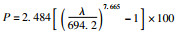

式中,λ为A或B谱峰位(nm),P为压力(GPa),T为温度(K),a=19.99,b=6.75+b1(T-298)+b2(T-298)2,R=(A+B)/2。
红宝石作为压标具有化学性质稳定、易获得、峰强度大、用量少、图谱信噪比高、标定的压力范围大等优点,并且该方法简单、快速,因而红宝石荧光法是常用的压力标定方法。为此,针对红宝石荧光法特征,结合扩展的水平光路显微红外光学系统,设计并构建了红宝石荧光测压系统(图 4a, b)。该系统包括:(1)XYZ移动平台;(2)可见光光源(S);(3)具有长工作距离的物镜;(4)照相系统;(5)激光器;(6)聚焦镜(L1、L2);(7)反射镜(M1、M2、M3);(8)分束器(BS1、BS2);(9)可变光阑(IRIS)。

|
图 4 红宝石测压系统光路示意图(a)和测压系统(b) Fig. 4 Optical layout of the ruby system (a) and the pressure calibration system (b) |
(3) 构建了外加温的温控系统。通过一系列的试验和摸索,并针对金刚石压腔装置(diamond anvil cell, DAC)外加温的特点,研制了适用于BX-90型DAC的外加温电阻炉。该炉是由一种特殊的陶瓷、Cr-Ni丝组成(图 5a),被设计用来在一个受限制的空间内缠绕线圈以便最大限度地增加功率,同时缩小与金刚石压砧的最小距离。将电阻炉安装在金刚石压砧的周围,其原理就是通过控制加载电流进行精确地温度调节,样品温度的获得是通过热辐射来实现的,加热均匀且温度梯度小,样品的加热可以从室温到1200K。样品温度是通过不同的独立测量的组合来监测的。选配了恰当的k型热电偶(Omega Chal-005),在实验前建立并通过校准曲线它与样品的温度直接相关(图 5b),然后将其固定在一金刚石压砧的台面来实测并报告金刚石压砧台面(样品)的温度变化(图 5b),从而可以在原位的实验过程中进行实时的控温和测温。

|
图 5 外加温电阻炉(a)和温控仪(b) Fig. 5 External heater (a) and the temperature controller (b) |
在上述的基础上,将构建的测压系统、测温系统与扩展的水平光路红外显微系统相连,从而实现了在实验过程中原位的测温、测压及红外光谱分析与观察的化学动力学研究(图 2)。
3 原位高温高压下绿帘石实验模拟绿帘石由于稳定于较宽的压力和温度范围内,可以作为重要的造岩矿物或副矿物出现在近地表、岩浆岩、高压-超高压变质作用的各种岩石中,它不仅被认为是典型的变质矿物,也是非常重要的火成岩热液矿物,以及沉积物中的碎屑重矿物。Yao et al. (2000)在苏鲁榴辉岩的绿帘石中发现了柯石英包裹体,表明绿帘石是超高压岩石中的稳定矿物;Poli and Schmidt (2004)的研究表明黝帘石可以稳定在压力=7.0GPa的范围内,常见的绿帘石-斜黝帘石固溶体分解发生在T=600~700℃,且实验数据显示这些矿物可以通过载入稀土元素而稳定(Nagasaki and Enami, 1998; Hermann, 2002; Enami et al., 2004)。Carbonin and Molin (1980)首先利用单晶X射线衍射对天然绿帘石族矿物进行了系统研究,特别是晶体几何形态和结构伴随着Al-Fe3+(成分范围XEp=0.30~0.86)替代的变化。随后,Bonazzi and Menchetti (1995)、Giuli et al. (1999)分别扩展这项工作,并对天然和合成的富含REE、Fe2+绿帘石(成分范围XEp=0.24~1.14、0.66~1.09)进行了有关Fe3+与Fe2+的替代以及稀土元素的加入在晶体结构中作用的研究。他们的研究数据表明含Fe3+绿帘石衍射谱峰的强弱对晶体结构的有序度非常敏感即Z轴 > Y轴 > X轴(Taran and Langer, 2000);同时发现Al-Fe3+固溶体体系中随着Fe含量的增加,晶体结构中光谱谱峰会向低波数漂移,直至最终分解(Burns and Strens, 1967; Giuli et al., 1999; Taran and Langer, 2000)。Parkin and Burns (1980)、Taran and Langer (2000)分别研究了在T=573K、600K条件下,温度对绿帘石中Fe3+的影响,发现结构中谱峰的强度随着温度的增加而增强。Taran and Langer (2000)在室温、P=4.6、9.6GPa条件下,研究了天然绿帘石(XEp=0.78)的非偏振吸收光谱,发现随着压力的增加,仅仅16500cm-1谱峰向低波数漂移,且其半高宽也随之降低;P=5GPa时,随着压力进一步的增加吸收光谱的强度略有降低;P=9.6GPa时,新的谱峰出现。基于2次实验数据,Taran and Langer (2000)计算出绿帘石结构中M3位随温度变化的热膨胀系数,认为绿帘石中M3位保留了畸变或随温度增加而更加扭曲;由于M3位与压力间没有明显的变化,所以认为绿帘石结构中的M3位在高压下有较高的压缩性。Della Ventura et al. (1996)的研究认为绿帘石中OH结构的变化伴随着M3位上Mn3+和A位上Sr的载入而改变,并且与晶体结构中Fe含量呈线性关系(Langer and Raith, 1974; Langer et al., 1976; Heuss-Aßbichler, 2000)。Bradbury and Williams (2003)、Liebscher and Gottschalk (2004)也分别利用红外光谱研究了在不同压力条件下绿帘石单斜结构的变化、以及黝帘石在不同温度条件下的演化行为。并结合早期研究数据,证实了晶体中质子O10—H…O4键的存在(Belov and Rumanova, 1954)。同时Bradbury and Williams (2003)通过对斜黝帘石晶体结构中OH的研究发现,在常压下氢原子优先与O10链接,但随着压力的增加,H缓慢地向远离O10方向移动,表明O10—H键长的增加可能伴随能量的降低,这与所观察到的OH谱峰随着压力的增加移向低波数的现象一致。然而,原位高温高压条件下绿帘石族矿物晶体结构和水溶解度的数据是严重缺乏的,因此,利用构建的具有水平光路的显微红外系统展开了有关原位高温高压条件下绿帘石的稳定与水溶解度之间关系的初步研究。
3.1 样品描述绿帘石晶体样品来自非洲马达加斯加,岩石呈现墨绿色,为中粗粒-粗粒不等粒结构,块状构造,岩石主要由绿帘石组成(图 6a)。绿帘石晶体较大( > 2mm)且内部包裹体较少(图 6b),依据电子探针分析结果,获得其晶体化学式为Ca2(Al2.15Fe0.8Ti0.05)Si3O12(OH)。

|
图 6 绿帘石样品及显微照片 (a)马达加斯加绿帘石岩,由绿帘石组成,手标本;(b)绿帘石(Epi)显微照片,揭示了绿帘石单晶几乎不含包裹体,单偏光 Fig. 6 Epidosite sample and micro-photograph (a) photograph of the epidosite sample from the Madagascar. Hand specimen; (b) micro-photograph reveal that epidote (Epi) crystal contains almost no inclusions |
将绿帘石晶体分别进行平行于或垂直于C轴的切片,然后进行单晶的磨制,磨至单晶的厚度为16μm时,待实验备用。
3.2 实验条件本次实验采用DAC装置结合扩展显微红外透射光谱系统和拉曼光谱技术对绿帘石进行了原位高温高压下晶体结构及其结构水的研究。DAC装置为BX-90型(图 7a),金刚石砧面直径为400μm;高压密封垫片为耐高温的铼片,预压前厚度为250μm,预压后厚度为76μm(红外光谱)和44μm(拉曼光谱),使用激光在压痕中心打孔直径为190μm的样品腔体。将预先处理好的绿帘石单晶样品与红宝石小球粒一起放置在样品腔体内,绿帘石单晶样品在红外透射光谱和拉曼光谱实验中的厚度为16μm和直径分别约105×38μm(图 7b)、86×20μm(图 7c)。传压介质分别为KBr(红外光谱)、4: 1配比的甲醇、乙醇混合溶液(拉曼光谱)(杜建国等, 2011),样品腔体内压力的标定均采用红宝石荧光法(Mao et al., 1986(室温下); Rekhi et al., 1999; Wei et al., 2011(高温下))。使用外加温电阻炉,热电偶放入样品腔内,在炉子的上下分别垫上云母片+石棉片的绝热层。实验采用恒压升温的方法,首先在室温下加压至3.8GPa,然后以20℃/min的速率升温至一定温度,稳定20min后,进行红宝石谱峰压力标定、以及原位光谱测量;然后以相同的升温速率继续加热到下一个温度点,稳定20min后,再进行压力标定、及光谱测量,如此反复进行,直至达到所需温度压力标定及光谱测量才结束。红外光谱测试在新构建的具有水平光路的显微红外系统完成,分束器是KBr,使用的是液氮冷却的宽带MCT检测器,样品和背景的扫描次数均为640次,分辨率是8cm-1,测量的波数范围是4000~400cm-1。拉曼光谱分析在北京高压科学研究中心拉曼光谱实验室完成,分析所用仪器为英国Renishaw公司生产的Invia共焦显微激光拉曼光谱仪,激发光源波长488nm,光栅2400,物镜为三丰20倍长工作距离,50%激光强度(50mW),采集时间为50s,累计次数2。

|
图 7 原位实验设备以及绿帘石样品 (a) BX90型金刚石压机;(b)绿帘石高温高压红外光谱实验样品腔图,样品腔内装有绿帘石、红宝石,传压介质为KBr;(c)绿帘石高温高压拉曼光谱实验样品腔图,样品腔内有绿帘石、红宝石,传压介质为甲、乙醇混合溶液 Fig. 7 In situ experimental equipment and epidote sample (a) BX90 type DAC; (b) photo shows epidote, ruby ball and KBr of pressure transfer medium for IR; (c) photo shows epidote, ruby ball and mixed solution of methanol and ethanol of pressure transfer medium for Raman |
在新构建的水平光路红外显微系统上,运用BX-90型DAC原位模拟了不同温、压条件下(T=室温-873K,P=1.14~11.87GPa)绿帘石中OH演化的初步实验。
图 8a显示了绿帘石在常温常压下、以及高温高压下的红外吸收光谱图谱。常温常压下绿帘石OH吸收峰谱大致可分为五组(图 8a):3357cm-1(a)、3397cm-1(b)、3461cm-1(c)、3660cm-1(d)、3762cm-1(e),在室温条件下增加压力,其所有的OH峰谱强度整体大幅度地减弱,当持续加压,a、b谱峰开始向低波数位移,但整体未发生明显的变化。但当加温373K,P=4.81GPa时,c、d、e谱峰强度突然增加;随着温压的增加,c、d、e谱峰强度逐步降低,在P=8.83GPa、T=673K完全消失;而a、b谱峰在室温、P=1.18GPa时合并为单一谱峰后,又随着温压的增加不仅向低波数位移,而且分裂为3312~3326cm-1、3348~3352cm-1谱峰(图 8a)。将不同温压条件下绿帘石中所有的OH谱峰进行其OH含量的面积积分,发现其与温压呈负相关(图 9),表明绿帘石晶体中OH含量随着温压的增加而逐步降低,而图 9有2次的突变分别与压力、温度有关。

|
图 8 绿帘石原位高温高压下红外光谱分析图谱 (a) 3000~4000cm-1;(b) 400~1200cm-1 Fig. 8 Infrared spectrum experiments of epidote at high temperature and high pressure in situ |

|
图 9 绿帘石中OH红外光谱谱峰强度与温压关系图解 Fig. 9 Diagram of temperature and pressure dependence of IR spectral intensity of OH species |
图 8b为实验获得的绿帘石在不同温压条件下的红外光谱数据,图 8b中的878cm-1红外波谱被指派为Si-O振动,其随着温压的增加逐步向低波数位移,并没有新的谱峰产生。
图 10则显示绿帘石在不同温压条件下拉曼光谱的实验数据,从图中可以看出拉曼振动的信号主要集中在200~1200cm-1之间,约有13组的拉曼活性谱峰位,分别为288cm-1(ν1)、326cm-1(ν2)、438cm-1(ν3)、465cm-1(ν4)、511cm-1(ν5)、533cm-1(ν6)、576cm-1(ν7)、612cm-1(ν8)、880cm-1(ν9)、937cm-1(ν10)、996cm-1(ν11)、1041cm-1(ν12)、1105cm-1(ν13)(图 10、表 1)。其中ν1的拉曼振动谱峰与Ca-O键的振动有关,ν2、ν4~ν6的拉曼振动谱峰则主要是与晶体结构中八面体位置的M-O键(M为Al或Fe)的伸缩振动有关,而ν3、ν7~ν10、ν13的拉曼振动谱峰是由于Si-O键的对称伸缩振动导致的(Langer and Raith, 1974; Qin et al., 2003, 2016)(表 1)。随着温压的增加,所有的谱峰均发生了演化。从常温常压逐步加温加压到T=623K、P=12.33GPa过程中,几乎所有的谱峰逐步向高波数位移(图 10),期间并未出现有新的振动谱峰产生,但是一些振动谱峰随着温压的增加其强度逐步变弱,值得注意的是ν6、ν11、ν12在T=373K、P=5.11GPa时突然消失(图 10),这与在绿帘石中OH红外光谱谱峰(c-e组)在同一温度-压力区间突然增加有偶合关系,可能与Fe有关(Su et al., 2009)。随着温压的进一步增加,所有的谱峰开始逐步向低波数位移(图 10),而ν19和ν10则合并为单个谱峰(图 10)。通过简单计算可以获得绿帘石在高温高压下Ca-O键、M-O键以及Si-O键拉曼振动谱峰的平均位移率分别为:0.68cm-1/GPa×100℃、0.58cm-1/GPa×100℃、0.91cm-1/GPa×100℃,这可能暗示了Si-O键具有比Ca-O键、M-O键较强的压缩性。

|
图 10 绿帘石原位高温高压下拉曼光谱分析图谱 Fig. 10 Raman spectrum experiments of epidote at high temperature and high pressure in situ |
|
|
表 1 绿帘石拉曼光谱、红外光谱谱峰及其指派 Table 1 Raman and Infrared spectrum of epidote |
(1) 搭建了红宝石荧光测压系统。
(2) 构建了外加温的温控系统。
(3) 在布鲁克Vertex 70v真空型红外光谱仪基础上,扩展、构建了一套适用于高压、高/低温测量、水平光路的傅立叶变换红外显微系统,并与红宝石测压系统联用,实现原位条件下观察与研究在地球内部含水物质的性质和结构特征的实验平台。
(4) 绿帘石原位高温高压下的拉曼光谱和红外光谱实验研究表明,从常温常压条件加温加压至P=11.87~12.73GPa和T=773~873K时,虽然谱峰发生了演化,但是绿帘石的结构仍然稳定存在。特别是M-O键具有较弱的压缩性由于Fe-Al在八面体中的替代有关,而红外光谱中OH振动谱峰的突然增加、消失也与此Fe的氧化还原有关。为此绿帘石的温压条件至少可以稳定P=11.87~12.73GPa和T=773~873K范围内,可以将水携带到地幔的深处(350~380km)。
4.2 存在问题金刚石压砧的保护。众所周知,金刚石在温度> 650℃时会发生氧化而导致石墨化,而要真实地模拟俯冲带中物质在深俯冲过程中物质组成的变化、元素迁移、挥发份物性等动力学过程,尤其是达到地幔深度环境下,其温度均要>650℃,如何在超高温的实验过程中保护金刚石压砧不受氧化作用,将是下一步需考虑和解决的关键问题。
致谢 感谢北京高压科学研究中心郑海燕、林小欢、王丽娟在拉曼光谱实验中的帮助;特别感谢矿物收藏家钟国宾先生提供的绿帘石晶体样品。
祝贺叶大年院士八十寿诞,感谢叶老师多年来在工作中给予的悉心指导与帮助。
Anderson OL, Isaak DG and Yamamoto S. 1989. Anharmonicity and the equation of state for gold. Journal of Applied Physics, 65(4): 1534-1543. DOI:10.1063/1.342969 |
Aske N, Kallevik H and Sjöblom J. 2002. Water-in-crude oil emulsion stability studied by critical electric field measurements:Correlation to physico-chemical parameters and near-infrared spectroscopy. Journal of Petroleum Science and Engineering, 36(1-2): 1-17. DOI:10.1016/S0920-4105(02)00247-4 |
Belov NV and Rumanova JM. 1954. The crystal structure of epidote. Trudy Inst. Kryst. Akad. Nauk. SSSR, 9: 103-164. |
Beran A. 2002. Infrared spectroscopy of micas. In: Mottana A, Sassi EP, Thompson JB Jr and Guggenheim S (eds.). Reviews in Mineralogy and Geochemistry. Volume 46: Micas: Crystal Chemistry & Metamorphic Petrology. Washington: Mineralogical Society of America, 17-28
|
Birch F. 1947. Finite elastic strain of cubic crystals. Physics Review, 71(11): 809-824. DOI:10.1103/PhysRev.71.809 |
Bonazzi P and Menchetti S. 1995. Monoclinic members of the epidote group:Effects of the AI⇌Fe3+⇌Fe2+ substitution and of the entry of REE3+. Mineralogy and Petrology, 53(1-3): 133-153. DOI:10.1007/BF01171952 |
Bradbury SE and Williams Q. 2003. Contrasting bonding behavior of two hydroxyl-bearing metamorphic minerals under pressure:Clinozoisite and topaz. American Mineralogist, 88(10): 1460-1470. DOI:10.2138/am-2003-1010 |
Braithwaite JS, Wright K and Catlow CRA. 2003. A theoretical study of the energetics and IR frequencies of hydroxyl defects in forsterite. Journal of Geophysical Research:Solid Earth, 108(B6): 2284. |
Burns RG and Strens RGJ. 1967. Structural interpretation of polarized absorption spectra of the Al-Fe-Mn-Cr epidotes. Mineralogical Magazine, 36(278): 204-226. DOI:10.1180/minmag.1967.036.278.04 |
Busigny V, Cartigny P, Philippot P and Javoy M. 2003. Ammonium quantification in muscovite by infrared spectroscopy. Chemical Geology, 198(1-2): 21-31. DOI:10.1016/S0009-2541(02)00420-5 |
Carbonin S and Molin G. 1980. Crystal-chemical considerations on eight metamorphic epidotes. Neues Jahrbuch fur Mineralogie-Abhandlungen, 139: 205-215. |
Decker DL. 1971. High-pressure equation of state for NaCl, KCl, and CsCl. Journal of Applied Physics, 42(8): 3239-3244. DOI:10.1063/1.1660714 |
Della Ventura G, Mottana A, Parodi GC and Griffin WL. 1996. FTIR spectroscopy in the OH-stretching region of monoclinic epidotes from Praborna (St. Marcel, Aosta Valley, Italy). European Journal of Mineralogy, 8(4): 655-665. |
Diaz M, Farmer VC and Prost R. 2000. Characterization and assignment of far infrared absorption bands of K+ in muscovite. Clays and Clay Minerals, 48(4): 433-438. DOI:10.1346/CCMN |
Du JG, He DW, Gao XC, Gong ZZ, Wu XY, Zhou WG, Wang DJ, Zhai SM, Ji GF and Wei DQ. 2011. Experimental and Theoretical Studies of Mineral and Rock at High Pressure and Temperature. Beijing: Seismological Press.
|
Enami M, Liou JG and Mattinson CG. 2004. Epidote minerals in high P/T metamorphic terranes:Subduction zone and high-to ultrahigh-pressure metamorphism. Reviews in Mineralogy and Geochemistry, 56(1): 347-398. |
Fei YW, Ricolleau A, Frank M, Mibe K, Shen GY and Prakapenka V. 2007. Toward an internally consistent pressure scale. Proceedings of the National Academy of Sciences of the United States of America, 104(22): 9182-9186. DOI:10.1073/pnas.0609013104 |
Forman RA, Piermarini GJ, Barnett JD and Block S. 1972. Pressure measurement made by the utilization of ruby sharp-line luminescence. Science, 176(4032): 284-285. DOI:10.1126/science.176.4032.284 |
Funamori N and Jeanloz R. 1997. High-pressure transformation of Al2O3. Science, 278(5340): 1109-1111. DOI:10.1126/science.278.5340.1109 |
Giuli G, Bonazzi P and Menchetti S. 1999. Al-Fe disorder in synthetic epidotes; A single-crystal X-ray diffraction study. American Mineralogist, 84(5-6): 933-936. DOI:10.2138/am-1999-5-629 |
Grasset O. 2001. Calibration of the R ruby Fluorescence lines in the pressure range (0~1GPa) and the temperature range (250~300K). High Pressure Research, 21(3-4): 139-157. DOI:10.1080/08957950108201020 |
Han L, Zhou YS and He CR. 2013. Water-enhanced plastic deformation in felsic rocks. Science China (Earth Sciences), 56(2): 203-216. DOI:10.1007/s11430-012-4367-6 |
Hermann J. 2002. Allanite:Thorium and light rare earth element carrier in subducted crust. Chemical Geology, 192(3-4): 289-306. DOI:10.1016/S0009-2541(02)00222-X |
Heuss-Aßbichler S. 2000. Ein neues Ordnungsmodell für die Mischkristallreihe Klinozoisit-Epidot und das Granat-Epidot-Geothermometer. Munich: Habilitationthesis: 105.
|
Hirose K, Sata N, Komabayashi T and Ohishi Y. 2008. Simultaneous volume measurements of Au and MgO to 140GPa and thermal equation of state of Au based on the MgO pressure scale. Physics of the Earth and Planetary Interiors, 167(3-4): 149-154. DOI:10.1016/j.pepi.2008.03.002 |
Holmes NC, Moriarty JA, Gathers GR and Nellis WJ. 1989. The equation of state of platinum to 660GPa (6.6Mbar). Journal of Applied Physics, 66(7): 2962-2967. DOI:10.1063/1.344177 |
Langer K and Raith M. 1974. Infrared spectra of Al-Fe(Ⅲ)-epidotes and zoisites, Ca2(Al1-pFe3+p)Al2O(OH)[Si2O7]. American Mineralogist, 59(11-12): 1249-1258. |
Langer K, Abu-Eid RM and Anastasiou P. 1976. Absorptionsspektren synthetischer Piemontite in den Bereichen 43000~11000cm-1 (232, 6~909, 1nm) und 4000~250cm-1 (2, 5~40μm). Zeitshrift fur Kristallographie, 144: 434-436. |
Liebscher A and Gottschalk M. 2004. The T-X dependence of the isosymmetric displacive phase transition in synthetic Fe3+-Al zoisite:A temperature-dependent infrared spectroscopy study. American Mineralogist, 89(1): 31-38. |
Mao HK. 1978. High-pressure physics:Sustained static generation of 1.36 to 1.72 megabars. Science, 200(4346): 1145-1147. DOI:10.1126/science.200.4346.1145 |
Mao HK, Xu J and Bell PM. 1986. Calibration of the ruby pressure gauge to 800kbar under quasi-hydrostatic conditions. Journal of Geophysical Research:Solid Earth, 91(B5): 4673-4676. DOI:10.1029/JB091iB05p04673 |
Matsui M, Parker SC and Leslie M. 2000. The MD simulation of the equation of state of MgO:Application as a pressure calibration standard at high temperature and high pressure. American Mineralogist, 85(2): 312-316. DOI:10.2138/am-2000-2-308 |
Nagasaki A and Enami M. 1998. Sr-bearing zoisite and epidote in ultra-high pressure (UHP) metamorphic rocks from the Su-Lu province, eastern China:An important Sr reservoir under UHP conditions. American Mineralogist, 83(3-4): 240-247. DOI:10.2138/am-1998-3-407 |
Nazzareni S, Skogby H and Zanazzi PF. 2011. Hydrogen content in clinopyroxene phenocrysts from Salina mafic lavas (Aeolian arc, Italy). Contributions to Mineralogy and Petrology, 162(2): 275-288. DOI:10.1007/s00410-010-0594-z |
Parkin KM and Burns RG. 1980. High temperature crystal field spectra of transition metal-bearing minerals: Relevance to remote-sensed spectra of planetary surfaces. In: Proceedings of the 11th Lunar and Planetary Science Conference. Houston: NASA, 731-755
|
Poirier JP. 2000. Introduction to the Physics of the Earth's Interior. Cambridge: Cambridge University Press: 63-109.
|
Poli S and Schmidt MW. 2004. Experimental subsolidus studies on epidote minerals. Reviews in Mineralogy and Geochemistry, 56(1): 171-195. DOI:10.2138/gsrmg.56.1.171 |
Qin F, Wu X, Wang Y, Fan DW, Qin S, Yang K, Townsend JP and Jacobsen SD. 2016. High-pressure behavior of natural single-crystal epidote and clinozoisite up to 40GPa. Physics and Chemistry of Minerals, 43(9): 649-659. DOI:10.1007/s00269-016-0824-7 |
Qin S, Wu X, Liu J, Liu J, Wu ZY, Li XD and Lu AH. 2003. Compressibility of epidote up to 20GPa at 298K. Chinese Physics Letters, 20(7): 1172-1174. DOI:10.1088/0256-307X/20/7/358 |
Rekhi S, Dubrovinsky L and Saxena S. 1999. Temperature-induced ruby fluorescence shifts up to a pressure of 15GPa in an externally heated diamond anvil cell. High Temperature-High Pressure, 31(3): 299-305. DOI:10.1068/htrt161 |
Rosén P, Vogel H, Cunningham L, Reuss N, Conley DJ and Persson P. 2010. Fourier transform infrared spectroscopy, a new method for rapid determination of total organic and inorganic carbon and biogenic silica concentration in lake sediments. Journal of Paleolimnology, 43(2): 247-259. DOI:10.1007/s10933-009-9329-4 |
Rossman GR. 2006. Analytical methods for measuring water in nominally anhydrous minerals. In: Keppler H and Smyth JR (eds.). Water in Nominally Anhydrous Minerals. Reviews in Mineralogy and Geochemistry. Chantilly: Mineralogical Society of America, 62: 1-28
|
Shim SH, Duffy TS and Kenichi T. 2002. Equation of state of gold and its application to the phase boundaries near 660km depth in Earth's mantle. Earth and Planetary Science Letters, 203(2): 729-739. DOI:10.1016/S0012-821X(02)00917-2 |
Shuai K and Yang XZ. 2017. Quantitative analysis of H-species in anisotropic minerals by polarized infrared spectroscopy along three orthogonal directions. Contributions to Mineralogy and Petrology, 172(2-3): 14. DOI:10.1007/s00410-017-1336-2 |
Speziale S, Zha CS, Duffy TS, Hemley RJ and Mao HK. 2001. Quasi-hydrostatic compression of magnesium oxide to 52GPa:Implications for the pressure-volume-temperature equation of state. Journal of Geophysical Research:Solid Earth, 106(B1): 515-528. DOI:10.1029/2000JB900318 |
Su W, Ji ZP, Ye K, You ZD, Liu JB, Yu J and Cong BL. 2004. Distribution of hydrous components in jadeite of the Dabie Mountains. Earth and Planetary Science Letters, 222(1): 85-100. DOI:10.1016/j.epsl.2004.02.028 |
Su W, Zhang M, Redfern SAT and Bromiley GD. 2008. Dehydroxylation of omphacite of eclogite from the Dabie-Sulu. Lithos, 105(1-2): 181-190. DOI:10.1016/j.lithos.2008.03.006 |
Su W, Zhang M, Redfern SAT, Gao J and Klemd R. 2009. OH in zoned amphiboles of eclogite from the western Tianshan, NW-China. International Journal of Earth Sciences, 98(6): 1299-1309. DOI:10.1007/s00531-008-0379-z |
Su W, Zhou Q and Liu X. 2013. Characteristic of pargasitic amphibole at high pressure by in situ μ-Raman, μ-FTIR spectroscopy and synchrotron X-ray diffraction. In: American Geophysical Union, Fall Meeting 2013. Washington: AGU
|
Taran MN and Langer K. 2000. Electronic absorption spectra of Fe3+ in andradite and epidote at different temperatures and pressures. European Journal of Mineralogy, 12(1): 7-15. |
Tateyama H, Shimda S and Sudo T. 1977. Estimation of K-O distance and tetrahedral rotation angle of K-micas from far-infrared absorption spectral data. American Mineralogist, 62(5-6): 534-539. |
Tokiwai K and Nakashima S. 2010. Dehydration kinetics of muscovite by in situ infrared microspectroscopy. Physics and Chemistry of Minerals, 37(2): 91-101. |
Vennari CE, O'Bannon EF and Williams Q. 2017. The ammonium ion in a silicate under compression:Infrared spectroscopy and powder X-ray diffraction of NH4AlSi3O8-buddingtonite to 30GPa. Physics and Chemistry of Minerals, 44(2): 149-161. DOI:10.1007/s00269-016-0844-3 |
Wei QG, Dubrovinskaia N and Dubrovinsky L. 2011. Ruby and Sm:YAG fluorescence pressure gauges up to 120GPa and 700K. Journal of Applied Physics, 110(4): 043513. DOI:10.1063/1.3624618 |
Xu JA, Mao HK and Bell PM. 1986. High-pressure ruby and diamond fluorescence:Observations at 0.21 to 0.55 terapascal. Science, 232(4756): 1404-1406. DOI:10.1126/science.232.4756.1404 |
Yang XZ and Keppler H. 2011. In-situ infrared spectra of OH in olivine to 1100℃. American Mineralogist, 96(2-3): 451-454. DOI:10.2138/am.2011.3717 |
Yao YP, Ye K, Liu JB, Cong BL and Wang QC. 2000. A transitional eclogite-to high pressure granulite-facies overprint on coesite-eclogite at Taohang in the Sulu ultrahigh-pressure terrane, Eastern China. Lithos, 52(1-4): 109-120. DOI:10.1016/S0024-4937(99)00087-0 |
Zhang M, Redfern SAT, Salje EKH, Carpenter MA and Hayward CL. 2010a. Thermal behavior of vibrational phonons and hydroxyls of muscovite in dehydroxylation:In situ high-temperature infrared spectroscopic investigations. American Mineralogist, 95(10): 1444-1457. DOI:10.2138/am.2010.3472 |
Zhang M, Redfern SAT, Salje EKH, Carpenter MA and Wang L. 2010b. H2O and the dehydroxylation of phyllosilicates:An infrared spectroscopic study. American Mineralogist, 95(11): 1686-1693. |
杜建国, 贺端威, 高春晓, 龚自正, 伍向阳, 周文戈, 王多君, 翟双猛, 姬广富, 魏冬青. 2011. 矿物岩石高温高压实验与理论研究. 北京: 地震出版社.
|