


2. Department of Neurosurgery, Beijing Anzhen Hospital, Capital Medical University, Beijing 100029, China
Nan Zhang is an Associate Clinical Professor in the Department of Neurosurgery of Beijing Anzhen Hospital, Capital Medical University. Dr. Zhang specializes in neurovascular surgery, cranial base surgery, and the surgical treatment of chronic pain. His research interests focus on cranial base microsurgical anatomy and surgical approaches, neurointerventional techniques, and neural stem cell treatments for stroke. He has served as a principal investigator in many research projects, including the National High-Technology Research and Development Program (863 program). He has published over 20 papers and 7 works, and he has obtained 4 Ministerial and Provincial-Level Science and Technology Awards. In addition, he won the Beijing Outstanding Young Physician Award in 2012.
Nan Zhang, E-mail:dr_zhangnan@163.com.
Cardiovascular and cerebrovascular diseases are serious threats to human health and are often the first of many causes of death. At present, the diagnosis and treatment of cardiovascular and cerebrovascular diseases rely on biomedical imaging technology. The major imaging techniques include ultrasound imaging (UI)[1], computed tomography (CT)[2], magnetic resonance imaging (MRI)[3], and digital subtraction angiography (DSA)[4], all of which have inevitable limitations. UI has a low spatial resolution and poor signal-to-noise ratio. The contrast of CT is too low, MRI involves complex image reconstruction, and neither of them allow real-time imaging. DSA has played a vital role in the diagnosis and treatment of vascular diseases in recent years. However, it simply reflects the shape of the lumen rather than the intravascular flow, and it is especially difficult to evaluate each layer of the vascular wall and the composition of atherosclerotic plaque. The motion artifacts produced by the heartbeat, breathing, and otherinevitable movements are also a major disadvantage of DSA and make it difficult to diagnose and analyze the angiogram. In addition, during a DSA examination, patients are exposed to a high dose of radiation and administered a high dose of contrast agents that greatly increase the risk of allergic reactions and renal dysfunction. Moreover, the composition of atherosclerotic plaque is closely related to plaque rupture[5], but none of the aforementioned methods can determine the plaque composition. Therefore, it is important to focus on new theories and methods for medical imaging.
Ghost imaging (GI)[6-8] can obtain information about the target by measuring the second-order correlation of the two optic paths. In 1994, Shih et al. of Maryland University obtained anentangled light source through parametric down-conversion and quantum geometrical imaging, during which quantum interference phenomena of two-photon entanglement were observed[9, 10]. Thus, entangled light correlation imaging was realized for the first time.
Because the preparation of the entangled light source is difficult, researchers have considered whether a hot light source can be used instead of an entangled light source. In 2002, Boyd et al. of Rochester University simulated a hot light source using a small-angle swivel mirror, and thermal-light correlation imaging was achieved for the first time in a quantum imaging architecture[11]. The hot light is easy to prepare, which is convenient for use in the medical field.
In 2008, Shapiro of the Massachusetts Institute of Technology first proposed computational ghost imaging (CGI)[12]. The ground glass used in conventional correlation imaging was replaced by a spatial light modulator, which greatly simplifies the complexity of the optical path and makes the correlation imaging technique more practical. CGI has the strong advantages of a high sensitivity and anti-interference, making it suitable for use in the medical field.
As indicated by the research status and the development trend of GI from entangled light imaging and thermal imaging to CGI, GI is developing towards the practical direction. Additionally, it is more suitable for practical applications because the computed correlation has the characteristics of a controllable matrix. Therefore, we use CGI to realize the integrated imaging.
With the continuous progress of technology and the urgent needs of clinical diagnosis and treatment, GI is drawing interest in the medical field. GI is used for experiments in the hard-X-ray energy range[13]. We can improve the imaging quality by using the strong anti-interference ability of GI. This paper presents a new technique for the online imaging of blood vessels based on GI for the first time, which can achieve the real-time online imaging of the intravascular flow, the structure of the vascular wall, and the atherosclerotic plaque composition by CGI.
2 Theory of GIThe optical path of pseudothermal GI is shown in Figure 1. The pseudothermal light source is a random light field generated by ground glass irradiated by a laser, and we can obtain the image by correlating the information collected by two detectors. The bucket detector, which has non-spatial resolution, collects the total intensity of the light passing through the object or reflected by it. A charge-coupled device with spatial resolution collects the light-field distribution, where the optical path does not contain the object to be measured.

|
| Figure 1 Principle of traditional optical path in intensity-dependent imaging. |
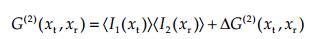
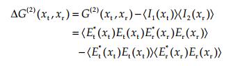
In Figure 1, E(x1) and E(x2) represent the light field at any two points of the light source S. h(x1, xt) and h(x2, xr) represent the transfer functions for the light source passing through different optical systems and reaching the bucket detector and point detector, respectively. Et(xt) represents the light field on the bucket-detector plane. Er(xr) represents the light field on the point-detector plane. Equations (1) and (2) are the second-order correlation function and the fluctuation correlation function of the second-order correlation imaging system, respectively, which describe the imaging result of second-order correlation imaging. The angle brackets represent ensemble averages.
|
(1) |
|
(2) |
In pseudo-thermal GI, the two optical arms must be equal to ensure the accuracy of the reference arm field information, which has many restrictions and is not suitable for medical imaging.
3 Angiography method using CGICGI is a single-arm GI method that is suitable for angiography. CGI has a high sensitivity and ease of use in angiography and can use the spectral information about the vessel wall and atherosclerotic plaque. Therefore, we propose wide spectrum CGI (SCGI) for online angiography.
3.1 Principle of CGI using spatial light modulatorFigure 2 shows the optical path in SCGI. In the CGI system, the wide laser beam emitted from the lasers irradiates the surface of the spatial light modulator, which uses Digital Micromirror Device (DMD), and the modulated light field irradiates the surface of the object, whose reflected light field is collected by the bucket detector. The random data loaded into the spatial light modulator are correlation-calculated with the measured value of the bucket detector, which can obtain the imaging result. Because the modulation information is controllable, the information about the light-field distribution on the object can be designed. Therefore, the reference optical path is not needed, and the imaging can be completed by using the single-arm scheme combined with the single-pixel bucket detector[14, 15].

|
| Figure 2 Principle of CGI. |
Correlation measurement is the theoretical and experimental basis of GI. It involves the second-order correlation function theory and the measurement method. In contrast to two-path imaging, a modulated matrix is used instead of Er(xr) in Eq. (2). The second-order correlation between the random light field generated by the modulator and the light field at the bucket detector is measured.
3.2 Advantages of CGI in medical fieldQuantum correlation imaging is non-local and has a high resolution, a high sensitivity, a low sampling rate, no lens imaging, and strong anti-interference. Compared with other technologies, its high sensitivity and anti-interference make it advantageous in the medical field. In the imaging process, a bucket detector with non-spatial resolution is needed. A single photodetector can be used, which significantly improves the sensitivity of the imaging system. In addition, the scatter mediums affect the imaging result. Scatter mediums exist both in the emission path and the reception path which is closest to the actual condition in medical-field imaging. The influence of the scattering medium on the total light-intensity fluctuation is not significant. Therefore, the scattering medium in the path does not significantly affect the final imaging result. However, in traditional direct imaging, the scattered light field, whose interference is difficult to eliminate via a simple mechanism, greatly interferes with the imaging result[16]. The blood in the vessel is a turbid scattering medium, and the feature of GI provides a theoretical basis to reduce the interference for applications in medical angiography.
3.3 ApproachThe absorption lines differ for different tissues in the blood vessel. Via comparative analysis of imaging at different wavelengths, we can acquire information that cannot be obtained in the normal view. Moreover, combined with GI, real-time online imaging can be achieved to distinguish the structure of vascular tissue. Figure 3 shows a diagram of the system used to obtain a image of the intravascular flow, vascular wall structure, and atherosclerotic plaque composition using GI.

|
| Figure 3 System diagram. |
The system consists of a broadband light source, a spatial light modulator, an array flexible fiber bundle, a collecting lens, a single-point light-intensity detector, a data-acquisition card, and a data control and processing unit. The data-acquisition card is used for collecting light-intensity information. The datasynchronization unit is used for synchronous control of the transmission module and the collection module. The data control and processing unit is used for transmitting the modulation information to the transmitting module and processing the collected light-intensity information.
3.3.1 Online intravascular wall lumen imaging based on SCGIThe optical signal irradiates the spatial light modulator. The two-dimensional matrix of the spatial light modulator simulates the binarized light field, whose resolution determines the resolution of the endoscopic imaging. The modulated light field irradiates the vessel, and the end of the optical fiber is aligned with the imaging object to couple the light reflected by the object into the optical fiber. The reflected light is received by a single-pixel detector. By maintaining the bucket detector and the random array synchronization generated by the spatial light modulator, the array generated by the spatial light modulator is known. Using the total light intensity of the object image information measured by the single-pixel detector, we can achieve real-time imaging via a reconstruction algorithm.
3.3.2 Vascularwall structure and atherosclerotic plaque composition analysis based on GIThe current technology can only realize imaging in the shape of the lumen and does not have good performance in the detection of vascular wall structures and atherosclerotic plaque compositions. To solve this problem, we propose the use of SCGI for angiography.
Vascular wall structures can be imaged via spatial three-dimensional GI. The modulated light irradiates the vessel wall tissue. The arrival times of the reflected light at different depths of the tissue differ, and we can distinguish the different arrival times of the two beams of light to obtain the depth information about the object.
Atherosclerotic plaque consists of fat, fibrous tissue, calcium, red blood cells, macrophages, platelets, and other components, and the composition of the plaque is closely related to its susceptibility to rupturing. It is critical to determine the plaque composition, which can be done via four-dimensional imaging—including spatial 3D and spectrum imaging—because treatment regimens differ for different degrees of vulnerability. Additionally, the absorption lines differ for different tissues in the blood vessel. We can obtain the required information by combining the comparative analysis of imaging at different wavelengths with GI.
4 ConclusionsWe propose SCGI for online angiography to image the intravascular flow, vascular wall structure, and components of atherosclerotic plaque. It contributes to more accurate and effective diagnosis of cardiocerebral vascular diseases. SCGI enhances the endoscope spectral resolution and detection depth. SCGI can also be extended to endoscopy, colonoscopy, laparoscopy, etc., which can image the tissue and structure of pelvic lesions in real time. It also fills the gaps in the application of GI in medical imaging and has important social and economic benefits.
Conflict of interests
All contributing authors have no conflict of interests.
| [1] | Dahl JJ. Diagnostic ultrasound:Imaging and blood flow measurements (second edition). Ultrasound Med Biol, 2015, 41(12): 3259–3260. DOI:10.1016/j.ultrasmedbio.2015.07.017 |
| [2] | Sun ZH, Al Moudi M, Cao Y. CT angiography in the diagnosis of cardiovascular disease:A transformation in cardiovascular CT practice. Quant Imaging Med Surg, 2014, 4(5): 376–396. |
| [3] | Lindsay AC, Biasiolli L, Knight S, CunningtonC, Robson MD, Neubauer S, Kennedy J, Handa A, Choudhury RP. Non-invasive imaging of carotid arterial restenosis using 3T cardiovascular magnetic resonance. J Cardiovasc Magn Reson, 2014, 16: 5. DOI:10.1186/1532-429X-16-5 |
| [4] | Turski PA, Strother CM, Mistretta CA, Sackett JF, Crummy AB, Stieghorst MF, Lieberman RP. Current applications of digital subtraction angiography in clinical neuroradiology. Proc Annu Symp Comput Appl Med Care, 1982(2): 943–950. |
| [5] | Mughal MM, Khan MK, DeMarco JK, Majid A, Shamoun F, Abela GS. Symptomatic and asymptomatic carotid artery plaque. Expert Rev Cardiovasc Ther, 2011, 9(10): 1315–1330. DOI:10.1586/erc.11.120 |
| [6] | Ryczkowski P, Barbier M, Friberg AT, Dudley JM, Genty G. Ghost imaging in the time domain. Nat Photonics, 2016, 10(3): 167–170. DOI:10.1038/nphoton.2015.274 |
| [7] | Padgett M, Aspden R, Gibson G, Edgar M, Spalding G. Ghost imaging. Opt Photonics News, 2016, 27(10): 38–45. DOI:10.1364/OPN.27.10.000038 |
| [8] | Wang CL, Gong WL, Shao XH, Han SS. The influence of the property of random coded patterns on fluctuation-correlation ghost imaging. J Opt, 2016, 18(6): 065703. DOI:10.1088/2040-8978/18/6/065703 |
| [9] | Meyers R, Deacon K, Shih Y. A new two-photon ghost imaging experiment with distortion study. J Mod Opt, 2007, 54(16-17): 2381–2392. DOI:10.1080/09500340701400117 |
| [10] | Pittman TB, Shih YH, Strekalov DV, Sergienko AV. Optical imaging by means of two-photon quantum entanglement. Phys Rev A, 1995, 52(5): R3429. DOI:10.1103/PhysRevA.52.R3429 |
| [11] | Bennink RS, Bentley SJ, Boyd RW. "Two-Photon" coincidence imaging with a classical source. Phys Rev Lett, 2002, 89(11): 113601. DOI:10.1103/PhysRevLett.89.113601 |
| [12] | Shapiro JH. Computational ghost imaging. In Proceedings of 2009 Conferenceon Lasersand Electro-optics and 2009 Conferenceon Quantumelectronics and Laser Science Conference, Baltimore, MD, USA, 2009: pp 1–2. |
| [13] | Pelliccia D, Rack A, Scheel M, Cantelli V, Paganin DM. Experimental X-ray ghost imaging. Phys Rev Lett, 2016, 117(11): 113902. DOI:10.1103/PhysRevLett.117.113902 |
| [14] | Yuan S, Liu XM, Zhou X, Li ZY, Yang YR. Multipleobject ghost imaging with a single-pixel detector. J Opt, 2016, 45(1): 92–98. DOI:10.1007/s12596-015-0302-5 |
| [15] | Welsh SS, Edgar MP, Bowman R, Jonathan P, Sun BQ, Padgett MJ. Fast full-color computational imaging with single-pixel detectors. Opt Express, 2013, 21(20): 23068–23074. DOI:10.1364/OE.21.023068 |
| [16] | Liu BL, Yang ZH, Qu SF, Zhang AX, Wu LA. Influence of turbid media at different locations in computational ghost imaging. Acta Opt Sinica, 2016, 36(10): 1026017. DOI:10.3788/AOS |