2. 上海海洋大学 农业农村部鱼类营养与环境生态研究中心, 上海 201306;
3. 上海海洋大学 水产科学国家级实验教学示范中心, 上海 201306
中华绒螯蟹(Eriocheir sinensis)属软甲纲(Malacostraca)十足目(Decapoda),主要分布在亚洲北部、朝鲜西部和中国,是我国水产养殖业主要的淡水蟹类养殖品种之一,经济价值较高[1]。蟹类在个体的生长发育过程中会蜕去旧外壳,形成新外壳以适应不断生长的身体[2]。中华绒螯蟹一生中一般蜕壳15 ~ 20次[3-4]。蟹在蜕壳时容易遭受生物敌害,对环境较敏感,因此蜕壳的中华绒螯蟹存活率较低[5]。如何提高蜕壳存活率,降低养殖成本,提升经济效益,仍然是中华绒螯蟹养殖业亟待解决的问题之一。蜕壳是一个复杂的生理过程,目前已经有蜕壳行为和形态[6-7]、营养生化指标[8-12]、环境胁迫处理[13]和蜕壳相关基因[14-15]等方向的研究。
目前,甲壳动物常被作为研究钙离子交换的模型物种[16]。中华绒螯蟹作为甲壳动物,其蜕壳过程与钙离子交换密切相关,因而极具有研究钙离子交换的价值。中华绒螯蟹蜕壳时,体内的钙离子随着旧外壳的脱落而迅速流失,体表软而脆弱,蜕壳后机体迅速从水体、食物等获取钙质,以便新外壳的形成及硬化[17]。在人工养殖中,蟹出现大面积蜕壳时,为防止缺乏钙质导致蟹蜕壳不遂,养殖户往往会向水体和饲料中添加钙制剂以帮助蟹顺利蜕壳[18]。钙离子主要通过2种方式进入蟹的机体内,一种是通过摄入饲料经过消化系统进入机体,另一种则是通过鳃从水体中吸收[19]。鳃不仅是蟹的呼吸器官,还具有调节渗透压平衡和离子交换的功能[20]。中华绒螯蟹的鳃共有6对,分为前鳃(前3对)和后鳃(后3对),前鳃主要负责呼吸,后鳃负责调节渗透压平衡和离子交换[21]。已经有研究表明,在重金属胁迫条件下,鳃的结构会受到不同程度的损伤[23-24],并且鳃的组织结构状况决定着鳃能否正常发挥其生理功能[22]。目前,在中华绒螯蟹的不同蜕壳时期,已经有肝胰腺、肌肉、Y器官等组织的显微与超微结构的研究[29-33],但对鳃的组织结构变化的研究几乎没有。而在其他甲壳动物上,已经有较多对鳃的组织结构的相关研究[25-28]。
因此,在中华绒螯蟹的蜕壳周期内,对不同蜕壳时期鳃和外壳中的钙含量进行测定,并观察其组织结构的变化,为完善中华绒螯蟹蜕壳方面的研究,进一步研究如何提高中华绒螯蟹的蜕壳存活率,开展健康养殖,提高经济效益提供理论支持。
1 材料与方法 1.1 实验材料实验中所用到的中华绒螯蟹取自上海海洋大学崇明基地养殖场,在室内养殖系统中驯化、养殖及采样。蟹为一年生幼蟹,背甲宽20~30 mm,平均体质量为(5.82±2.47) g。蟹养殖在配置有循环水系统的双层玻璃养殖缸内,养殖缸内部用亚克力板分隔为多个隔间,1个隔间内放置1只蟹。养殖期间,每天更新约1/4的水,自然水温(20~25 ℃),持续充氧,自然光节律,持续时间约为60 d。每天傍晚喂食(基地配制的饲料),喂食前清理残饵。根据康现江等[34]的方法确定蟹的蜕壳时期,各个蜕壳时期均采集10只蟹用于实验。本研究均采集中华绒螯蟹的后鳃。考虑到蟹壳不同位置的钙含量和组织特征可能有差异[35],因此均采集蟹背甲中间位置的外壳。蜕壳期的蟹取刚蜕壳半小时以内的蟹,超过半小时但外壳还未硬化的则归为蜕壳后期。解剖前4 ℃低温麻醉,取蟹的后鳃和外壳样品分别置于-40 ℃冰箱保存用于钙含量的测定及固定液中固定用于显微和超微结构的观察。
1.2 实验方法鳃样品测定前经冰浴匀浆,然后离心得到匀浆上清液测定钙含量(钙测试盒购自南京建成),用mmol/gprot表示。外壳样品在硝化反应后进行钙含量测定(电感耦合等离子放射光谱法),用mg/kg表示。
光学显微镜观察用样品预处理方法:样品置于Bouin式液里固定24 h,采用梯度乙醇脱水,然后用二甲苯透明,石蜡进行包埋,切片厚度为5 μm,最后用苏木精和伊红进行常规H.E染色。
电镜样品预处理方法:样品置于多聚甲醛固定液中24 h,用1%锇酸和磷酸缓冲液PB(pH 7.4)室温下固定2 h,然后用梯度乙醇和丙酮脱水。鳃样品用812包埋剂包埋,切片厚度为60 ~ 80 nm,经铀铅双染色用于透射电镜观察。外壳样品经临界点干燥仪干燥,然后喷金30 s进行导电处理,用于扫描电镜观察。
1.3 数据分析数据采用平均值±标准差表示,P < 0.05为统计学差异显著。采用Levene检验法检测方差齐次性,采用ANOVA对实验结果进行方差分析,采用Duncan氏法进行多重比较,采用t检验法检测样本含量较小的数据。
2 结果 2.1 中华绒螯蟹钙含量的变化 2.1.1 鳃中钙含量的变化在1个蜕壳周期内,蜕壳前期的钙含量最高,蜕壳期的钙含量最低(P < 0.05)。从蜕壳间期到蜕壳前期,蜕壳期到蜕壳后期,中华绒螯蟹鳃中的钙含量均略有升高(P>0.05)。见图 1。

|
不同字母代表差异性显著(P < 0.05),相同字母代表差异不显著(P>0.05)。 Different letters show significant difference (P < 0.05), the same letter shows the difference is not significant (P>0.05). 图 1 不同蜕壳时期中华绒螯蟹鳃中的钙含量变化 Fig. 1 Changes of calcium content in gills of Eriocheir sinensis in different molting stages |
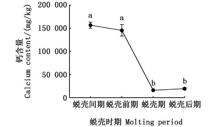
在蜕壳周期内,蜕壳间期外壳中的钙含量最高,蜕壳期的钙含量最低(P < 0.05)。蜕壳间期和蜕壳前期的钙含量差异不显著,蜕壳期和蜕壳后期的钙含量差异不显著(P>0.05)。见图 2。

|
不同字母代表差异性显著(P < 0.05),相同字母代表差异不显著(P>0.05)。 Different letters show significant difference (P < 0.05), the same letter shows the difference is not significant (P>0.05). 图 2 不同蜕壳时期中华绒螯蟹外壳中的钙含量变化 Fig. 2 Changes of calcium content in carapaces of Eriocheir sinensis in different molting stages |
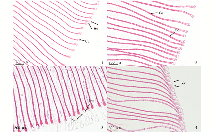
中华绒螯蟹在蜕壳间期和蜕壳前期的鳃差异不显著,都有完整的鳃结构,鳃丝排列整齐,每个鳃丝都有中央血腔,血腔中内容物丰富,柱细胞将血腔分隔,鳃丝末端的边缘管空腔明显(图版Ⅰ-1, 2)。在蜕壳期和蜕壳后期,鳃的中央血腔中均有内容物,但是蜕壳后期的内容物染色较浅。在蜕壳期,内外角质层离散明显,边缘管处的空腔较大(图版Ⅰ-3),直到蜕壳后期,鳃的内外角质层开始聚集(图版Ⅰ-4)。

|
1.蜕壳间期;2.蜕壳前期;3.蜕壳期;4.蜕壳后期;Mv.边缘管;Cu.角质层;Pc.柱细胞;Co.血腔内容物;Ocu.外角质层;Icu.内角质层。 1.Intermolt; 2.Premolt; 3.Ecdysis; 4.Postmolt; Mv. Marginal vessel; Cu.Cuticle; Pc.Pillar cell; Co.Haemocoele content; Ocu.Outer cuticle; Icu.Innter cuticle. 图版 Ⅰ 不同蜕壳时期中华绒螯蟹鳃的显微结构观察 Plate Ⅰ Microstructure of gills of Eriocheir sinensis in different molting stages |
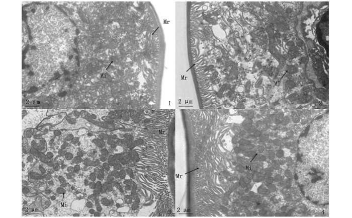
以蜕壳间期为参照,鳃腔中的线粒体丰富,角质层附近的微绒毛结构密集,鳃腔中充满内容物,染色较深(图版Ⅱ-1)。在蜕壳前期,核仁及其周围的内容物的染色加深(图版Ⅱ-2)。在蜕壳期,相较于前期和间期,鳃腔中的线粒体数量更为丰富,微绒毛数量较多,鳃腔中的内容物含量减少,染色颜色较浅(图版Ⅱ-3)。在蜕壳后期,相较于蜕壳期,鳃腔中的线粒体和微绒毛数量均增多,并且内容物含量增多(图版Ⅱ-4)。

|
1.蜕壳间期;2.蜕壳前期;3.蜕壳期;4.蜕壳后期;Mr.微绒毛;Mi.线粒体。 1.Intermolt; 2. Premolt; 3.Ecdysis; 4.Postmolt; Mr.Microvilli; Mi.Mitochondria. 图版 Ⅱ 不同蜕壳时期中华绒螯蟹鳃的超微结构观察 Plate Ⅱ Ultrastructural observation of gills of Eriocheir sinensis in different molting stages |
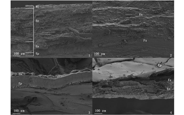
如图版Ⅲ所示,以蜕壳间期为参照,中华绒螯蟹的外壳具有4层结构,分别是上表皮、外表皮、内表皮和膜层,其中外表皮和内表皮较厚,可见层状结构。与内表皮相比,外表皮的层状结构更为细密(图版Ⅲ-1)。在蜕壳前期,可以观察到除膜层以外的其他3层结构,内表皮最厚(图版Ⅲ-2)。在蜕壳期,蟹的外壳明显变薄,可以观察到上表皮和外表皮已经开始形成(图版Ⅲ-3)。在蜕壳后期,蟹的外壳仍然较薄,外表皮开始形成,外表皮和内表皮的界面不清晰(图版Ⅲ-4)。

|
1.蜕壳间期;2.蜕壳前期;3.蜕壳期;4.蜕壳后期;Ml.膜层;En.内表皮;Ex.外表皮;Ep.上表皮。 1.Intermolt; 2.Premolt; 3.Ecdysis; 4.Postmolt; Ml.Membranous layer; En.Endocuticle; Ex.Exocuticle; Ep.Epicuticle. 图版 Ⅲ 不同蜕壳时期中华绒螯蟹外壳的超微结构观察 Plate Ⅲ Ultrastructural observation of carapaces of Eriocheir sinensis in different molting stages |
不同生境下的蟹鳃具有不同的结构特征[22]。由于中华绒螯蟹是两栖蟹,其鳃的边缘管处具有空腔,这种结构有利于蟹灵活地调节渗透压和交换离子,而三疣梭子蟹的生活水域较稳定,其鳃的边缘管处没有发现这种空腔结构。这一现象不仅解释了不同生境会导致蟹鳃的组织结构不同,也表明蟹鳃的组织结构可以反映其适应的功能,不同功能的鳃具有不同的组织结构特征。鳃是中华绒螯蟹主要的离子转运器官,与蟹的钙离子交换过程有密切关联,因此,观察蟹在不同蜕壳时期鳃的组织结构特征,区分不同蜕壳时期的特征差异,可以从组织学的角度解释中华绒螯蟹的钙含量变化。
3.1 中华绒螯蟹鳃和外壳的钙含量变化中华绒螯蟹在蜕壳前需要吸收储存大量能量为蜕壳做准备[4],鳃中的钙含量在蜕壳前期增加。有研究[36]表明蟹在蜕壳前会吸收旧外壳中的钙用于蜕壳后新外壳的矿化,在蜕壳前期,中华绒螯蟹外壳中的钙含量在蜕壳期降低,蟹机体内的部分钙随着旧壳的蜕去而流失,因此在鳃和外壳中的钙离子含量均减少,外壳中的钙大量流失。到蜕壳后期,随着蟹对钙的摄取和吸收,鳃和外壳中的钙含量逐渐增加[37-38]。田志环等[28]的结果显示,外壳中的钙含量在蜕壳期最高,在蜕壳后期较低,这与本研究的结果不一致,推测可能是蜕壳时期确定方法及取样位置的不同导致的差异。
3.2 中华绒螯蟹鳃和外壳的组织结构变化中华绒螯蟹鳃的边缘管空腔对蟹运输血液、离子交换和调节渗透压具有重要作用[26]。在蜕壳期,鳃的边缘管空腔尚未恢复正常形态,鳃的离子交换功能可能受到影响,钙含量较低。到蜕壳后期,鳃的边缘管空腔形成,钙含量增加。电镜观察结果表明,鳃腔中的内容物含量在不同蜕壳时期发生变化,推测内容物可能是血液或脂类等营养物质。微绒毛与线粒体参与细胞的生长与分裂等活动,其数量与细胞的活跃程度密切相关。在蜕壳期,鳃腔中的线粒体和微绒毛数量较多,表明鳃的细胞活动较为活跃。到蜕壳后期,线粒体和微绒毛趋于密集,线粒体在细胞核和微绒毛周围聚集,可能表明蟹在蜕壳后进行活跃的生理活动,以快速恢复身体各项机能。在蜕壳期,相较其他3个蜕壳时期,外壳非常薄,仅有上表皮和外表皮,在蜕壳前期,外壳内表皮的层状结构明显。有研究[32]表明,在蜕壳前期,外壳中的糖类、蛋白质和胶原纤维等物质都被重新吸收,并且蟹在蜕壳前已经形成部分新外壳,因此,外壳的钙含量在蜕壳前期降低。本文初步研究了中华绒螯蟹幼蟹在不同蜕壳时期的钙含量及鳃和外壳的形态特征变化,后续将继续在分子水平研究不同蜕壳时期鳃和外壳的钙分布及转运机制,以促进中华绒螯蟹在蜕壳方面的基础生物学研究。
| [1] |
张超. 中华绒螯蟹幼蟹蜕壳与生长的初步研究[J]. 水利渔业, 2007, 27(2): 58-60. ZHANG C. Studies on the molting and growth of the young mitten crab[J]. Reservoir Fisheries, 2007, 27(2): 58-60. DOI:10.3969/j.issn.1003-1278.2007.02.025 |
| [2] |
ROER R D, DILLAMAN R M. The initiation and early stages of postmolt mineralization in the Blue Crab, Callinectes sapidus[J]. Frontiers in Marine Science, 2018, 5: 151. DOI:10.3389/fmars.2018.00151 |
| [3] |
张列士, 陆锦天. 中华绒螯蟹(Eriocheir sinensis)蜕壳和生长的研究进展[J]. 水产科技情报, 2001, 28(6): 246-250. ZHANG L S, LU J T. Research progress in molting and growth of Eriocheir sinensis[J]. Fisheries Science & Technology Information, 2001, 28(6): 246-250. DOI:10.3969/j.issn.1001-1994.2001.06.002 |
| [4] |
陶尚春. 河蟹的脱壳与生长[J]. 科学养鱼, 2010(8): 78. TAO S C. Molting and growth of Eriocheir sinensis[J]. Scientific Fish Farming, 2010(8): 78. |
| [5] |
翟基文. 影响河蟹蜕壳生长及致死原因初析[J]. 内陆水产, 2001(10): 35. ZHAI J W. Preliminary analysis of the causes of molting growth and death of Eriocheir sinensis[J]. Current Fisheries, 2001(10): 35. |
| [6] |
严生良, 俞泰济. 中华绒螯蟹蜕壳(Ecdysis)的初步观察[J]. 淡水渔业, 1990(6): 8-10, 20. YAN S L, YU T J. Preliminary observation on ecdysis of Eriocheir sinensis[J]. Freshwater Fisheries, 1990(6): 8-10, 20. |
| [7] |
黄姝, 王中清, 茅海成, 等. 中华绒螯蟹在实验室条件下的成蟹阶段蜕壳与生长观察[J]. 上海海洋大学学报, 2014, 23(3): 359-365. HUANG S, WANG Z Q, MAO H C, et al. Observation on molting and growth of adult Chinese mitten crab reared in the laboratory condition[J]. Journal of Shanghai Ocean University, 2014, 23(3): 359-365. |
| [8] |
舒磊, 母昌考, 王春琳, 等. 蜕壳后不同时间的软壳三疣梭子蟹常规营养成分含量变化[J]. 应用海洋学学报, 2013, 32(2): 278-282. SHU L, MU C K, WANG C L, et al. Variations of the common nutritional composition contents of soft shell swimming crab (Portunus trituberculatus) after molting[J]. Journal of Applied Oceanography, 2013, 32(2): 278-282. DOI:10.3969/J.ISSN.2095-4972.2013.02.019 |
| [9] |
TIAN Z H, KANG X J, MU S M. The molt stages and the hepatopancreas contents of lipids, glycogen and selected inorganic elements during the molt cycle of the Chinese mitten crab Eriocheir sinensis[J]. Fisheries Science, 2012, 78(1): 67-74. DOI:10.1007/s12562-011-0426-8 |
| [10] |
马明君, 王春, 吴旭干, 等. 中华绒螯蟹幼蟹蜕皮周期中肝胰腺和肌肉脂类的动态变化[J]. 水产学报, 2014, 38(11): 1889-1898. MA M J, WANG C, WU X G, et al. Dynamic changes of lipids in hepatopancreas and muscle during the molting cycle of young Chinese mitten crab (Eriocheir sinensis)[J]. Journal of Fisheries of China, 2014, 38(11): 1889-1898. |
| [11] |
SPINDLER-BARTH M. Changes in the chemical composition of the common shore crab, Carcinus maenas, during the molting cycle[J]. Journal of Comparative Physiology, 1976, 105(2): 197-205. DOI:10.1007/BF00691122 |
| [12] |
张文军, 贺诗水, 成永旭, 等. 蜕壳前后中华绒螯蟹幼蟹肝胰腺脂肪酸组成变化[J]. 上海师范大学学报(自然科学版), 2002, 45(S1): 24-27. ZHANG W J, HE S S, CHENG Y X, et al. The changes of fatty acids composition in hepatopancreas of juvenile Chinese mitten crab (Eriocheir sinensis) before and after molt[J]. Journal of Shanghai Teachers University (Natural Science), 2002, 45(s1): 24-27. |
| [13] |
路允良, 王芳, 赵卓英, 等. 盐度对三疣梭子蟹生长、蜕壳及能量利用的影响[J]. 中国水产科学, 2012, 19(2): 237-245. LU Y L, WANG F, ZHAO Z Y, et al. Effects of salinity on growth, molt and energy utilization of juvenile swimming crab Portunus trituberculatus[J]. Journal of Fishery Sciences of China, 2012, 19(2): 237-245. |
| [14] |
王瑶, 杨志刚, 沈城, 等. 中华绒螯蟹EcR基因全长cDNA克隆及表达分析[J]. 水产学报, 2014, 38(5): 651-661. WANG Y, YANG Z G, SHEN C, et al. The full length cDNA cloning and expression analysis of EcR from the Chinese mitten crab (Eriocheir sinensis)[J]. Journal of Fisheries of China, 2014, 38(5): 651-661. |
| [15] |
王瑶, 杨志刚, 郭子好, 等. 中华绒螯蟹RXR基因全长cDNA克隆及表达分析[J]. 水产学报, 2013, 37(12): 1761-1769. WANG Y, YANG Z G, GUO Z H, et al. The full length cDNA cloning and expression analysis of RXR from the Chinese mitten crab (Eriocheir sinensis)[J]. Journal of Fisheries of China, 2013, 37(12): 1761-1769. |
| [16] |
LEITE V P, ZANOTTO F P. Calcium transport in gill cells of Ucides cordatus, a mangrove crab living in variable salinity environments[J]. Comparative Biochemistry and Physiology Part A: Molecular & Integrative Physiology, 2013, 166(2): 370-374. |
| [17] |
WHEATLY M G, ZANOTTO F P, HUBBARD M G. Calcium homeostasis in Crustaceans: subcellular Ca dynamics[J]. Comparative Biochemistry and Physiology Part B: Biochemistry and Molecular Biology, 2002, 132(1): 163-178. DOI:10.1016/S1096-4959(01)00520-6 |
| [18] |
周金华. 补钙在养殖池塘和河蟹养殖过程中作用[J]. 水产养殖, 2011, 32(6): 52. ZHOU J H. The role of calcium supplement in pond and crab culture[J]. Journal of Aquaculture, 2011, 32(6): 52. DOI:10.3969/j.issn.1004-2091.2011.06.025 |
| [19] |
陈立侨, 堵南山, 赖伟. 水体和配合饲料中钙、磷含量对河蟹生长的影响[J]. 淡水渔业, 1994, 24(1): 13-15. CHEN L Q, DU N S, LAI W. Effect of calcium and phosphorus content in water and compound feed on the growth of Eriocheir sinensis[J]. Freshwater Fisheries, 1994, 24(1): 13-15. |
| [20] |
FARRELLY C A, GREENAWAY P. Morphology and ultra-structure of the gills of terrestrial crabs (Crustacea, Gecarcinidae and Grapsidae): adaptations for air-breathing[J]. Zoomorphology, 1992, 112(1): 39-49. DOI:10.1007/BF01632993 |
| [21] |
TORRES G, CHARMANTIER-DAURES M, CHIFFLET S, et al. Effects of long-term exposure to different salinities on the location and activity of Na+-K+-ATPase in the gills of juvenile mitten crab, Eriocheir sinensis[J]. Comparative Biochemistry and Physiology Part A: Molecular & Integrative Physiology, 2007, 147(2): 460-465. |
| [22] |
赵艳民, 王新华, 孙慧. Hg2+在中华绒螯蟹幼蟹鳃内的积累及其对组织结构的影响[J]. 动物学杂志, 2008, 43(1): 1-7. ZHAO Y M, WANG X H, SUN H. Accumulation of waterborne mercury in gills of juvenile Eriocheir sinensis and its effect on gills structure[J]. Chinese Journal of Zoology, 2008, 43(1): 1-7. |
| [23] |
卢敬让, 赖伟. 镉对中华绒螯蟹鳃组织及其亚显微结构的影响[J]. 海洋与湖沼, 1991, 22(6): 566-570. LU J R, LAI W. Effects of cadmium on tissue and ultrastructure of gill of Eriocheir sinensis (M. EDW.)[J]. Oceanologia et Limnologia Sinica, 1991, 22(6): 566-570. |
| [24] |
佟蕊, 成永旭, 吴旭干, 等. 3种不同栖息环境下蟹鳃的超微结构、脂类组成及含量的比较[J]. 水产学报, 2011, 35(9): 1426-1435. TONG R, CHENG Y X, WU X G, et al. Ultrastructure and lipid profile of gills in three crabs living in different habitats[J]. Journal of Fisheries of China, 2011, 35(9): 1426-1435. |
| [25] |
田志环, 康现江, 焦传珍. 中华绒螯蟹蜕皮周期中肝胰腺细胞组成的变化[J]. 中国水产科学, 2013, 20(6): 1175-1181. TIAN Z H, KANG X J, JIAO C Z. Changes in cell type composition in the hepatopancreas of Chinese mitten crab Eriocheir sinensis during the molting cycle[J]. Journal of Fishery Sciences of China, 2013, 20(6): 1175-1181. |
| [26] |
田志环, 成永旭, 吴旭干, 等. 中华绒螯蟹蜕壳过程中肌肉的组织学、超微结构及主要蛋白质含量的变化[J]. 水生生物学报, 2017, 41(5): 1036-1041. TIAN Z H, CHENG Y X, WU X G, et al. Changes of Histology, Ultrastructure and main protein in muscles of Chinese mitten crab, Eriocheir sinensis during the molt cycle[J]. Acta Hydrobiologica Sinica, 2017, 41(5): 1036-1041. |
| [27] |
赵维信, 陆剑峰. 中华绒螯蟹Y-器官在蜕皮周期中的超微结构[J]. 中国水产科学, 2004, 11(1): 74-77. ZHAO W X, LU J F. Ultrastructure of Y-organ in Eriocheir sinensis during molt cycle[J]. Journal of Fishery Sciences of China, 2004, 11(1): 74-77. DOI:10.3321/j.issn:1005-8737.2004.01.014 |
| [28] |
田志环, 康现江, 焦传珍. 中华绒螯蟹蜕皮过程中体壁结构和主要成分的变化[J]. 水生生物学报, 2013, 37(5): 899-904. TIAN Z H, KANG X J, JIAO C Z. Structural and constituent changes in integument during the molt cycle of Chinese mitten crab Eriocheir sinensis[J]. Acta Hydrobiologica Sinica, 2013, 37(5): 899-904. |
| [29] |
堵南山, 薛鲁征, 赖伟. 中华绒螯蟹(Eriocheir sinensis)雄性生殖系统的组织学研究[J]. 动物学报, 1988, 34(4): 329-333, 389-390. DU N S, XUE L Z, LAI W. Histology of female reproductive system in Chinese mitten-handed crab, Eriocheir sinensis (Crustacea, Decapoda)[J]. Acta Zoologica Sinica, 1988, 34(4): 329-333, 389-390. |
| [30] |
顾志峰, 王文, 杜开和, 等. 中华绒螯蟹鳃的组织及超微结构[J]. 湖泊科学, 2000, 12(2): 182-184. GU Z F, WANG W, DU K H, et al. Histological and ultrastructural studies on the gill in Eriocheir sinensis[J]. Journal of Lake Sciences, 2000, 12(2): 182-184. DOI:10.3321/j.issn:1003-5427.2000.02.014 |
| [31] |
王巧伶. 中华绒螯蟹呼吸系统的组织学和组织化学[J]. 重庆师范学院学报(自然科学版), 1995, 12(1): 56-61. WANG Q L. Histology and histochemistry of the respiratory system in Eriocheir sinensis[J]. Journal of Chongqing Teachers College (Natural Science Edition), 1995, 12(1): 56-61. |
| [32] |
BARRA J A, PEQUEUX A, HUMBERT W. A morphological study on gills of a crab acclimated to fresh water[J]. Tissue and Cell, 1983, 15(4): 583-596. DOI:10.1016/0040-8166(83)90009-5 |
| [33] |
FREIRE C A, ONKEN H, MCNAMARA J C. A structure-function analysis of ion transport in crustacean gills and excretory organs[J]. Comparative Biochemistry and Physiology Part A: Molecular & Integrative Physiology, 2008, 151(3): 272-304. |
| [34] |
康现江, 田志环, 吴江立, 等. 中华绒螯蟹蜕皮周期及蜕皮过程中肝胰腺消化酶活性的变化[J]. 中国水产科学, 2012, 19(5): 806-812. KANG X J, TIAN Z H, WU J L, et al. Molt stages and changes in digestive enzyme activity in hepatopancreas during molt cycle of Chinese mitten crab, Eriocheir sinensis[J]. Journal of Fishery Sciences of China, 2012, 19(5): 806-812. |
| [35] |
徐新章. 河蟹蜕壳前后蟹体与蟹壳中钙磷变化的研究[J]. 江西水产科技, 1995(2/3): 21-27. XU X Z. Changes of calcium and phosphorus in crab body and shell before and after molting[J]. Jiangxi Fishery Science and Technology, 1995(2/3): 21-27. |
| [36] |
WHEATLY M G. Crustacean models for studying calcium transport: the journey from whole organisms to molecular mechanisms[J]. Journal of the Marine Biological Association of the United Kingdom, 1997, 77(1): 107-125. DOI:10.1017/S0025315400033816 |
| [37] |
黄朝辉, 母昌考, 胡丹丹, 等. 三疣梭子蟹蜕壳周期肝胰腺、外壳和鳃中钙含量的变化[J]. 生物学杂志, 2012, 29(5): 22-24. HUANG C H, MU C K, HU D D, et al. Variation of calcium content of hepatopancreas, exoskeleton and gill in molting cycle of Portunus trituberculatus[J]. Journal of Biology, 2012, 29(5): 22-24. DOI:10.3969/j.issn.2095-1736.2012.05.022 |
| [38] |
王顺昌, 魏亦军, 申德林. 中华绒螯蟹蜕皮过程中肌肉、肝胰脏和甲壳中钙和磷含量的变动[J]. 水产学报, 2003, 27(3): 219-224. WANG S C, WEI Y J, SHEN D L. Calcium and phosphorus levels in the muscle, hepatopancreas and carapace of Eriocheir sinensis in different stages of moulting cycle[J]. Journal of Fisheries of China, 2003, 27(3): 219-224. |
2. Centre for Research on Environmental Ecology and Fish Nutrition, Ministry of Agriculture and Rural Affairs, Shanghai Ocean University, Shanghai 201306, China;
3. National Demonstration Center for Experimental Fisheries Science Education, Shanghai Ocean University, Shanghai 201306, China
 2021,
Vol. 30


