 2015, Vol. 34
扩展功能
文章信息
- 高丽芳, 王宏元, 梁刚
- GAO Lifang, WANG Hongyuan, LIANG Gang
- 中华大蟾蜍蝌蚪后肢骨的发育及骨化顺序研究
- Development and Ossification Sequence of the Hind Limb during Metamorphosis of Bufo gargarizans Tadpoles
- 四川动物, 2015, 34: 345-351
- Sichuan Journal of Zoology, 2015, 34: 345-351
- 10.3969/j.issn.1000-7083.2015.03.004
-
文章历史
- 收稿日期:2014-09-13
- 接受日期:2014-12-26


变态是无尾两栖动物幼体生活史中的一个重要组成环节。 水中栖息生活的无尾两栖类蝌蚪在变态过程中会进行骨骼重构和四肢的发育,成为适应陆生环境的四足两栖类。四足动物的四肢肢体由近端至远端分别为近端(stylopodium)、中段(zeugopodium)和远端(autopodium)肢体3个部分。近端肢体一般由1根长骨构成,即前肢附着在肩带上的肱骨或后肢附着在腰带上的股骨。中段肢体是由2根长骨构成,分别是前肢的桡骨、尺骨或后肢的胫骨、腓骨。远端肢体则由两部分组成,近端的腕骨或跗骨和远端的掌骨或蹠骨、趾骨(Wagner et al.,2001)。一般认为,四足类动物的近端和中段肢体起源于肉鳍鱼类偶鳍近端的内骨骼,即陆生脊椎动物的四肢是从古总鳍鱼的偶鳍演化而来的(朱敏,于小波,1999)。 相对而言,对于四足类的远端肢体,例如指、趾的起源,依旧存在巨大的争议(Janvier,1997; Fröbisch,2008)。一些学者认为四足类的指(趾)也是从肉鳍鱼类偶鳍远端的内骨骼演化形成的,其指(趾)是沿近端、中段肢体向前延伸分叉的构造,而不是四足类起源时出现的新构造。另一些学者则认为四足类指(趾)是四足类特有的新构造,起源于新生的指弓,其主要依据是"指弓学说"(Shubin et al,1997;朱敏,于小波,1999)。
从鱼类的偶鳍演化至四足动物的四肢,是脊椎动物从水生向陆生进化的一个关键环节。四肢的出现有利于脊椎动物在进化中从水生到陆生生活方式的过渡。另一方面,两栖动物变态过程中骨骼的重构还包括骨骼的骨化,以增加骨密度来增强支撑能力,因为无尾目动物由水生到陆栖首先要解决身体重量担负问题。桑洲娴等(2014)已对中华大蟾蜍Bufo gargarizans变态过程中脊椎骨化次序进行了研究。本研究拟对中华大蟾蜍蝌蚪不同发育阶段的后肢骨骼进行双染色,来探究其后肢各部分的出现模式和骨化顺序,旨在推演无尾目动物在由水生到陆栖的变态过程中,后肢的生态适应和系统进化历程,进而为四足类远端肢体——指(趾)的起源提供新的数据资料。 1 材料与方法
中华大蟾蜍蝌蚪采自秦岭北坡的陕西省西安市长安区大峪水库周围的小水潭(34°01'00″N,109°06'52″E,海拔700~730 m)。样本采用Gosner(1960)中无尾目动物发育分期方法进行发育时期划分。取中华大蟾蜍蝌蚪各时期(G38~G46)各6尾样本进行骨骼染色,采用改良后的Taylor和van Dyke(1985)的方法进行骨骼双染色,标本经茜素红(Alizarin red)和阿利新蓝(Alcian blue)染色,再经甘油透明,硬骨呈紫红色,软骨呈蓝色。
用Zeiss Discovery V12型体视显微镜观察,佳能7D数码相机进行图像采集。本实验中后肢骨骼名称均采用Pugener和Maglia(2009)的术语进行描述。 2 结果
中华大蟾蜍成体的腰带(pelvic girdle)由髂骨(ilium)、坐骨(ischium)和耻骨(pubis)3部分构成。髂骨即肠骨,为1对杆状硬骨,其背面具嵴棱,前端拗在荐椎(sacral)横突的下面,后端则呈三角状庞大,与耻骨前部作合掌式对接。坐骨似扇形,亦作合掌式对接,前缘与髂骨后方连接,后缘与耻骨后方对位连接。耻骨似三角形,且作合掌式对接,前接髂骨而后连坐骨,并与之结合成圆盘状。圆盘中央凹陷成髂臼(acetabulum),与股骨头结合形成髂关节。
Gosner 36期,髂骨已经出现,呈杆状,为软骨。Gosner 37期,髂骨长度有所增加,未骨化。至Gosner 38期,髂骨骨干处开始骨化,染色呈红色。此外,软骨性的坐骨与耻骨出现(图版Ⅰ:1)。Gosner 40期,两侧髂骨在耻骨前方作合掌式对接愈合(图版Ⅰ:3)。在Gosner 43期,髂骨已完全骨化(图版Ⅰ:5)。直到Gosner 46期,坐骨已骨化,但耻骨仍未发现骨化。

|
| 图版Ⅰ 中华大蟾蜍蝌蚪变态过程中腰带骨化顺序示意图(背面观) PlateⅠ Ossification sequence of the pelvic girdle during metamorphosis of Bufo gargarizans tadpoles(dorsal view) 中华大蟾蜍蝌蚪腰带骨骼双染图(Double staining of larval pelvic girdle skeleton of Bufo gargarizans):1. 38期,髂骨、坐骨及耻骨都已出现,但两侧髂骨没有作合掌式对接愈合(Stage 38,ilium,ischium and pubis appear,and both halves of the pelvis hasn’t come into contact);2. 39期,髂骨骨化部分为整体的30%,两侧髂骨没有作合掌式对接愈合(Stage 39,the ossified section takes 30% of the whole ilium. Both halves of the pelvis hasn’t come into contact);3. 40期,两侧髂骨已作合掌式对接愈合(Stage 40,both sides of the ilium have been fused);4. 42期,髂骨骨化部分约为整体的50%,坐骨开始骨化(Stage 42,the ossified section takes 50% of the whole ilium. Ischium begins to ossify);5. 43期,髂骨几乎骨化完全,坐骨未完全骨化,耻骨仍保持软骨化(Stage 43,ilium ossifies almost completely,ischium ossifies incomplete,and the pubis remains cartilaginous);比例尺(scale bars)=1 mm。 |
中华大蟾蜍蝌蚪的后肢(hind limb)是由股骨(femur)、胫腓骨(tibio-fibula)、跗骨(tarsus)、末端跗骨3-2(distal tarsale 3-2)、末端跗骨1(distal tarsale 1)、蹠骨(metatarsus)V-I 、趾V-I以典型模式3-4-3-2-2排列的趾骨(phalanges)和前拇趾(prehallux)组成(图版Ⅱ:7,图版Ⅲ:12)。

|
| 图版Ⅱ 中华大蟾蜍蝌蚪变态过程中后肢骨化次序 PlateⅡ Ossification sequence of the hind limb during metamorphosis of Bufo gargarizans tadpoles 中华大蟾蜍蝌蚪36~43期后肢骨骼双染图(Double staining of larval hind limb skeleton of Bufo gargarizans at stage 36~43):1. 36期,股骨、胫骨、腓骨、跗骨出现,趾Ⅴ、Ⅳ、Ⅲ的蹠骨及趾Ⅳ的近端趾骨出现(Stage 36,femur,tibia,fibula,fibulare,metatarsale Ⅳ,metatarsale Ⅲ,metatarsaleⅤ and the proximal phalanx of toe Ⅳ can be observed);2. 37期,趾Ⅱ、趾Ⅰ的蹠骨及趾Ⅳ的第二趾骨、趾Ⅴ与趾Ⅲ的第一趾骨出现(Stage 37,metatarsaleⅡ and metatarsaleⅠappear together with the second phalanx of toe Ⅳ and the first phalanxes of toes Ⅲ and toesⅤ);3. 38期末,趾Ⅳ的第三趾骨、趾Ⅴ与趾Ⅲ的第二趾骨、趾Ⅱ和趾Ⅰ的第一趾骨及远端跗骨3-2和前拇趾的第一趾骨出现,并且股骨的骨干部分开始轻微的骨化(At the end of stage 38,the third phalanx of toe Ⅳ,the second phalanx of toeⅤ and toe Ⅲ,the proximal phalanx of toeⅡ and toeⅠappear together with distal tarsale 3-2 and the proximal phalanges of the prehallux. And ossification of diaphyses begins in the femur slightly);4. 39期,远端跗骨1出现,而且胫骨和腓骨两端逐渐有愈合现象,骨干开始骨化(stage 39,distal tarsale 1 appears,and the fusion of the distal ends of the fibula and the tibia begins at this stage. Their diaphyses begin to ossify);5. 40期,趾Ⅴ[KG-0.5mm]-[KG-0.5mm]Ⅰ的趾骨以3-4-3-2-2的排列模式出现,而且可观察到前拇趾的第二趾骨(stage 40,phalangeal formulae for the hindlimb,i.e. 3-4-3-2-2 from toeⅤ to toeⅠis formed. And the second phalanges of the prehallux can be observed);6. 41期,胫腓骨完全愈合,而且骨干已完全骨化;跗骨开始有轻微的骨化(stage 41,the tibio-fibula is completely fused,and their diaphyses are fully ossified. Ossification of diaphyses begins in the tarsus slightly);7. 42期,趾Ⅴ、Ⅳ、Ⅲ的蹠骨已有骨化现象(stage 42,metatarsale Ⅲ-Ⅴbegin to ossify);8. 43期,趾Ⅱ、趾Ⅰ的蹠骨及趾Ⅴ-Ⅰ的趾骨已完全骨化,但前拇趾及远端跗骨仍未骨化(stage 43,metatarsaleⅡ,metatarsaleⅠ and all phalanges ossify completely,but the prehallux and distal tarsale remain chondrify);比例尺(scale bars)=1 mm。 |
股骨为棒状硬骨,其近端与髂臼连接,近端和远端处略有弯曲。股骨在Gosner 36期已出现,呈短棒状(图版Ⅱ:1)。发育至Gosner 37期,股骨的长度有所增加(图版Ⅱ:2)。Gosner 38期,软骨性股骨的骨干开始骨化(图版Ⅱ:3,表 1)。从Gosner 39期到Gosner 42期,股骨骨干的骨化由中央的骨干向两端逐渐延伸(图版Ⅱ:4~7)。到Gosner 43期,股骨的骨干已完全骨化,但两端骨骺仍未骨化(图版Ⅱ:8)。
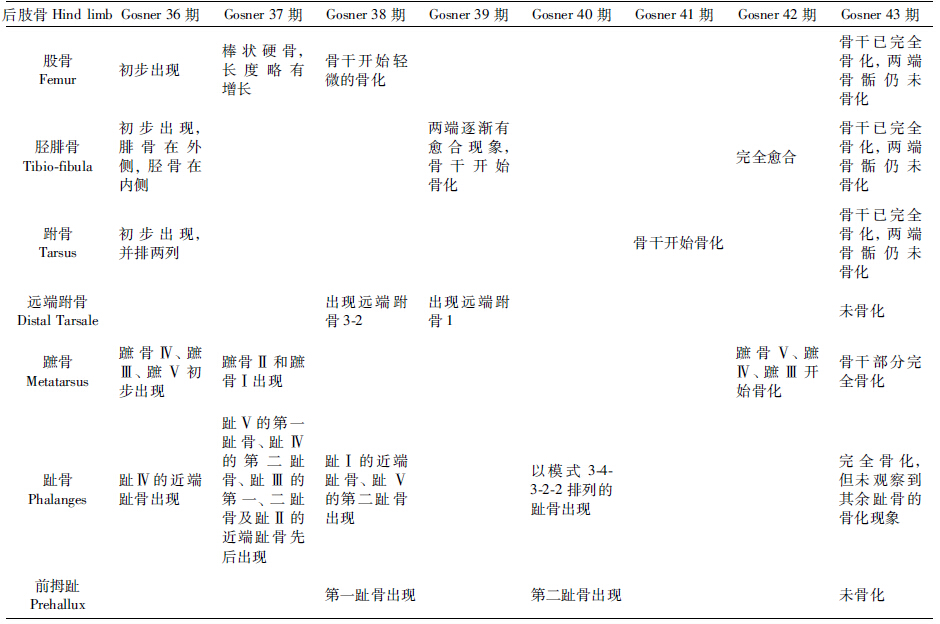
胫腓骨由胫骨和腓骨愈合而成,略长于股骨。在Gosner 36期,胫骨和腓骨出现,腓骨位于外侧,胫骨在内侧(图版Ⅱ:1)。到Gosner 39期,胫骨和腓骨两端的骨骺出现愈合现象,但是骨干之间仍有较大空隙,并且胫骨和腓骨的骨干开始骨化(图版Ⅱ:4,表 1)。在Gosner 40期和Gosner 41期,胫腓骨愈合和骨化程度都在逐渐加强(图版Ⅱ:5~6)。到Gosner 42期,胫腓骨骨干之间的间隙消失,提示胫腓骨完全愈合(图版Ⅱ:7)。到Gosner 43期,胫腓骨的骨干部位已完全骨化,但两端骨骺仍为软骨(图版Ⅱ:8)。
跗骨由腓附骨(fibulare)和胫侧骨(tibiale)并列构成,二者无愈合现象。在Gosner 36期,跗骨已经出现(图版Ⅱ:1,图版Ⅲ:1,表 1)。到Gosner 37期,腓跗骨和胫侧骨的两端融合,主轴保持独立(图版Ⅱ:2,图版Ⅲ:3~4)。与股骨、胫腓骨相比较,跗骨开始骨化的时间较晚,在Gosner 41期才开始骨化(图版Ⅱ:6,图版Ⅲ:9)。至Gosner 43期,跗骨的骨干完全骨化,但直到Gosner 43期末,两端骨骺仍未骨化(图版Ⅱ:8,图版Ⅲ:12)。

|
| 图版Ⅲ 中华大蟾蜍蝌蚪后肢骨各部分软骨形成和骨化的顺序示意图 PlateⅢ Schematic sequence of chondrification and ossification of hind limb in Bufo gargarizans tadpole 白色表示软骨成分,黑色表示骨化成分(White color indicates cartilaginous elements,black color denotes ossified elements);1,2. 36期,在初期(图 1)胫侧骨和腓跗骨原基发育良好,而且趾Ⅳ和趾Ⅲ的蹠骨出现;到末期(图 2)趾Ⅴ的蹠骨和趾Ⅳ的近端趾骨出现(Stage 36,during initial period,fibulare and tibiale are well developed,as well as the primordium of metatarsale Ⅳ and metatarsale Ⅲ; to the end,the condensation of metatarsale V appears together with the condensation of the proximal phalanx of toe Ⅳ);3,4. 37期,在初期时(图 3)胫侧骨和腓附骨的远端开始愈合,趾Ⅱ和趾Ⅰ的蹠骨、趾Ⅴ的近端趾骨、趾Ⅳ的第二趾骨及趾Ⅲ的第一趾骨出现;到末期(图 4)胫侧骨和腓附骨的两端完全愈合,趾Ⅲ的第二趾骨和趾Ⅱ的第一趾骨出现(Stage 37,the fusion of the distal ends of the tibiale and the fibulare begins in the initial. And metatarsaleⅡ and metatarsaleⅠappear together with the proximal phalanx of toe V and the second phalanx of toe Ⅳ and the first phalanx of toe Ⅲ. By the end of this stage,the tibiale and fibulare are completely fused at both ends. And the second phalanx of toe Ⅲ and the first phalanx of toe Ⅱappear);5,6. 38期,在初期(图 5),趾Ⅴ的第二趾骨和趾Ⅳ的第三趾骨出现;到末期(图 6),趾Ⅰ的近端趾骨及末端跗骨3-2、前拇指的近端趾骨出现(Stage 38,the second phalanx of toeⅤ and the third phalanx of toe Ⅳappear in the initial. By the end of this stage,the proximal phalanx of toeⅠappears together with distal tarsale 3-2 and the proximal phalanges of the prehallux);7. 39期,末端跗骨1出现(Stage 39,distal tarsale 1 appears);8. 40期,趾Ⅴ-Ⅰ的末端趾骨形成(Stage 40,the distal phalanges of toe Ⅴ-Ⅰform);9. 41期,跗骨开始骨化(Stage 41,ossification of diaphyses begins in the tarsus slightly);10. 42期,蹠骨Ⅴ、Ⅳ、Ⅲ开始骨化(Stage 42,metatarsale Ⅲ-Ⅴbegin to ossify);11,12. 43期,骨干的骨化由近端到远端,由轴后到轴前的方向有所进展,到43期末,除了各成分两端骨骺及末端跗骨和前拇指未骨化,其余部分已完全骨化(Stage 43,ossification in diaphyses progresses in proximo-distal and postaxial-preaxial direction. By the end of this stage,hind limb skeletons have ossified compeletly except epiphysises,distal tarsale and prehallux);到变态末端,所有的组成成分都被骨化(At the end of metamorphosis all elements are fully ossified);1. 远端跗骨1distal tarsale 1,3-2. 远端跗骨3-2 distal tarsale 3-2,Ⅰ-Ⅴ. 趾Ⅰ-Ⅴ toes Ⅰ-Ⅴ,f. 腓跗骨fibulare,mt. 蹠骨metatarsus,p. 趾骨phalanges,Ph. 前拇趾prehallux,t. 胫侧骨tibiale。 |
远端跗骨由远端跗骨3-2和远端跗骨1组成(表 1)。从Gosner 36期到Gosner 38期初均未观察到远端跗骨。直到Gosner 38期末,远端跗骨3-2才出现,位于蹠骨Ⅱ和蹠骨Ⅲ近侧端之间(图版Ⅱ:3,图版Ⅲ:6)。到Gosner 39期,可观察到远端跗骨1出现,位于蹠骨Ⅰ近端下方(图版Ⅱ:4,图版Ⅲ:7)。远端跗骨的骨化较晚,在Gosner 43期末仍未骨化(图版Ⅱ:8,图版Ⅲ:12)。
蹠骨(跖骨)为5支细长骨节,第4支最长,第3支和第5支次之,第2支又次之,第1支最短。在Gosner 36期,出现蹠骨Ⅳ和蹠骨Ⅲ,然后出现蹠骨Ⅴ(图版Ⅱ:1,图版Ⅲ:1~2,表 1)。在Gosner 37期,蹠骨Ⅱ和蹠骨Ⅰ分别先后出现(图版Ⅱ:2,图版Ⅲ:3)。到Gosner 42期,蹠骨Ⅴ、蹠骨Ⅳ、蹠骨Ⅲ开始骨化(图版Ⅱ:7,图版Ⅲ:10)。至Gosner 43期时,蹠骨骨干全部骨化,经茜素红染色呈红色(图版Ⅱ:8,图版Ⅲ:11)。
趾骨是与蹠骨相连的骨节,趾节向远端逐渐细小,每个蹠、趾各关节两端都覆以软骨骺。在Gosner 36期,趾Ⅳ的近端趾骨出现(图版Ⅱ:6,图版Ⅲ:2,表 1)。到Gosner 37期,趾Ⅴ的近端趾骨、趾Ⅳ的第二趾骨、趾Ⅲ的第一、二趾骨及趾Ⅱ的近端趾骨一起[KG(2x]出现,趾Ⅰ的趾骨仍未出现(图版Ⅱ:2,图版Ⅲ:3~4)。到Gosner 38期初,趾Ⅴ的第二趾骨出现(图版Ⅲ:5),到Gosner 38期末,趾Ⅰ的近端趾骨出现(图版Ⅱ:3,图版Ⅲ:6)。到Gosner 40期,趾Ⅴ-Ⅰ以模式3-4-3-2-2排列的趾骨出现(图版Ⅱ:5,图版Ⅲ:8)。趾骨骨干的骨化比较晚,到Gosner 43期才观察到趾Ⅴ、趾Ⅳ、趾Ⅲ、趾Ⅱ的近端趾骨有骨化现象,随后趾Ⅰ的趾骨也开始骨化。直到Gosner 43期末,趾骨骨干部分完全骨化,经茜素红染色呈红色,但未观察到两端软骨骺的骨化现象(图版Ⅱ:8,图版 Ⅲ:12)。
前拇趾,在拇趾内侧尚有一个前拇趾或超常拇指。在Gosner 38期,可观察到前拇趾的突起,但不是很明显,到Gosner 38期末,前拇趾的第一趾骨出现(图版Ⅱ:3,图版Ⅲ:6,表 1)。直到Gosner 40期,第二趾骨出现(图版Ⅱ:5,图版Ⅲ:8,表 1)。前拇趾的骨化比较晚,到43期末仍未骨化(图版Ⅱ:8,图版Ⅲ:12)。 3 讨论
本研究结果显示,在Gosner 36期,中华大蟾蜍蝌蚪后肢的股骨、胫腓骨、跗骨、蹠骨Ⅳ、蹠骨Ⅲ、蹠骨Ⅴ及趾Ⅳ的近端趾骨均已出现。多指节蟾属Pseudis platensis后肢骨中的股骨、胫腓骨、跗骨首先出现,远端蹠骨、趾骨随后出现(Fabrezi & Goldbery,2009)。古生代鳃龙科幻螈Branchiosaurid Apateon及现存蝾螈的后肢发育中,近端肢体出现后,中段肢体及远端肢体才呈先后顺序依次出现(Fröbisch et al.,2007)。以上可见,两栖动物肢体的发育模式基本相似,都遵循着由近端到远端发育的顺序。
然而,两栖动物在远端肢体的发育模式上差异明显。中华大蟾蜍的蹠骨Ⅳ首先出现,随后蹠骨Ⅲ或者蹠骨Ⅴ产生,趾出现的顺序为Ⅳ-(Ⅴ/Ⅲ)-Ⅱ-Ⅰ。古生代鳃龙科幻螈及现存蝾螈显示蹠骨Ⅱ优先出现,趾形成的顺序是Ⅱ-Ⅰ-Ⅲ-Ⅳ-(Ⅴ)(Wake & Shubin,1998;Fröbisch et al.,2007)。可见,在后肢远端肢体发育模式上,无尾类呈轴后支配的特征,与四足动物肢体发育的一般模式保持一致;而有尾类则呈现出明显的轴前支配,是唯一与四足动物肢体发育和软骨化轴后支配保守模式不同的例外(Fröbisch et al.,2007)。Fröbisch et al.,2007推测现存有尾目蝾螈的轴前支配可能与幼体适应爬行,依附水生植被有关。根据Shubin和Wake(2003)的四足动物保守发育轨迹,蝾螈远端肢体和其他四足动物的远端肢体为同源结构。无尾目蛙类与羊膜动物肢体发育呈轴后支配模式,提示在两栖动物中,无尾类在进化地位上为主系。
豹纹蛙Rana pipiens后肢的近端股骨首先开始骨化,随后是胫腓骨、跗骨及更远端肢体成分的骨化(Kemp & Hoyt,1965,2005)。多指节蟾属后肢骨化的发生是按从近端到远端、从轴后到轴前的方向进行(Fabrezi et al.,2009)。欧螈Triturus vulgaris、美国隐鳃鲵Cryptobranchus alleganiensis和极北小鲵Salam and rella keyserlingii后肢的骨化均首先发生在股骨,然后是胫骨和腓骨(Fröbisch et al.,2007)。本文显示中华大蟾蜍后肢的股骨首先在Gosner 38期开始骨化,随后胫腓骨在Gosner 39期开始骨化,而后是跗骨及更远端肢体的骨化。由此可见,两栖动物后肢的骨化顺序基本相同,都遵循着由近端到远端的模式。
然而,两栖动物在中段及远端肢体的骨化模式上却存在明显的差异。中华大蟾蜍蹠骨Ⅳ、 蹠骨Ⅲ优先骨化。中国林蛙Rana temporaria腓跗骨首先骨化,然后胫侧骨和蹠骨Ⅳ骨化,蹠骨的骨化序列是Ⅳ-(Ⅱ/Ⅲ)-Ⅴ-Ⅰ,这反映了林蛙肢体骨化的轴后支配模式(Fröbisch,2008)。而古生代鳃龙科幻螈在肢体骨化中建立了轴前支配,由轴前到轴后的顺序进行骨化,即胫骨先于腓骨骨化,并执行趾Ⅱ-(Ⅰ/Ⅲ)-Ⅳ-Ⅴ的骨化顺序(Fröbisch et al.,2007;Fröbisch,2008)。墨西哥钝口螈Ambystoma mexicanum的胫骨较腓骨优先骨化,趾骨的骨化顺序通常遵循趾Ⅱ-Ⅰ-Ⅲ-Ⅳ-Ⅴ的顺序(Nye et al.,2003;Fröbisch,2008)。此外,幻螈的末端趾骨骨化较早,此模式也在原始的蝾螈和一些衍生的蝾螈类群中发现(Vorobyeva et al.,1997;Vorobyeva & Hinchliffe,2011)。综上比较可知,两栖动物中有尾类和无尾类的后肢骨化模式存在一定的差异,无尾类遵循着轴后支配的模式,而有尾类则相反。同时比较后肢软骨形成模式和骨化模式,发现在无羊膜动物四肢的软骨形成和骨化序列之间存在较强的相关性,这也许代表了四足动物的一个祖征。
在现代四足类,腰带皆与脊柱相连,充当脊柱和后肢之间的桥梁,起着支持身体的作用。两栖类变态过程中,蛙类蝌蚪两侧髂骨的延伸及融合是对登陆的一种适应。终身水栖的无尾两栖类铃蟾属Bombina和非洲爪蟾属Xenopus,两侧髂骨早在其延伸为最终长度之前就已开始连接;而可进行陆地栖息的无尾两栖类如蟾蜍属Bufo、铲足蟾属Pelobates和林蛙属Rana,其两侧髂骨只有达到最终长度时才开始连接(Roková & Roek,2005)。本文结果显示,中华大蟾蜍蝌蚪在Gosner 40期,两侧髂骨在耻骨前方作合掌式对接融合,此时髂骨已发育为最终长度。这提示髂骨融合为无尾类由水生到陆生栖息环境的转换做准备,同时标志着中华大蟾蜍适应陆地栖息生活。其次耻骨的骨化是一个原始的特征,在非洲爪蟾属、尾蟾属Ascaphus及滑跖蟾属Leiopelma的耻骨才进行骨化(Stephenson,1952;Ritland ,1955;van Dijk,1955;Jarvik,1996)。本研究发现,中华大蟾蜍蝌蚪发育至变态期,耻骨仍未骨化。以上证据提示中华大蟾蜍的进化地位在无尾两栖类中可能比较高级。
四肢作为四足动物的一个典型特征,其在探索大量多样的栖息地和各种生活方式方面起了重要的作用(Fröbisch,2008)。典型多支点杠杆的五趾型附肢出现,使附肢不仅可依躯体运动,而且附肢各部可作相对应的转动,有利于沿地面爬行。中华大蟾蜍的2个后肢在驱动其前进时起了主要作用(Wang et al.,2010)。变态过程中后肢股骨与胫腓骨相对长度的变化趋势是幼蛙登陆的重要指标。研究结果显示,中华大蟾蜍后肢股骨和胫腓骨在Gosner 36期保持水平,在Gosner 37期股骨与胫腓骨连接处有弯曲出现,随后弯曲度加大,到Gosner 42期,股骨与胫腓骨垂直,与成体基本一致。而且股骨与胫腓骨的相对长度也逐渐变大,直至最后股骨与胫腓骨长度接近。这种长度的变化体现了后肢在中华大蟾蜍运动方面起着主要的驱动力;同时作为一个杠杆,为多支点杠杆的五趾型附肢的运动起了一定的作用。两栖类蝌蚪的形态是多样的,但在变态过程中,后肢胫腓骨的愈合体现了无尾类骨骼发育的保守性,以及由水栖到陆栖环境转换的骨骼系统形态改变和生态适应。另一方面,两栖动物变态过程中骨骼的骨化增加了骨密度,导致支撑能力增强,也进一步解决了无尾目动物由水生到陆栖身体重量担负问题。
| 桑洲娴, 王宏元, 王秋梦, 等. 2014. 中华大蟾蜍蝌蚪变态过程中脊椎骨化次序[J]. 动物学杂志, 49(1): 87-93. |
| 朱敏, 于小波. 1999. 新兴的"进化-发育生物学"[J]. 科学, 51(5): 14-18. |
| Fabrezi M, Goldberg J. 2009. Heterochrony during skeletal development of Pseudis platensis (Anura, Hylidae) and the early offset of skeleton development and growth[J]. Journal of Morphology, 270(2): 205-220. |
| Frbisch NB, Carroll RL, Schoch RR. 2007. Limb ossification in the Paleozoic Branchiosaurid Apateon (Temnospondyli) and the early evolution of preaxial dominance in tetrapod limb development[J]. Evolution & Development, 9(1): 69-75. |
| Frbisch NB. 2008.Ossification patterns in the tetrapod limb-conservation and divergence from morphogenetic events[J]. Biological Reviews, 83(4): 571-600. |
| Gosner KL. 1960. A simplified table for staging anuran embryos and larvae with notes on identification[J]. Herpetologica, 16(3): 183-190. |
| Janvier P. 1997. Early vertebrates[M]. Oxford: Oxford University Press. |
| Jarvik E. 1996. The devonian tetrapod Ichthyostega[J]. Lethaia, 29(1): 76. |
| Kemp NE, Hoyt JA. 1965. Influence of thyroxine on ossification of the femur in Rana pipiens[J]. J Cell Biol, 27: 518. |
| Kemp NE, Hoyt JA. 1969. Sequence of ossification in the skeleton of growing and metamorphosing tadpoles of Rana pipiens[J]. Journal of Morphology, 129(4): 415-443. |
| Nye HLD, Cameron JA, Chernoff EAG, et al. 2003. Extending the table of stages of normal development of the axolotl: limb development[J]. Developmental Dynamics, 226(3): 555-560. |
| Pugener LA, Maglia AM. 2009. Skeletal morphogenesis of the vertebral column of the miniature hylid frog Acris crepitans, with comments on anomalies[J]. Journal of Morphology, 270(1): 52-69. |
| Ritland RM. 1955. Studies on the post-cranial morphology of Ascaphus truei. I. skeleton and spinal nerves[J]. Journal of Morphology, 97(1): 119-177. |
| Rocková H, Rocek Z. 2005. Development of the pelvis and posterior part of the vertebral column in the Anura[J]. Journal of Anatomy, 206(1): 17-35. |
| Shubin N, Tabin C, Carroll S. 1997. Fossils, genes and the evolution of animal limbs[J]. Nature, 388: 639-648. |
| Shubin NH, Wake DB. 2003. Morphological variation, development, and evolution of the limb skeleton of salamanders[J]. Amphibian Biology, 5: 1782-1808. |
| Stephenson EM. 1952. The vertebral column and appendicular skeleton of Leiopelma hochstetteri Fitzinger[C].// Transactions of the Royal Society of New Zealand, 179: 601-613. |
| Taylor WR, van Dyke GC. 1985. Revised procedures for staining and clearing small fishes and other vertebrates for bone and cartilage study[J]. Cybium, 9(2): 107-119. |
| van Dijk DE. 1955. The 'tail' of Ascaphus: a historical resume and new histological-anatomical details[J]. Ann Univ Stellenbosch, 31: 1-71. |
| Vorobyeva EI, Hinchliffe JR. 2011. Developmental pattern and morphology of Salamandrella keyserlingii limbs (Amphibia, Hynobiidae) including some evolutionary aspects[J]. Russian Journal of Herpetology, 3(1): 68-81. |
| Vorobyeva EI, Ostrovskaya OP, Hinchliffe RJ. 1997. Specific features of development of the paired limbs in Ranodon sibiricus Kessler (Hynobiidae, Caudata)[J]. Russian Journal of Developmental Biology, 28(3): 150-158. |
| Wagner GP, Chiu CH. 2001. The tetrapod limb: a hypothesis on its origin[J]. Journal of Experimental Zoology, 291(3): 226-240. |
| Wake DB, Shubin N. 1998. Limb development in the pacific giant salamanders, Dicamptodon (Amphibia, Caudata, Dicamptodontidae)[J]. Canadian Journal of Zoology, 76(11): 2058-2066. |
| Wang CF, Tong J, Sun JY. 2010. Kinematics of Chinese toad Bufo gargarizans[J]. Science China Technological Sciences, 53(11): 2936-2941. |