文章信息
- 赵猛, 亢晶.
- Zhao Meng, Kang Jing.
- 漆树科4种植物次生韧皮部的解剖比较
- Anatomical Comparisons of the Secondary Phloem of Four Species of Anacardiaceae
- 林业科学, 2019, 55(6): 167-175.
- Scientia Silvae Sinicae, 2019, 55(6): 167-175.
- DOI: 10.11707/j.1001-7488.20190620
-
文章历史
- 收稿日期:2018-11-09
- 修回日期:2019-01-10
-
作者相关文章
木本植物次生韧皮部的研究大多集中在松属(Pinus)植物茎的次生韧皮部解剖结构方面,如马尾松(P. massoniana)、油松(P. tabulaeformis)、白皮松(P. bungeana)、华山松(P. armandii) (范六民等, 1994)。对韧皮部解剖结构的研究有助于了解其演化趋势,在对白杜(Euonymus maackii)(林金安等, 1993)以及鹅掌楸属(Liriodendron)鹅掌楸(L. chinensis)、北美鹅掌楸(L. tulipifera)和杂交鹅掌楸(L. chinensis×tulipifera)的次生韧皮部研究中(王丰等, 2013),不仅将植物进行了种类的区分,并对韧皮部的演化趋势进行了探讨。在漆树科(Anacardiaceae)植物分类学的研究中,形态学与解剖学的证据推动了漆树科植物系统分类的发展,依据韧皮部中乳汁道的结构特征、木质部中射线的特征以及合成双黄酮的能力证明了漆树科与橄榄科(Burseraceae)的姊妹类群的地位(Wannan et al., 1988; 1990; 1991; Wannan, 1986),并且对atpB和rbcL基因序列的分子系统学研究结果也支持了形态学与解剖学的研究结论(Gadek et al., 1996; APG Ⅳ, 2016; Savolainen et al., 2000a; 2000b)。
漆树科植物在全世界约有82个属,分属于槟榔青亚科(Spondiadoideae)和腰果亚科(Anacardioideae) (APG Ⅳ, 2016; Pell, 2004),已有研究发现漆树科植物各器官中普遍存有乳汁道(Metcalfe et al., 1950)。我国分布的漆树科植物有盐肤木属(Rhus)、黄连木属(Pistacia)、黄栌属(Cotinus)和漆属(Toxicodendron)等17个属(引种1属)(Ming, 2008),漆树(Toxicodendron vernicifluum)乳汁道的发生和发育过程以及次生韧皮部结构等已有研究(赵猛等, 2012),其余各属间植物的乳汁道及次生韧皮部的比较解剖学方面尚缺乏系统性研究。
漆树科植物的乳汁道在次生韧皮部纵切面上呈长管状,绝大多数无分枝,但在漆树科盐肤木族(Trib. Rhoeae)盐肤木属(Rhus)植物火炬树(Rhus typhina)和光漆树(Rhus glabra)乳汁道解剖结构的研究中发现乳汁道产生分枝并互相联结(马淑英等, 1997; Fahn et al., 1974)。本研究以盐肤木族3属4个种——盐肤木(Rhus chinensis)、青麸杨(Rhus potaninii)、黄连木(Pistacia chinensis)和黄栌(Cotinus coggygria)的次生韧皮部为研究对象,比较其结构特点,阐明次生韧皮部各组成细胞的发育和分布规律,并比较乳汁道的结构特征,为植物次生韧皮部组成细胞的发育与分泌结构之间的关系研究提供理论依据,并为漆树科盐肤木族植物系统进化研究提供参考。
1 材料与方法 1.1 试验材料研究树种为盐肤木、青麸杨、黄连木和黄栌。2016年8月在山西省临汾市尧都区和乡宁县云丘山,选取树龄10~15年的生长健康的植株,用锋利裁纸刀取地上主干高约1~1.5 m处树皮样品,每个样品块约2 cm×2 cm大小,取样后去除最外侧落皮层。样品含形成层、次生韧皮部及周皮,不含次生木质部。
1.2 研究方法将所获取的树皮材料由形成层一侧至周皮按不同切面分割为2 mm×2 mm左右的组织块,戊二醛(2.5%)、锇酸(1%)固定,酒精脱水,环氧丙烷过渡,Spi 812环氧树脂包埋,Reichert-Jung超薄切片机切片,制成厚度为1~2 μm的半薄切片,甲苯胺蓝染色。Nikon Eclipse 50 i显微镜观察,DS-Fil成像系统拍照,图片采用Photoshop CS5处理。采用Image Pro Plus 6.0测量显微数据,根据韧皮部各组成细胞的显微结构特征进行测量,其中具功能韧皮部测量横切面由形成层外侧至无功能韧皮部之间的厚度;筛管长度测量筛管纵切面两侧筛板间的长度,筛管面积测量横切面筛管面积;薄壁细胞测量横切面细胞面积和直径;韧皮纤维测量成束的韧皮纤维的横切面面积;乳汁道测量横切面直径及面积。在所制成的切片上每种次生韧皮部组成细胞测量20个样本,采用Microsoft Office Excel 2010处理数据。
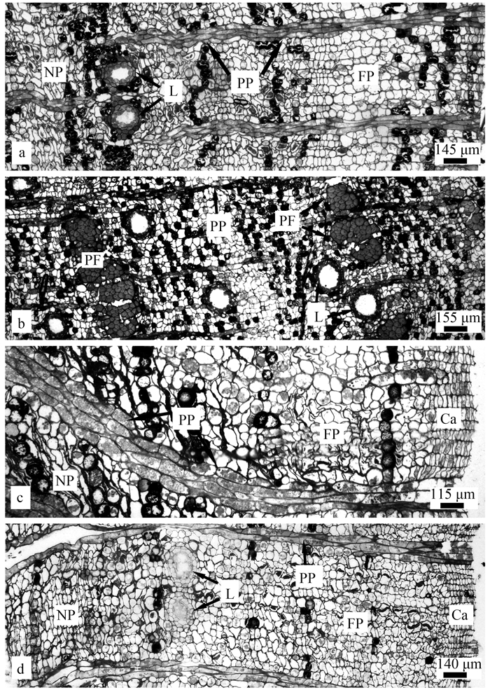
2 结果与分析 2.1 韧皮部组成细胞及其乳汁道的变化在所观察的4种漆树科植物次生韧皮部显微结构中发现,黄连木、青麸杨和盐肤木能够观察到具功能韧皮部和无功能韧皮部之间的界限,黄栌界限不明显;4种植物次生韧皮部的组成细胞基本相同,均由筛管分子和伴胞、韧皮薄壁组织细胞、射线细胞和乳汁道构成,其中盐肤木次生韧皮部中无韧皮部纤维分布(图 1)。
|
图 1 漆树科4个种的次生韧皮部横切面 Fig. 1 The secondary phloem cross sections of four species of Anacardiaceae a, b, c, d分别为黄连木、黄栌、青麸杨和盐肤木次生韧皮部横切面。Ca:维管形成层;FP:功能韧皮部;L:乳汁道;NP:无功能韧皮部;PE:薄壁细胞;PF:韧皮纤维;PP:韧皮射线。 a, b, c, and d are secondary phloem cross sections of Pistacia chinensis, Cotinus coggygria, Rhus potaninii, and Rhus chinensis, respectively. Ca: Cambium; FP: Conducting phloem; L: Resin canal; NP: Non-conducting phloem; PE: Parenchymal cell; PF: Phloem fibres; PP: Phloem pith. |
对分布在具功能韧皮部和无功能韧皮部的各组成细胞进行比较后发现,无功能韧皮部中薄壁细胞体积增大,而筛管因受到薄壁细胞的挤压,横切面直径和面积均显著减小;无功能韧皮部中乳汁道直径及横切面面积增加;另外,韧皮纤维仅在无功能韧皮中分布。次生韧皮部各组成细胞长度、直径以及面积等方面的变化见表 1。
|
|
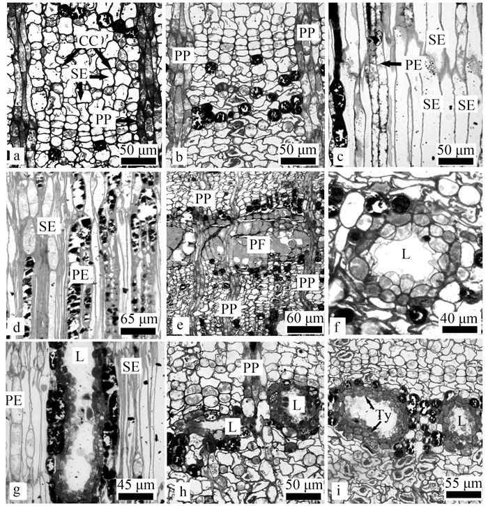
通过观察黄连木次生韧皮部显微结构发现,黄连木的次生韧皮部结构由径向的韧皮射线和轴向的筛管、伴胞、韧皮薄壁组织细胞、乳汁道和韧皮纤维共同组成,黄连木射线细胞多为3列,偶见4列(图 1a)。次生韧皮部可分为具功能韧皮部和无功能韧皮部2部分。由形成层当年分裂分化产生的次生韧皮部为具功能韧皮部(图 2a)。具功能韧皮部的外侧为往年分化形成的无功能韧皮部。黄连木具功能韧皮部和无功能韧皮部之间界限明显。具功能韧皮部筛管分子横切面呈不规则形或多角形,饱满,占具功能韧皮部区域较大面积比例(图 2a);无功能韧皮部横切面中筛管因受到挤压细胞壁弯曲(图 2b)。黄连木具功能韧皮部筛管分子纵切面中呈长梭形,筛板处有明显P-蛋白颗粒;薄壁组织细胞体积较小,细胞内无内含物(图 2c)。无功能韧皮部纵切面中筛管被挤压为狭长型细胞,细胞壁堆叠;薄壁组织细胞为生活细胞,形态饱满,近圆形或椭圆形,内含物增加(图 2d)。黄连木无功能韧皮部分布有大量的韧皮纤维(图 2e)。
|
图 2 黄连木次生韧皮部结构 Fig. 2 Secondary phloem structure of Pistacia chinensis a:具功能韧皮部横切面;b:无功能韧皮部横切面,示筛管受到挤压变形;c:具功能韧皮部纵切面,示筛管显微结构;d:无功能韧皮部纵切面,示筛管受到挤压变形,薄壁细胞体积增大;e:无功能韧皮部韧皮纤维;f:乳汁道横切面;g:乳汁道纵切面;h:示不同发育阶段的乳汁道;i:示乳汁道拟侵填体。CC:伴胞;L:乳汁道;PE:薄壁细胞;PP:韧皮射线;SE:筛管;PF:韧皮纤维;Ty:拟侵填体。 a: Cross section of conducting phloem; b: Cross section of non-conducting phloem, show the sieve tubes shrinked; c: Longitudinal section of conducting phleom, show the micro-structure of sieve tubes; d: Longitudinal section of non-conducting phloem, show that sieve tubes were crushed and parenchymal cells volume increased; e: Phloem fibres in secondary phloem; f: Cross section of resin canal; g: Longitudinal section of resin canal; h: The developing resin canal; i: Tylosids in resin canal. CC: Companion cell; L: Resin canal; PE: Parenchymal cell; PP: Phloem pith; SE: Sieve elements; PF: Phloem fibres; Ty: Tylosids. |
对黄连木次生韧皮部显微结构观察发现,乳汁道分布于2列射线细胞之间;成熟乳汁道是由1层原生质浓厚的分泌细胞(12~22个)和外围1~2层稍扁平的鞘细胞组成的近圆形或椭圆形腔道(图 2f)。径向纵切面观,乳汁道为长管道状,其延伸方向与茎长轴平行(图 2g)。处于不同阶段的乳汁道分别呈狭缝状和椭圆形,直至最终形成近圆形的完全成熟的乳汁道(图 2h)。黄连木乳汁道的腔道内会形成拟侵填体,能够观察到有多层细胞增生的现象,逐渐堵塞大部分的乳汁道腔(图 2i)。次生韧皮部最外层乳汁道受挤压呈椭圆形,分泌细胞已开始失去合成和分泌化合物的功能,萎缩破毁。
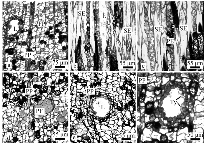
2.3 黄栌次生韧皮部结构黄栌的次生韧皮部结构与黄连木茎内次生韧皮部结构存在一定的差别。黄栌次生韧皮部横切面观也可分为具功能韧皮部和无功能韧皮部2部分,但具功能韧皮部和无功能韧皮部之间的分层现象不明显,无清晰界限,韧皮部射线细胞多为双列(图 1b);维管形成层、筛管伴胞和韧皮薄壁细胞呈径向列排布(图 3a)。黄栌纵切筛管分子纵向伸长,径向壁上筛管之间以倾斜的端壁相连(图 3b)。随着发育的进行韧皮薄壁细胞体积增大(图 3c)。大量的韧皮纤维呈带状排列分布在无功能韧皮部(图 3d)。
|
图 3 黄栌次生韧皮部结构 Fig. 3 Secondary phloem structure of Cotinus coggygria a:维管形成层及功能韧皮部结构;b:功能韧皮部及乳汁道纵切面;c:韧皮部纵切面,示膨大的韧皮薄壁细胞;d:示韧皮部韧皮纤维;e:乳汁道横切面;f:乳汁道横切面及少量拟侵填体细胞。Ca:维管形成层;L:乳汁道;PE:薄壁细胞;PP:韧皮射线;SE:筛管;PF:韧皮纤维;Ty:拟侵填体。 a: Cambium and conducting phloem; b: Longitudinal section of conducting phloem and resin canal; c: Longitudinal section of secondary phleom, show the parenchymal cells increased in size; d: Phloem fibres in phloem; e: Cross section of resin canal; f: Cross section of resin canal, show the tylosids in resin canal. Ca: Cambium; L: Resin canal; PE: Parenchymal cell; PP: Phloem pith; SE: Sieve elements; PF: Phloem fibres; Ty: Tylosids. |
黄栌乳汁道散布于射线细胞之间,无分枝;乳汁道由1层原生质浓厚的分泌细胞(15~20个)和扁平的鞘细胞(1~2层)环绕腔道组成。成熟乳汁道近圆形(图 3e)。黄栌乳汁道也能够观察到拟侵填体的现象,但细胞数量较少,仅形成少量的增生细胞(图 3f)。
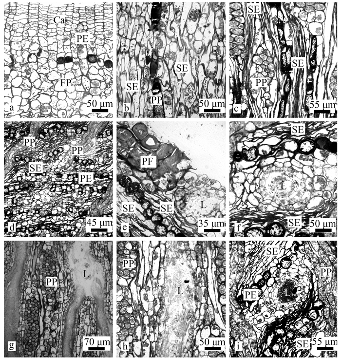
2.4 青麸杨次生韧皮部结构青麸杨具功能韧皮部与无功能韧皮部之间分层明显,具功能区域内的韧皮射线整齐辐射状排列,无功能韧皮部内倾斜状弯曲排列(图 1c)。筛管、伴胞与韧皮薄壁细胞在具功能韧皮部呈整齐径向列排布(图 4a)。具功能韧皮部筛管细胞近方形,比无功能韧皮部筛管细胞饱满,内含颗粒状P-蛋白(图 4b);无功能韧皮部内筛管细胞为不规则多边形,被韧皮薄壁细胞挤压扁平直至重叠。具功能韧皮部中的韧皮薄壁组织细胞有少量内含物存在,从纵切面观察,具功能韧皮部中的薄壁组织细胞为狭而短的细胞,与筛管细胞相邻;无功能韧皮部内薄壁组织细胞形态饱满,近圆形。具功能韧皮部韧皮薄壁细胞小于无功能韧皮部韧皮薄壁细胞(图 4c)。青麸杨射线在具功能韧皮部横切面观多为三列,偶见双列或四列射线细胞,为同型细胞,辐射状排列(图 4d)。
|
图 4 青麸杨次生韧皮部结构 Fig. 4 Secondary phloem structure of Rhus potaninii a:维管形成层及功能韧皮部横切面;b:功能韧皮部纵切面;c:无功能韧皮部切向切面,示筛管受到挤压变形及韧皮射线结构;d:无功能韧皮部横切面,韧皮射线倾斜分布;e:无功能韧皮部韧皮纤维;f:乳汁道横切面;g:示具分枝乳汁道切向切面;h:乳汁道切向切面,示乳汁道内分泌物;i:无功能韧皮部横切面,示衰老乳汁道。Ca:维管形成层;L:乳汁道;PE:薄壁细胞;PF:韧皮纤维;PP:韧皮射线;SE:筛管。 a: Cross section of cambium and conducting phloem; b: Longitudinal section of conducting phloem; c: Tangential section of non-conducting phloem, show the shrinked sieve tubes and phloem ray cells; d: Cross section of non-conducting phloem, show the slanting phloem ray; e: Phloem fibres in non-conducting phloem; f: Cross section of resin canal; g: Tangential section of resin canal, show the branch of resin canal; h: Tangential section of resin canal, show the secretions in the canal; i: Cross section of non-conducting phloem, show the senescent resin canal. Ca: Cambium; L: Resin canal; PE: Parenchymal cell; PF: Phloem fibres; PP: Phloem pith; SE: Sieve elements. |
在青麸杨植株次生韧皮部中仅存在少量的韧皮纤维,且未完全木化(图 4e)。无功能韧皮部中韧皮纤维排列稀疏松散。青麸杨次生韧皮部乳汁道散布于2列射线细胞间。青麸杨成熟乳汁道为由1层原生质浓厚的分泌细胞(13~20个)和稍扁平的鞘细胞(2~3(5)层)环绕组成的近圆形的腔(图 4f)。青麸杨的乳汁道与其他3种植物乳汁道不同,具有明显的分枝现象(图 4g),且乳汁道内充满了分泌物(图 4h)。无功能韧皮部区域内乳汁道腔隙直径显著增大,分泌细胞原生质变少,乳汁道横切面近圆形(图 4i);靠近周皮处的乳汁道,分泌细胞与鞘细胞均为扁平细胞,分泌细胞内的原生质逐渐消失,腔道内分泌物也在逐渐减少直至消失。同时由于受到内部维管形成层分裂分化的影响,近周皮处的乳汁道会由圆形腔道逐渐变为长形或椭圆形腔道。青麸杨乳汁道内无拟侵填体存在。
2.5 盐肤木次生韧皮部结构盐肤木具功能韧皮部和无功能韧皮部间发育生长分界清晰(图 1d)。盐肤木次生韧皮部结构与前面所研究的3种植物相似,维管形成层、筛胞和韧皮薄壁细胞呈切向带交替排布。具功能韧皮部筛管细胞饱满,横切面上伴胞紧随筛管而生(图 5a);无功能韧皮部内筛管受到严重挤压堆叠(图 5b),且外侧筛管细胞受挤压更为明显(图 5c)。具功能韧皮部纵切面上同样能够观察到伴胞紧随筛管而生(图 5d);韧皮射线在无功能韧皮部内会出现弯曲倾斜现象(图 5e)。
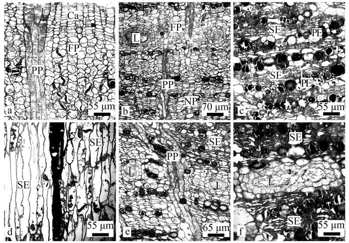
|
图 5 盐肤木次生韧皮部结构 Fig. 5 Secnodary phloem structure of Rhus chinensis a:维管形成层及功能韧皮部;b:无功能韧皮部;c:无功能韧皮部,示筛管受到挤压至细胞壁堆叠;d:具功能韧皮部纵切面;e:无功能韧皮部倾斜射线;f:示乳汁道融合。Ca:维管形成层;FP:具功能韧皮部;L:乳汁道;NP:无功能韧皮部;PE:薄壁细胞;PP:韧皮射线;SE:筛管。 a: Cross section of cambium and conducting phloem; b: Non-conducting phloem; c: Cross section of non-conducting phloem, show the sieve tubes were squeezed and cell wall overlapped; d: Longitudinal section of conducting phloem; e: Cross section of non-conducting phloem, show the slanting phloem ray; f: Cross section of non-conducting phloem, show the resin canals fused. Ca: Cambium; FP: Conducting phloem; L: Resin canal; NP: Non-conducting phloem; PE: Parenchymal cell; PP: Phloem pith; SE: Sieve elements. |
盐肤木乳汁道结构与其他3种植物相似,由1层分泌细胞(13~22个)和2~3层扁平鞘细胞环绕形成近圆形或椭圆形腔道。盐肤木乳汁道不具分枝,横切面上可观察到有些乳汁道细胞具有融合现象的发生(图 5f)。
3 讨论 3.1 漆树科4种植物次生韧皮部的结构特征以及“树皮年轮”现象对落叶树木的韧皮部结构的研究表明,可利用次生韧皮部组成细胞及其排列方式进行类型的划分(Takeshi,1990)。树木解剖学的研究结果可为植物系统演化提供证据,如白杜、鹅掌楸、北美鹅掌楸和杂交鹅掌楸等温带地区树木的韧皮部组成细胞的解剖学特征为卫矛科(Celastraceae)、鹅掌楸属系统性演化过程提供了理论支持(林金安等, 1993; 王丰等, 2013)。本次研究所观察的漆树科4种植物的次生韧皮部组成细胞的排列较为规则,筛管分子细胞和伴胞、韧皮薄壁组织细胞呈切向带状交替排列,这些特征符合温带分布双子叶树木次生韧皮部的共同特征(赵猛等, 2012)。但是这4种植物的次生韧皮部射线细胞的排列方式不同,韧皮纤维的数量以及分泌道是否具有分枝等存在差异;黄连木、青麸杨和盐肤木的具功能韧皮部和不具功能韧皮部的分界现象明显,黄栌的分界现象不明显;青麸杨的射线细胞在无功能韧皮部中呈倾斜状分布,韧皮纤维数量较少,最突出的特征就是其乳汁道具分枝。
温带地区树木具有韧皮部分层现象,即每年产生的次生韧皮部之间可以划分出层次,类似次生木质部中形成的生长轮,即“树皮年轮”(Esau, 1969;Metcalfe et al., 1979;Trockenbrodt,1990)。在本研究中黄连木、青麸杨和盐肤木次生韧皮部的分层现象较为明显,具有“树皮年轮”现象,黄栌不甚明显。次生韧皮部的组成细胞以及各自的排列方式在分类学上有一定的理论价值,也能为树木年轮学的研究提供部分资料。
3.2 漆树科4种植物次生韧皮部中乳汁道的结构特征漆树科80余属植物中,已发现有大约40属植物中存在乳汁道(Pell, 2004)。同一科属植物的乳汁道的发生方式及解剖结构基本一致。漆树科火炬树根和茎切向切面上乳汁道分枝联结成网状(马淑英等, 1997);光漆树的乳汁道在茎的切向面上产生分枝并互相联结(Fahn et al., 1974)。本研究所观察的漆树科4种植物乳汁道为近圆形或椭圆形的腔道,腔道周围是由1层分泌细胞和多层鞘细胞包被而成,这些特征与漆树(Toxicodendron vernicifluum)(傅淑颖等, 2007)、光叶黄栌(Cotinus coggygria var. cinerea) (马淑英等, 1992)等的乳汁道结构是相同的;但是不同种分泌细胞的数目、鞘细胞层数以及分泌腔的腔道直径、乳汁道的分枝情况等均存在差异,青麸杨乳汁道具有分枝,这种分枝型乳汁道在漆树科植物中较为稀有,具有一定的分类学价值,黄连木、黄栌和盐肤木乳汁道不分枝。
臭椿(Ailanthus altissima)茎经机械损伤可诱导其茎形成层活动方式发生变化,形成创伤性分泌道,之后相邻的2个创伤性分泌道会呈现融合趋势(史宏勇等, 2011)。在观察自然生长的盐肤木次生韧皮部横切面时发现少量乳汁道有融合现象,但出现这种现象的比例较低。
4 结论漆树科4个种(黄栌、黄连木、青麸杨和盐肤木)次生韧皮部均是由筛管分子和伴胞、韧皮薄壁组织细胞、射线细胞、乳汁道和韧皮纤维构成,呈切向带状相间排列,但在次生韧皮部组成细胞及乳汁道结构等方面存在一定差异。黄连木、青麸杨和盐肤木的具功能韧皮部和无功能韧皮部分界明显;青麸杨的射线细胞在无功能韧皮部中倾斜分布,其余3种植物均为整齐纵向排列;青麸杨的乳汁道为分枝型乳汁道,其他3个种的乳汁道均无分枝,仅盐肤木有少量相邻乳汁道有融合现象发生;黄连木和黄栌次生韧皮部内分布有大量的韧皮纤维,青麸杨韧皮部中较少,而盐肤木韧皮部中无韧皮纤维分布。青麸杨乳汁道是目前为止漆树科植物发现的较为稀有的分枝型乳汁道。研究结果能够为漆树科系统进化研究提供参考依据。
范六民, 李焜章, 陈耀堂, 等. 1994. 15种松属植物茎次生韧皮部的解剖学研究. 应用基础与工程科学学报, 2(Z1): 267-276. (Fan L M, Li K Z, Chen Y T, et al. 1994. Study on the secondary phloem anatomy of the stem of 15 species in Pinus. Journal of Basic Science & Engineering, 2(Z1): 267-276. [in Chinese]) |
傅淑颖, 魏朔南, 胡正海. 2007. 漆树各器官中乳汁道的分布与结构特征研究. 西北植物学报, 27(4): 651-656. (Fu S Y, Wei S N, Hu Z H. 2007. Distributed regularity and anatomic feature of laticiferous canals of Toxicodendron vernicifluum(Stokes) F. A. Barkley. Acta Botanica Boreali-Occidentalia Sinica, 27(4): 651-656. DOI:10.3321/j.issn:1000-4025.2007.04.002 [in Chinese]) |
林金安, 高信曾. 1993. 白杜次生韧皮部的解剖学研究. 植物学报, 35(7): 506-512. (Lin J A, Gao X Z. 1993. Anatomical studies on secondary phloem of Euonymus bungeanus. Chinese Bulletin of Botany, 35(7): 506-512. [in Chinese]) |
马淑英, 胡正海. 1992. 西北地区漆树科四属植物分泌道的解剖学研究. 西北植物学报, 12(3): 180-187. (Ma S Y, Hu Z H. 1992. Anatomical studies on the secretory canals of four genus of Anacadiaceae in northwest China. Acta Botanica Boreali-Occidentalia Sinica, 12(3): 180-187. DOI:10.3321/j.issn:1000-4025.1992.03.002 [in Chinese]) |
马淑英, 胡正海. 1997. 火炬树分泌道的发育解剖学研究. 西北植物学报, 17(5): 112-117. (Ma S Y, Hu Z H. 1997. Development anatomical studies on the secretory ducts of Rhus typhina. Acta Botanica Boreali-Occidentalia Sinica, 17(5): 112-117. DOI:10.3321/j.issn:1000-4025.1997.05.022 [in Chinese]) |
史宏勇, 周亚福, 郭建胜, 等. 2011. 臭椿茎中分泌道的发育及其组织化学研究. 西北植物学报, 31(7): 1291-1296. (Shi H Y, Zhou Y F, Guo J S, et al. 2011. Development and cytochemistry of secretory ducts in Ailanthus altissima. Acta Botanica Boreali-Occidentalia Sinica, 31(7): 1291-1296. [in Chinese]) |
王丰, 潘彪, 李伊乐. 2013. 鹅掌楸属3个树种次生韧皮部显微构造的比较. 南京林业大学学报:自然科学版, 37(5): 113-118. (Wang F, Pan B, Li Y L. 2013. Comparison of micro-structure of secondary phloem in Liriodendron. Journal of Nanjing Forestry University:Natural Science Edition, 37(5): 113-118. [in Chinese]) |
赵猛, 魏朔南, 胡正海. 2012. 漆树韧皮部的结构与发育. 林业科学, 48(9): 36-41. (Zhao M, Wei S N, Hu Z H. 2012. Structure and development of phloem in Toxicodendron vernicifluum. Scientia Silvae Sinicae, 48(9): 36-41. [in Chinese]) |
Angiosperm Phylogeny Group Ⅳ[APG Ⅳ]. 2016. An update of the Angiosperm Phylogeny Group classification for the orders and families of flowering plants:APG Ⅳ. Botanical Journal of the Linnean Society, 181: 1-20. DOI:10.1111/boj.2016.181.issue-1 |
Esau K. 1969. Handbuch der Pflanzenanatomie. Borntraeger: 363-372. |
Fahn A, Evert R F. 1974. Ultrastructure of the secretory ducts of Rhus glabra L. American Journal of Botany, 61(1): 1-14. DOI:10.1002/j.1537-2197.1974.tb06022.x |
Gadek P A, Fernando E S, Quinn C J, et al. 1996. Sapindales:Molecular delimitation and infraordinal groups. American Journal of Botany, 83(6): 802-811. DOI:10.1002/j.1537-2197.1996.tb12769.x |
Metcalfe C R, Chalk L. 1979. Anatomy of the dicotyledons:vol 1. Systematic anatomy of leaf and stem, with a brief history of the subject. Oxford: Clarendon Press.
|
Metcalfe C R, Chalk L. 1950. Anatomy of the dicotyledons. Oxford: Clarendon Press.
|
Ming T L. 2008. Flora of China:Vol 11. Beijing: Science Press, 335.
|
Pell S K. 2004. Molecular systematics of the Cashew Family(Anacardiaceae). PhD thesis of Louisiana State University.
|
Savolainen V, Chase M W, Hoot S B, et al. 2000a. Phylogenetics of flowering plants based on combined analysis of plastid atpB and rbcL gene sequences. Systematic Biology, 49(2): 306-362. DOI:10.1093/sysbio/49.2.306 |
Savolainen V, Fay M F, Albach D C, et al. 2000b. Phylogeny of the eudicots:a nearly complete familial analysis based on rbcL gene sequences. Kew Bulletin, 55(2): 257-309. DOI:10.2307/4115644 |
Takeshi F. 1990. Bark structure of deciduous broad-leaved trees grown in the San'in region, Japan. IAWA Journal, 11(3): 239-254. DOI:10.1163/22941932-90001181 |
Trockenbrodt M. 1990. Survey and discussion of the terminology used in bark anatomy. Iawa Bulletin, 11(2): 141-166. DOI:10.1163/22941932-90000511 |
Wannan B S. 1986. Systematics of the Anacardiaceae and its Allies. PhD thesis of University of New South Wales, Australia.
|
Wannan B S, Quinn C. 1988. Biflavonoids in the Julianiaceae. Phytochemistry, 27(10): 31-62. |
Wannan B S, Quinn C. 1990. Pericarp structure and generic affinities in the Anacardiaceae. Botanical Journal of the Linnean Society, 102(3): 225-252. DOI:10.1111/boj.1990.102.issue-3 |
Wannan B S, Quinn C. 1991. Floral structure and evolution in the Anacardiaceae. Botanical Journal of the Linnean Society, 107(4): 349-385. DOI:10.1111/boj.1991.107.issue-4 |
 2019, Vol. 55
