文章信息
- 刘苍伟, 苏明垒, 王思群, 王新洲, 赵荣军, 任海青, 王玉荣
- Liu Cangwei, Su Minglei, Wang Siqun, Wang Xinzhou, Zhao Rongjun, Ren Haiqing, Wang Yurong
- 不同生长期毛竹材细胞壁力学性能与微纤丝角
- Cell Wall Mechanical Properties and Microfibril Angle of Phyllostachys edulis in Different Growth Period
- 林业科学, 2018, 54(1): 174-180.
- Scientia Silvae Sinicae, 2018, 54(1): 174-180.
- DOI: 10.11707/j.1001-7488.20180120
-
文章历史
- 收稿日期:2016-07-25
- 修回日期:2017-03-09
-
作者相关文章
2. 田纳西大学可再生碳中心 诺克斯维尔 37996;
3. 南京林业大学材料科学与工程学院 南京 210037
2. Center for Renewable Carbon, University of Tennessee, Knoxville 37996;
3. School of Materials Science and Engineering, Nanjing Forestry University Nanjing 210037
毛竹(Phyllostachys edulis)是我国重要的竹类资源,广泛应用于制浆造纸和建筑等领域,适宜的采伐期对毛竹资源的高效利用具有重要意义(张齐生,1995;江泽慧,2002)。毛竹生长迅速,通常1~3月的幼龄竹即已达到成熟竹的尺寸,因此其组织比量、纤维长度和纤维细胞壁厚度等解剖参数在不同生长期表现差异较小(马灵飞等,1997)。张齐生等(2002)研究发现,毛竹化学成分含量在1~7年先增加后趋于稳定,到8年后开始明显下降,并依据其化学成分变化规律将毛竹的生长期分为增长期、成熟期和下降期。此外学者还发现毛竹的基本密度和抗压强度、抗拉强度等宏观力学性能都与竹龄呈二次抛物线关系,均为幼龄竹(0~1年)时最小,在4年左右达到最优,到10年后随着毛竹老化开始明显下降(周芳纯,1991;Wang et al., 2010;关明杰等,2010)。雷竹(Phyllostachys praecox)和苦竹(Pleioblastus amarus)的基本密度和宏观力学性能也同样表现为随着竹龄增大而增加至4~5年时趋于稳定(俞友明等,2004;2005)。
纳米压痕技术在木质材料领域的应用,使得在纳米尺度原位测定细胞壁的力学性能成为可能(Gindl et al., 2004;Wang et al., 2006;Tze et al., 2007)。近年来,纳米压痕技术也被用于探究竹材纤维细胞壁的力学性能,研究发现毛竹材薄壁基本组织细胞和厚壁纤维细胞壁层力学性能存在显著差异,厚壁纤维细胞弹性模量是薄壁组织细胞的3倍以上,硬度无明显差异(Yu et al., 2007;Zou et al., 2009),由此可见厚壁纤维细胞是决定竹材力学性能的主要成分。有学者采用纳米压痕技术及样品树脂包埋处理方法研究了0.5~4年竹龄毛竹纤维细胞壁力学性能(Yu et al., 2011),但对10年以上处于老化期的毛竹材纤维细胞壁力学性能以及系统综合对幼龄、成熟和过熟这3个生长发育阶段竹材纤维细胞壁力学性能变化趋势的研究较少。
微纤丝角是影响木材和竹材力学性能的关键因子,微纤丝角与木材力学强度呈负相关性(Cave et al., 1994;Barnett et al., 2004;Jäger et al., 2011)。国内学者对毛竹材微纤丝角的研究多采用X-射线衍射法,得到(002)面衍射图谱后经0.6T法(阮锡根等,1982)计算得出,所得结果表明毛竹材不同竹龄和部位的微纤丝角有差异但变化幅度较小,且浮动规律不明显(Yang et al., 2009;Wang et al., 2010)。国内目前还少有依据不同类型细胞衍射图谱进行高斯拟合曲线分析竹材细胞壁微纤丝角,并且对于不同生长期毛竹材纤维细胞壁的力学性能及微纤丝角特别是相同部位二者的相关性研究也少有报道。
为了在纳米尺度上深入探究竹材利用时为何通常选择4~5年的成熟竹材,而不选择已近成熟竹尺寸大小的幼龄竹和生长较长期的过熟竹,本文选择同一生境下的0.5年幼龄竹、4.5年成熟竹和10.5年过熟竹3个典型生长期的毛竹材为研究对象,在生材气干后未经包埋处理状态下应用纳米压痕技术原位获取厚壁纤维细胞壁力学性能,采用广角X-射线散射技术并结合高斯拟合分析方法测算相同部位毛竹材细胞壁的微纤丝角,从而阐明不同生长期毛竹材细胞壁的力学性能特征及其变化规律,以期为竹材的科学采伐和竹材分级、改性及重组研究与合理利用提供理论依据。
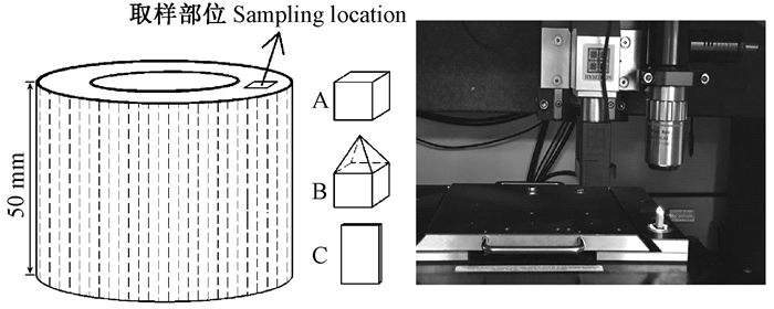
1 材料与方法 1.1 试验材料试验材料采自浙江省富阳庙山乌林场毛竹人工林。选取0.5年幼龄竹、4.5年成熟竹和10.5年过熟竹3个不同生长期的毛竹,每类选取3株,在茎杆中部截取3个50 mm高的竹筒用于测试。在每个竹环竹肉部分的中间相同部位上纵向截取A、B、C 3个试样分别用于显微结构观察、细胞壁力学性能测试和微纤丝角测量(图 1)。
|
图 1 试样加工示意及测试 Figure 1 The preparation of bamboo samples and testing |
1) 显微结构观察图 1所示试样A用于显微结构观察,尺寸为10 mm(R)×10 mm(T)×10 mm(L)。将试样置于80 ℃水中软化3天,再由Leica SM2010R滑走切片机制取20 μm厚切片,番红溶液染色,梯度酒精脱水,后用加拿大树胶封片,置于光学显微镜(Leica DMLB & Japan Q570)下观察并拍照(Zhao et al., 2014)。显微图像用于较大视野观察试样厚壁纤维细胞形态以及选取纳米压痕测试部位。
2) 纳米压痕测试图 1所示试样B用于纳米压痕测试,尺寸为10 mm(R)×10 mm(T)×15 mm(L)。将试件上端用滑走切片机切出金字塔形的塔尖,塔尖部位保证含有要测试的多个厚壁纤维细胞,再用装有钻石刀的半薄切片机将塔尖刨光。将试样保存在温度20 ℃、相对湿度60%±4%的纳米压痕仪试验空间内至少24 h,用于平衡试件温度和相对湿度。仪器设备为美国Hysitron公司的Triboindenter,采用针头半径小于100 nm的Berkovieh压头,荷载过程分加载、保载、卸载3个过程各5 s,最大荷载为400 μN,得到试样的加载卸载曲线,由Oliver-Pharr(Oliver et al., 1992)经验公式计算出不同竹龄毛竹材纤维细胞壁弹性模量和硬度。整个试验过程保持温度20 ℃、相对湿度(60±4)%恒定。
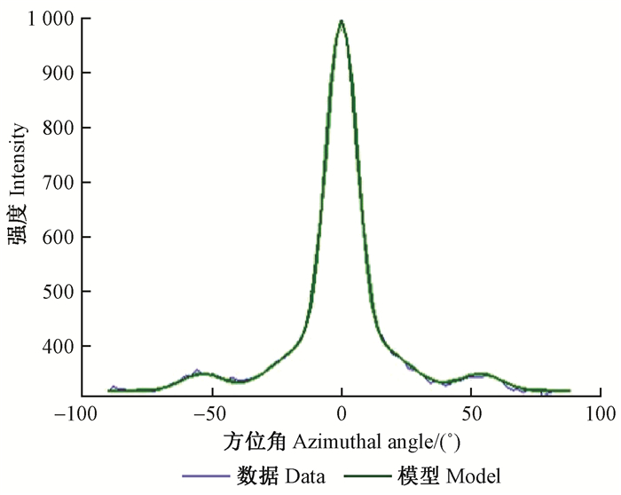
1.3 微纤丝角测算图 1所示试样C用于微纤丝角测试,尺寸为10 mm(T)×1.5 mm(R)×15 mm(L)。每个竹龄测试样品数是纵向3个竹环中间部位各取1片,3株共9个样品。采用广角X-射线散射法测量竹材的微纤丝角,将测试样品置于日本UltraX 18S型Rigaku阳极旋转X射线衍射仪上,获取毛竹材(002)面散射曲线,因竹材厚壁纤维细胞多为圆形,不同于木材晚材细胞形状,因此在分析其(002)面衍射图时采用高斯曲线拟合方法,通过MATLAB软件调整高斯方程参数可以将模型曲线与实测曲线基本吻合。然后参照已报道方法(Wang et al., 2012),计算得出竹材平均微纤丝角。
2 结果与分析 2.1 不同生长期毛竹材细胞壁力学性能1) 显微观察及纳米压痕定位由图 2A可知,毛竹材横切面具有典型的单子叶植物显微结构特征,星散状维管束分布于薄壁组织细胞中。维管束中有大量的厚壁纤维细胞包围着具有输导水分功能的大导管,这些厚壁纤维细胞是竹材力学性能的主要承担者(Zou et al., 2009)。本研究毛竹材细胞壁力学性能测试选取的也是维管束中的厚壁纤维细胞,且统一选取大导管外侧的厚壁纤维细胞,如图 2A和B所示。其中图 2B是未经包埋处理的毛竹材经钻石刀精刨光后在纳米压痕仪显微镜下拍摄。在试样上不同维管束及同一维管束不同部位进行定位并用探针扫描,可以清晰地看到毛竹材厚壁纤维细胞,在其细胞壁上选择压痕部位,如图 2B和C所示。

|
图 2 毛竹材横切面显微构造及纤维细胞壁压痕探针扫描 Figure 2 The cross-sectional microstructure and the scanning image of fiber cell walls by nanoindentation probe in moso bamboo A:横切面显微构造,B:在纳米压痕仪显微镜下维管束横切面微观构造,C:压痕探针扫描1部位厚壁纤维横切面。 A:The cross-sectional microstructure; B:The cross-sectional microstructure of vascular bundle under microscope of the nanoindentation; C:The scanning image of fiber cell walls in location 1 by nanoindentation probe. |
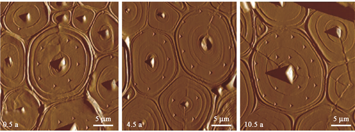
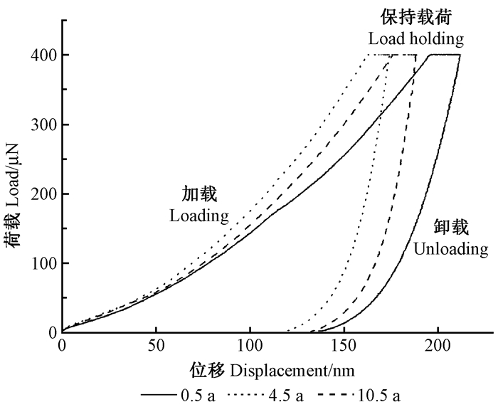
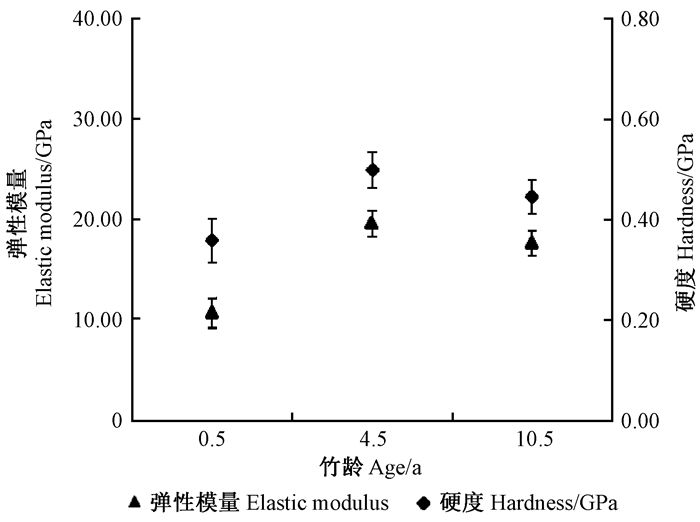
2) 细胞壁力学性能对选择的样品压痕部位进行测试,纳米压痕探针在纳米尺度上连续接触竹纤维细胞壁层表面,得到测试后残留变形及加载卸载曲线如图 3和4所示。依据压痕位点位置、形态和数据曲线特征,每个生长期最终选取33~36个有效压痕点数据进行分析。不同生长期毛竹材纤维细胞壁的弹性模量、硬度和最大加载深度结果如表 1所示。由表 1可知,幼龄毛竹、成熟毛竹和过熟毛竹3个生长期的细胞壁力学性能指标有较大不同,其中0.5年幼龄毛竹材的细胞壁弹性模量和硬度最小,为10.7 GPa和0.358 GPa;4.5年成熟毛竹材的细胞壁弹性模量和硬度均为最大,分别为19.6 GPa和0.498 GPa;10.5年过熟毛竹材的细胞壁硬度和弹性模量居二者之间,为17.6 GPa和0.445 GPa。由图 4和图 5可知,不同竹龄其弹性模量和硬度的变化趋势是一样的,当其弹性模量较大时其硬度也较大,而硬度与压痕深度呈负相关。由图 3所示,从左至右依次为0.5年、4.5年、10.5年毛竹纳米压痕残留变形,纳米压痕深度最深的是0.5年毛竹材,而纳米压痕深度最浅是4.5年毛竹材,10.5年毛竹材的纳米压痕深度居二者之间,图 4加载卸载曲线也表明了同样的结果,0.5年毛竹材位移最大。
|
图 3 不同生长期毛竹材纤维细胞壁纳米压痕测试后残留变形 Figure 3 The permanent displacement of the fiber cell walls in moso bamboo in different growth period after the nanoindentation test |
|
图 4 不同生长期毛竹材纤维细胞壁纳米压痕加载卸载曲线 Figure 4 Typical NI load-displacement curves of the fiber cell walls of moso bamboo in different growth period |
|
|
|
图 5 不同生长期毛竹材纤维细胞壁的硬度和弹性模量 Figure 5 The hardness and elastic modulus of the fiber cell walls of moso bamboo in different growth period |
综上所述,生长期对毛竹材纤维细胞壁力学性能是有影响的,起初随生长期的增大而增加,到成熟期达到最优,后又随着竹材老化细胞壁的力学性能有下降趋势。这与采用树脂包埋制样法测得不同竹龄毛竹材纤维细胞壁力学性能结果的不同可能与制样方法不同有关,后期已有文献报道用于包埋纳米压痕样品的包埋剂对测试结果会有一定影响(Meng et al., 2013)。本文采用非包埋制样法对不同竹龄毛竹纤维细胞壁力学性能进行测定,排除了包埋剂对测试值的干扰。此外对密度和木质素含量等影响材料力学性能的重要因子研究(Gindl et al., 2002;王传贵等,2012)发现,毛竹的基本密度和木质素含量在毛竹材质增长期均随竹龄增大而增加,但随着竹材进入老化期,毛竹的基本密度和木质素、综纤维素等化学成分含量表现出下降趋势,但仍大于早期幼龄竹材,与本研究中纤维细胞壁力学性能测试结果相符(马灵飞等,1997;房桂干等,1989;关明杰等,2010)。Wang等(2014)采用单根纤维微拉伸技术也得出4年生麻竹(Dendrocalamus latiflorus)纤维的刚度和强度比1年生大,同时毛竹和雷竹等其他竹材的宏观力学性能也在4~5年成熟期宏观力学性能较优(关明杰等,2010;俞友明等,2004;2005),这些研究结果均与不同生长期毛竹纤维壁力学性能的变化趋势是相一致的。
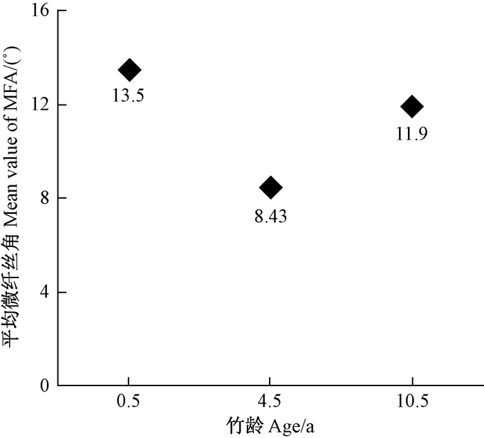
2.2 不同生长期毛竹材细胞壁微纤丝角本文在分析毛竹材细胞壁(002)面衍射图时采用高斯曲线拟合方法。如图 6所示,成熟材X-射线散射图实测曲线与模型曲线基本吻合。微纤丝角测算结果如图 7所示,3个不同竹龄毛竹材纤维细胞壁平均微纤丝角为11.3°,且差异明显,0.5年、4.5年、10.5年3个生长期毛竹材的微纤丝角分别为13.5°、8.43°和11.9°,变化趋势为先随生长期增加而降低,到成熟期后又随竹龄增大而升高。虽然10.5年毛竹材微纤丝角有所升高,但0.5年毛竹材微纤丝角仍是最大的。
|
图 6 X-射线散射图谱高斯拟合后强度分布曲线 Figure 6 Intensity distribution curve of X-ray scattering pattern fitted with Gauss function |
|
图 7 不同生长期毛竹材细胞壁平均微纤丝角 Figure 7 The mean values of MFA ofthe fiber cell walls of moso bamboo in different growth period |
有研究发现,竹龄对毛竹纤维壁厚的影响不大,纤维细胞在5个月时纤维壁厚已达到最大值(马灵飞等,1997;Huang et al., 2012),结合本研究结果表明,生长期对细胞壁微纤丝的排列有较大影响,微纤丝角随竹龄升高呈先降后增的变化规律。对木材尤其是针叶材次生壁微纤丝角变化规律和影响因素的研究表明,微纤丝角不仅受遗传因素影响,也与材料生长环境和营养状况相关(Donaldson,2008);同样对于竹材而言,微纤丝角也受多方面因素影响,因此应选择同一生境下且与细胞壁力学测试相同部位的竹片用于微纤丝角测试,以减少外界因素的干扰。2.3不同生长期毛竹材细胞壁力学性能与微纤丝角的相关关系随着纳米压痕技术在木材科学领域的不断应用,目前已从样品需要进行包埋处理发展到非包埋样品也可以用于原位测量木竹材细胞壁力学性能(Meng et al., 2013;Wang et al., 2016),本文采用非包埋处理样品方法直接进行纳米压痕测试,获得了不同生长期毛竹材细胞壁力学性能。广角X-射线散射法测量次生壁微纤丝角具有制样简单、无破坏和重复性高等优点,目前应用较广,但在对毛竹材微纤丝角测量时应用较少。国外在获取木竹材微纤丝角时多依据木竹材细胞壁形态特征选择测试方式及不同分析计算方法(Andersson et al., 2000;Andersson, 2006),本文在计算不同生长期毛竹材微纤丝角时采用的是国外常用的高斯拟合曲线计算方法。
应用以上2种测试方法获取不同生长期毛竹材细胞壁力学性能和微纤丝角,发现0.5年幼龄毛竹材的细胞壁弹性模量和硬度最小,分别是10.7 GPa和0.358 GPa,而其微纤丝角较大,达13.5°;4.5年成熟毛竹材的细胞壁弹性模量和硬度均为最大,分别为19.6 GPa和0.498 GPa,其微纤丝角却较小,为8.43°;10.5年过熟毛竹材的细胞壁硬度和弹性模量居二者之间,分别为17.6 GPa和0.445 GPa,其微纤丝角为11.9°,也位于0.5年和4.5年毛竹材微纤丝角之间。微纤丝角和纤维细胞壁力学性能测试结果呈现较好的负相关性,与前人研究微纤丝角与木材微力学性能关系得出的结论相吻合,微纤丝角越小其纵向力学性能越强(Cave et al., 1994;Jäger et al., 2011),表明微纤丝角也是评价竹材细胞壁力学性能的主要指标。
3 结论本研究采用纳米压痕技术结合非包埋法及广角X-射线散射仪结合高斯拟合分析方法能够准确获取不同生长期毛竹材纤维细胞壁力学性能指标值和微纤丝角,从而揭示二者在不同生长期的变化趋势,该研究方法是可行的。获得的结果表明,竹龄对毛竹纤维细胞壁力学性能和微纤丝排列均有影响,幼龄毛竹材纤维细胞壁力学性能与采伐期的成熟毛竹材纤维细胞壁力学性能仍有较大差别,随着竹龄增大,特别是达到成熟期时,细胞壁力学性能达到最大,其弹性模量和硬度分别为19.6 GPa和0.498 GPa。但毛竹材并不是生长期越长其细胞壁力学性能越好,而是随着竹材老化其力学性能呈下降状态。成熟期的毛竹材其纤维细胞壁微纤丝排列与主轴的夹角呈较小状态,也决定了其具有较优的力学性能。综合纤维细胞壁力学性能和微纤丝角测量结果,本研究在细胞壁水平阐明了毛竹材在4~5年成熟期时其微观力学性能优于幼龄毛竹和过熟毛竹。
房桂干, 王菊华, 任维羡, 等. 1989. 毛竹的形态学特性、超微结构及木素分布[J]. 中国造纸学报, 1(4): 42-49. (Fang G G, Wang J H, Ren W X, et al. 1989. A study on the morphology, ultrastructure and lignin distribution of bamboo[J]. Transaction of China Pulp and Paper, 1(4): 42-49. [in Chinese]) |
关明杰, 朱一辛, 莫弦丰, 等. 2010. 毛竹材质老化过程中几个基本性质变化与建筑原竹的选择[J]. 西南大学学报:自然科学版, 32(1): 144-147. (Guan M J, Zhu Y X, Mo X F, et al. 2010. The variety during aging process of bamboo and the selection of raw bamboo for construction[J]. Journal of Southwest University(Natural Science Edition), 32(1): 144-147. [in Chinese]) |
江泽慧. 2002. 世界竹藤[M]. 沈阳: 辽宁科学出版社. (Jiang Z H. 2002. World bamboo and rattan[M]. Shenyang: Liaoning Science and Technology Press. [in Chinese]) |
马灵飞, 马乃训. 1997. 毛竹材材性变异的研究[J]. 林业科学, 33(4): 356-364. (Ma L F, Ma N X. 1997. Study on the variation of wood properties of bamboo[J]. Scientia Silvae Sinicae, 33(4): 356-364. [in Chinese]) |
王传贵, 江泽慧, 费本华, 等. 2012. 化学成分对木材细胞壁纵向弹性模量和硬度的影响[J]. 北京林业大学学报, 34(3): 107-110. (Wang C G, Jiang Z H, Fei B H, et al. 2012. Effects of chemical components on longitudinal MOE and hardness of wood cell wall[J]. Journal of Beijing Forestry University, 34(3): 107-110. [in Chinese]) |
阮锡根, 尹思慈, 孙成志. 1982. 应用X射线衍射-(002)衍射弧法-测定木材纤维次生壁的微纤丝角[J]. 林业科学, 18(1): 64-71. (Ruan X G, Yin S C, Sun C Z. 1982. Using the X-ray diffraction(002) to determine the microfibril angle of the secondary wall of wood fiber[J]. Scientia Silvae Sinicae, 18(1): 64-71. [in Chinese]) |
俞友明, 金永明, 於琼华, 等. 2004. 雷竹竹材物理力学性质变异规律的研究[J]. 竹子研究汇刊, 23(2): 50-54. (Yu Y M, Jin Y M, Yu Q H, et al. 2004. A study on the varistion pattern of physical-mechanical properties of Phyllostachys praecox wood[J]. Journal of Bamboo Research, 23(2): 50-54. [in Chinese]) |
俞友明, 方伟, 林新春, 等. 2005. 苦竹竹材物理力学性能的研究[J]. 西南林学院学报, 25(3): 64-67. (Yu Y M, Fang W, Lin X C, et al. 2005. Study on physical and mechanical properties of bamboo (Pleioblastus amarus)[J]. Journal of Southwest Forestry College, 25(3): 64-67. [in Chinese]) |
周芳纯. 1991. 竹材的力学性质[J]. 竹类研究, (1): 45-57. (Zhou F C. 1991. Mechanical properties of bamboo[J]. Bamboo Research, (1): 45-57. [in Chinese]) |
张齐生. 1995. 竹材工业化利用[M]. 北京: 中国林业出版社. (Zhang Q S. 1995. The industrial utilization of bamboo[M]. Beijing: Chinese Forestry Publishing House. [in Chinese]) |
张齐生, 关明杰, 纪文兰. 2002. 毛竹材质生成过程中化学成分的变化[J]. 南京林业大学学报:自然科学版, 26(2): 7-10. (Zhang Q S, Guan M J, Ji W L. 2002. Variation of moso bamboo chemical compositions during mature growing period[J]. Journal of Nanjing Forestry University:Natural Science Edition, 26(2): 7-10. [in Chinese]) |
Andersson S, Serimaa R, Torkkeli M, et al. 2000. Microfibril angle on Norway spruce (Picea abies (L.) Karst.) compression wood:comparison of measuring techniques[J]. Journal of Wood Science, 46: 343-349. DOI:10.1007/BF00776394 |
Andersson S. 2006. A study of the nanostructure of the cell wall of the tracheids of conifer xylem by X-ray scattering. PhD Thesis of University of Helsinki. https://www.researchgate.net/publication/35394004_A_study_of_the_nanostructure_of_the_cell_wall_of_the_tracheids_of_conifer_xylem_by_X-ray_scattering
|
Barnett J R, Bonham V A. 2004. Cellulose microfibril angle in the cell wall of wood fibers[J]. Biological Reviews, 79(2): 461-472. DOI:10.1017/S1464793103006377 |
Cave I D, Walker J C F. 1994. Stiffness of wood in fast-grown plantation softwoods:the influence of microfibril angle[J]. Forest Products Journal, 44(5): 43-48. |
Donaldsom L. 2008. Microfibril angle:measurement, variation and relationships:a review[J]. IAWA, 29(4): 345-386. DOI:10.1163/22941932-90000192 |
Gindl W, Schöberl T. 2004. The significance of the elastic modulus of wood cell walls obtained from nanoindentation measurements[J]. Composites Part A:Applied Science and Manufacturing, 35(11): 1345-1349. DOI:10.1016/j.compositesa.2004.04.002 |
Huang Y H, Fei B H, Yu Y, et al. 2012. Plant age effect on mechanical propweties of moso bamboo (Phyllostachys heterocycle var. pubescens) single fibers[J]. Wood and Fiber Science, 44(2): 196-201. |
Jäger A, Bader T, Hofstetter K, et al. 2011. The relation between indentation modulus, microfibril angle, and elastic properties of wood cell walls[J]. Composites Part A Applied Science & Manufacturing, 42(6): 677-685. |
Meng Y J, Wang S Q, Cai Z Y, et al. 2013. A novel sample preparation method to avoid influence of embedding medium during nano-indentation[J]. Applied Physics A, 110(2): 361-369. DOI:10.1007/s00339-012-7123-z |
Oliver W C, Pharr G M. 1992. An improved technique for determining hardness and elastic modulus using load and displacement sensing indentation experiments[J]. Journal of Materials Research, 7(6): 1564-1583. DOI:10.1557/JMR.1992.1564 |
Tze W T Y, Wang S Q, Rials T G, et al. 2007. Nanoindentation of wood cell walls:continuous stiffness and hardness measurements[J]. Composite:Part A:Applied Science and Manufacturing, 38(3): 945-953. DOI:10.1016/j.compositesa.2006.06.018 |
Wang S, Lee S H, Tze W T Y, et al. 2006. Nanoindentation as a tool for understanding nano-mechanical properties of wood cell wall and biocomposites[J]. International Conference on Nanotechnology. |
Wang X Q, Li X Z, Ren H Q. 2010. Variation of microfibril angle and density in moso bamboo (Phyllostachys Pubescens)[J]. Journal of Tropical Forest Science, 22(1): 88-96. |
Wang Y R, Leppänen K, Andersson S, et al. 2012. Studies on the nanostructure of the cell wall of bamboo using X-ray scattering[J]. Wood Sci Technol, 46(1/3): 317-332. |
Wang H K, An X J, Li W J, et al. 2014. Variation of mechanical properties of single bamboo fibers (Dendrocalamus latiflorus Munro) with respect to age and location in culms[J]. Holzforschung, 68(3): 291-297. |
Wang Y R, Liu C W, Zhao R J, et al. 2016. Anatomical characteristics, microfibril angle and micromechanical properties of cottonwood (Populus deltoides) and its hybrids[J]. Biomass and Bioenergy, 93: 72-77. DOI:10.1016/j.biombioe.2016.06.011 |
Yang S M, Jiang Z H, Ren H Q, et al. 2009. Variations of the microfibril angle in developmental moso bamboo culms[J]. Journal of Nanjing Forestry University, 33(5): 73-76. |
Yu Y, Fei B, Zhang B, et al. 2007. Cell-wall mechanical properties of bamboo investigated by in-situ imaging nanoindentation[J]. Wood and Fiber Science, 39(4): 527-535. |
Yu Y, Tian G L, Wang H K, et al. 2011. Mechanical characterization of single bamboo fibers with nanoindentation and microtensile technique[J]. Holzforschung, 65(1): 113-119. |
Zou L H, Jin H, Lu W Y, et al. 2009. Nanoscale structural and mechanical characterization of the cell wall of bamboo fibers[J]. Materials Science and Engineering:C, 29(4): 1375-1379. DOI:10.1016/j.msec.2008.11.007 |
Zhao R J, Yao C L, Cheng X W, et al. 2014. Anatomical, chemical and mechanical properties of fast-growing Populus×euramericana cv. '74/76'[J]. IAWAI Journal, 35(3): 158-169. |
 2018, Vol. 54

