 2018, Vol. 38


钯(Pd)、铂(Pt)等贵金属常应用于工业领域, 其中, 零价钯纳米颗粒(Pd(0) NPs)因具有较好的选择性、较大的催化面积而被广泛研究(Hou et al., 2016).传统的化学法合成金属纳米颗粒需要高纯度的试剂、高温、复杂的技术和设备等, 除此以外, 还容易产生二次污染废物.近年来的研究表明, 微生物原位还原零价纳米颗粒无需极端的反应环境, 避免了有毒副产物的产生且易操控, 如Pd(Ha et al., 2016)、Pt(Ito et al., 2016)和Ag(Yang et al., 2014)等都可以在温和条件下被微生物还原制备.还原介体微生物是多样的, 如Pd(0) NPs可被Shewanella oneidensis(Windt et al., 2005)、Staphylococcus sciuri(Søbjerg et al., 2011)等制备.Pseudomonas putida(P. putida)是一种革兰氏阴性杆菌, 目前已有研究证明P. putida虽是好氧菌, 仍可用于有效还原Pd(0)(Rotaru et al., 2012), 但相关研究还较少.此外, 课题组前期的研究(Lv et al., 2017a;2017b)发现, P. putida可有效降解PBDEs等芳香化合物, 生物体系缓慢的电子传递速率是生物降解效率低的瓶颈, 内膜、周质和外膜的电子传递蛋白——细胞色素C是胞外和胞内电子传递链的关键组分.本实验通过在微生物体内生成纳米钯颗粒并嵌入细胞膜内直接参与电子传递过程, 加速电子转移, 有利于在后续的实验中进一步探究Pd(0) NPs的存在对微生物降解有机污染物的影响, 因此, 选择恶臭假单胞菌还原钯具有一定意义.
微生物合成金属纳米颗粒可以促进微生物修复环境中的金属污染, 并且含钯微生物能表现出良好的催化性能, 经过一系列氧化、还原、加氢等反应, 可应用在环保领域如多氯联苯的脱氯、铬的降毒等(Harrad et al., 2007).合成的纳米材料可以催化mmol·L-1级污染物的降解, 与此同时并不损害NPs的活性.根据文献报道, 微生物合成的Pd(0) NPs比化学合成的颗粒更有利于六氯环己烷的还原脱氯(Mertens et al., 2007).另外, 合成的Pd(0) NPs可以参与微生物中的直接电子传递(DET)过程(Wu et al., 2011).Sathish等(2011)证明, NADH变成NAD+所失的电子可供给H. anomala, 使Au(Ⅲ)还原为Au(0), 微生物还原Au(0)的过程加速了微生物系统内的电子流动.另一方面, 纳米材料已被证明会对微生物产生一定的影响(Lv et al., 2017b);金属NPs(Shirakawa et al., 2016)会对微生物产生不同程度的抑制作用;金属离子也类似, 杨居荣等(1982)证明了土壤中的As、Hg、Cd、Cr和Pb分别为10、0.7、3、50和100 mg·L-1时即会对微生物产生抑制, 而枯草杆菌在低量Cd的刺激下, 细菌总数会增加.在Guerra等(2005)的研究中, 2.08 μmol·L-1~0.2 mmol·L-1的不同Pd(Ⅱ)化合物会抑制E. coli的生长.贵金属的抗菌性能近几十年来备受关注, 但关于Pt和Pd NPs的研究鲜有报道, 在微生物原位还原金属NPs的领域中, 如果合成的颗粒及其离子态前体物质对微生物活性的影响不大, 在合成之后即可同步利用微生物的生物活性及钯的催化活性, 这对氯代苯系物等污染物的降解有重要意义.并且, 微生物活性的保持可促进贵金属纳米颗粒对微生物胞内酶、电子传递等的影响研究.基于此, 本研究选取恶臭假单胞菌(Pseudomonas putida, P. putida)为生物还原介体原位合成Pd(0) NPs, 采用SEM、TEM和XPS对合成后的微生物结构、颗粒形貌进行表征.合成之后, 通过电化学手段来探究金属NPs在微生物胞内的存在对微生物的作用, 并通过抑菌圈实验及相关表征来判断Pd(Ⅱ)和Pd(0) NPs对微生物活性的影响.
2 材料与方法(Materials and methods) 2.1 主要试剂和仪器试剂:四氯钯酸钠(Na2PdCl4, 98%, 麦克林)、乳酸(Lactic acid, 98%)、Luria-Bertani(LB)肉汤(广州环凯微生物科技有限公司)均为分析纯;配制溶液所用水皆为超纯水, 由Millipore-Q(USA)提供; 荧光试剂盒(FilmTracerTM LIVE/DEAD® Biofilm Viability Kit(Molecular Probes))
仪器:超净操作台(SW-CJ-IFD, 苏州安泰空气技术有限公司)、高压灭菌锅(MLS-3750, 日本三洋)、恒温培养箱(ZHWY-2102C, 上海智诚分析仪器制造有限公司)、电化学工作站(CHI 600E)、透射电子显微镜(Hitachi)、X射线光电子能谱分析仪(UPLVAC-PHI).
2.2 bio-Pd(0) NPs的制备 2.2.1 实验菌株的复苏与培养从保存于斜面的Pseudomonas putida菌株上挑取一环, 接种于100 mL LB(21 g·L-1)液体培养基中, 在30 ℃和150 r·min-1条件下恒温振荡培养至OD600约0.6, 5000 r·min-1离心5 min, 用50 mmol·L-1 pH=7.0的磷酸盐(PBS)缓冲溶液反复振荡洗涤, 离心3次以去除培养基;然后等量接种于各瓶100 mL LB培养基中扩大培养, 过程同上;最后得到洗涤后的菌液, 4 ℃下冷藏备用(Lv et al., 2017a).
2.2.2 Pd(0) NPs的微生物还原将上述待用菌液加入到100 mL血清瓶, 初始菌液OD600约为0.6.分别加入0.7、1和5 mL的Na2PdCl4(5 mmol·L-1)储备液使溶液中Pd(Ⅱ)的浓度最终分别为0.07、0.1和0.5 mmol·L-1;再各自加入2.5 mL乳酸溶液(0.1 mol·L-1)作电子供体, 使溶液最终的乳酸浓度为5 mmol·L-1.用50 mmol·L-1 PBS缓冲溶液补充至50 mL, 溶液pH调整为约7.0.向溶液中充N2 5 min, 盖紧, 于恒温培养箱中30 ℃、150 r·min-1下培养2 h(Wu et al., 2011).
2.3 测试及表征方法 2.3.1 Pd(0) NPs的表面形态及价态分析用场发射扫描电子显微镜(Field Emission Scanning Electron Microscope, FESEM)来观察微生物的表面形貌.操作如下:将菌液用2.5%的戊二醛固定2~4 h;随后用200 mmol·L-1的PBS缓冲液冲洗3次;再分别用30%、50%、70%、85%、95%的乙醇梯度脱水1次, 无水乙醇脱水2次, 每次浸泡15 min;梯度脱水后的样品冷冻干燥24 h;样品喷金处理后进行观察.
透射电子显微镜(Transmission Electron Microscope, TEM)可用于观察微生物合成纳米材料后的内部形貌.操作如下:将反应后的菌液滴在200目专用铜网上, 自然干燥后观察.
用X射线光电子能谱仪(X-ray photoelectron spectroscopy, XPS)进行元素分析, 确定纳米颗粒的价态, 测试样品结合能以C1s峰(284.7 eV)为基准进行校准.
2.3.2 Pd(0) NPs对微生物电子传递性能的影响分析微生物的电化学性能主要采用循环伏安法(CV)、恒电位法(I-t)和电子传递(Electron Transport System, ETS)速率的测定来表征.前两者使用电化学工作站及三电极体系进行实验:用50 mmol·L-1 PBS缓冲液作电解液, 三电极体系中使用铂丝作对电极、玻碳(GC)电极为工作电极、Ag/AgCl电极为参比电极, 扫速为10 mV·s-1.实验前要先对GC电极进行抛光, 随后将菌液滴在GC电极上, 在无菌操作台中自然风干.I-t实验中, 电压控制恒定在0.20 V, 0.2 mol·L-1的乳酸在稳定980 s后加入体系, 随后观察电流变化.
电子传递体系活性可以更深入地解析Pd(0) NPs对微生物活性和电子传递速率的影响(尹军等, 2008), TTC(红四氮唑)和INT(碘硝基氯化四氮唑蓝)是两种人工电子受体, 当菌体内发生氧化还原反应时, 它们可以接受氢原子, 从而被还原, 并且颜色发生变化(Zheng et al., 2011).测定TTC-ETS和INT-ETS(李红, 2010)两个指标, 更高的数值说明更多的人工电子受体在微生物呼吸链上接受了电子而被还原, 表示测试菌株有着更高的新陈代谢活性、电子传递活性.用二者表示的ETS可分别通过参考Packard(1971)和Kenner等(1975)的方法来测定, 被还原的量可以通过分光光度计测定, TTC-ETS和INT-ETS的计算公式如下:
|
(1) |
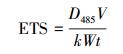
式中, ETS为电子传递效率(μg·mg-1·h-1);D485是在485 nm处的吸收值;V为反应后上清液的体积(mL);k为标准曲线的斜率(mL·μg-1);W为微生物干重(mg);t为反应时间(h).
2.3.3 Pd(Ⅱ)和Pd(0) NPs对微生物的影响通过抑菌圈实验来判断Pd(Ⅱ)对微生物生长的影响.取2.2.1节中的待用菌液稀释为105、106 CFU·mL-1, 涂布于LB琼脂平板上.将无菌滤纸裁剪成8 mm×8 mm的正方形, 然后分别滴上300 μL的Pd(Ⅱ)溶液(0.07、0.1、0.5、5 mmol·L-1), 分别平铺于平板中央.每个细菌浓度做3个平行实验.倒置培养皿, 放入恒温培养箱中, 在30 ℃下培养, 24 h后取出观察菌体生长情况及形成的抑菌圈大小.
采用激光共聚焦显微镜(Confocal Scanning Laser Microscopy, CSLM)来观测含不同量Pd(0) NPs的微生物活性.取一定合成反应前、后的微生物, 稀释至2×10⁶ CFU·mL-1, 用荧光试剂盒对细胞进行染色, 随后进行观察.
3 结果与讨论(Results and discussion) 3.1 合成生物纳米钯前后的P. putida表面形貌对比分析利用FESEM和TEM观察P. putida的微观形貌, 结果如图 1所示.从图中可以看出, P. putida是一类表面光滑的短杆菌, 无鞭毛, 长约为1~2 μm, 宽度约为0.5 μm.图 2b、d和f分别为2a、c和e的局部放大, 从图中清晰可见微生物边缘及周质空间中有均匀的黑色颗粒出现, 粒径约为10 nm.Bunge等(2010)的研究中观察到了类似的纳米颗粒或纳米颗粒聚合物, 最大粒径>500 μm, 可以印证本实验结果.
 |
| 图 1 P. putida的TEM(a)和SEM(b)图像 Fig. 1 TEM(a) and SEM(b) images of pure P. putida |
 |
| 图 2 含有不同量Pd(0) NPs的P. putida的TEM图像(3列图片分别来自初始反应Pd(Ⅱ)为0.07、0.1、0.5 mmol·L-1的体系;a、c、e.完整的含有Pd(0) NPs的微生物细胞TEM图像;b、d、f分别为a、c、e的相应放大TEM图像) Fig. 2 TEM images of the bacteria containing different amount of Pd(0) NPs |
为了证明Pd(0)的存在, 通过X射线光电子能谱分析(XPS)进行Pd的价态分析, 结果如图 3所示.图 3a为P. putida空白对照, 可以看到微生物中的C、N、O、P峰, 无钯.图 3b中出现了与3a中同样的C、N、O、P峰, 在插图中可以看到结合能在3d轨道的335.5 eV(3d5/2)和340.7 eV(3d3/2)两处出现, 为Pd(0)的特征峰, 这进一步确认了TEM图中所示的黑色颗粒为Pd(0), 说明通过微生物的还原, Pd(Ⅱ)变为零价态的钯.
 |
| 图 3 P. putida与Pd(Ⅱ)反应前(a)、后(b)的XPS全谱(b中插图为Pd的分峰谱图) Fig. 3 XPS of bacteria before(a) and after(b) the bioreduction of Pd(0) NPs(the illustration in b was the narrow spectrum of Pd(0)) |
利用CV分析可以清晰地体现出物质的氧化还原变化, 结果如图 4所示.由图 4a可知, P. putida在0.515 V(曲线2)处出现氧化峰, 随着初始反应Pd(Ⅱ)浓度由0.07 mmol·L-1上升到0.1 mmol·L-1, 明显的氧化峰出现在0.332 V(曲线3)和0.333 V(曲线4)处, 同时, 氧化电流由P. putida的1.101 μA上升到了2.193 μA(曲线3)和2.307 μA(曲线4).反向扫描的过程中, 在0.05 V处出现了微小的还原峰, 可能与细胞膜上的细胞色C的活动有关, P. putida的还原电流不明显(曲线2), 而初始反应Pd(Ⅱ)浓度为0.07 mmol·L-1和0.1 mmol·L-1时的还原电流分别为-0.7684 μA(曲线3)和-0.9023 μA(曲线4).说明Pd(0) NPs的存在对微生物内的氧化还原反应有强化作用.
 |
| 图 4 合成Pd(0) NPs前后的微生物的循环伏安曲线(a. 1:打磨干净的GC电极;2:P. putida;3:初始Pd(Ⅱ)为0.07 mmol·L-1;4:初始Pd(Ⅱ)为0.1 mmol·L-1;b.初始Pd(Ⅱ)为0.5 mmol·L-1) Fig. 4 Cyclic voltammograms of the bare GC electrode(1), washed pure P. putida coated on a GC electrode(2), cells(palladium-loaded) coated on a GC electrode after reacted with 0.07 and 0.1 mmol·L-1 initial Pd(Ⅱ)(3、4)(a) and cyclic voltammogram of cells(palladium-loaded) coated on a GC electrode after reacted with 0.5 mmol·L-1 initial Pd(Ⅱ)(b) |
然而, 溶液浓度升高至0.5 mmol·L-1时, 0.426 V处的催化电流为8.316 μA(图 4b), 高于含较低量Pd(0) NPs的微生物的情况, 并且还原电流迅速升高, 说明过量的Pd(0) NPs占据了大部分细胞周质空间, 其导电作用占据主导.Wu等(2011)的研究中, D. desulfuricans还原合成Pd(0)的CV曲线有类似结果.
3.3.2 I-t结果将与不同浓度Pd(Ⅱ)进行合成反应后的微生物用于恒电位实验, 电流变化的结果如图 5和表 1所示.加入乳酸之后电流的变化越大, 表示微生物对电子供体的响应越大, 说明微生物电子传递活性越大.在对照实验中, 空白GC电极和只有钯时, 加入乳酸之后电流并未出现大幅上升.含Pd(0) NPs的菌, 加入乳酸以后的上升电流可达(10.6±5.2) nA(反应初始Pd(Ⅱ)溶液为0.07 mmol·L-1), 并且随着钯的量增加为0.1 mmol·L-1, 稳定电流可继续上升至(22.5±9.7) nA, 二者分别是P. putida((5.7±2.8) nA)的约1.85~3.95倍, Windt等(2005)证明了生物纳米钯具有高的催化活性, 可能正是这一高活性促进了电子的流动.而当Pd(Ⅱ)上升至0.5 mmol·L-1时, 乳酸氧化电流无明显提高, 可能是因为过大的Pd(0) NPs负载量影响了微生物膜上的酶反应过程和微生物的电子传递活性.
 |
| 图 5 恒电位实验中加入乳酸后电流随时间的变化 Fig. 5 Lactate oxidation current changes in potentiostatic experiment |
| 表 1 恒电位实验中含不同量Pd(0) NPs的微生物的电流增加结果 Table 1 Summary of potentiostatic data for various working electrodes |
为了测试菌株的新陈代谢活性、电子传递活性, 选用指标TTC-ETS和INT-ETS, 通过人工电子受体TTC和INT得电子被还原的程度来表示菌体的电子传递活性, 进而体现出微生物的活性.实验结果如图 6所示, 随着Pd(Ⅱ)的浓度不断上升, ETS先增后减, 在0.1 mmol·L-1时最高, TTC-ETS可达89.02 μg·mg-1·h-1, INT-ETS为209.09 μg·mg-1·h-1, 相对于P. putida分别上升了63.97%和33.79%, 说明Pd(0) NPs促进了微生物的电子传递过程.
 |
| 图 6 合成Pd(0) NPs前后微生物ETS变化 Fig. 6 Changes of the two ETS values after the bioreduction of Pd(0) NPs |
进一步探究实验贵金属离子和纳米颗粒对菌株的影响, 进行了抑菌圈实验和激光共聚焦的表征.图 7显示了不同浓度Pd(Ⅱ)对P. putida菌落的抑菌表现, 观察可知, Pd(Ⅱ)浓度为0.07和0.1 mmol·L-1时, 在105和106 CFU·mL-1的P. putida菌落中皆无抑菌圈;Pd(Ⅱ)浓度提升至0.5 mmol·L-1时, 105 CFU·mL-1的P. putida在滤纸的下侧略有一点抑菌圈, 但当菌落增加为106 CFU·mL-1时, 无抑菌圈, 说明0.5 mmol·L-1的Pd(Ⅱ)有极弱抑菌能力.上述结果表明, 较低量的Pd(Ⅱ)进入微生物内并不会引起微生物失活, Li等(2013)证明革兰氏阴性菌对0.2 mmol·L-1的Pd(NO3)2有一定生物相容性, 这一结论可以印证以上实验结果.
 |
| 图 7 抑菌圈图 Fig. 7 Typical photographs of inhibition zone |
图 8为含不同量Pd(0) NPs的P. putida的CSLM图像.从图 8a、b可以看出, 当初始Pd(Ⅱ)反应浓度为0.07和0.1 mmol·L-1时, 图像呈绿色, 表明微生物大都为活菌.当初始Pd(Ⅱ)浓度上升到0.5 mmol·L-1时, 图像中只有星点的红色(图 8c), 说明该浓度下菌内含的Pd(0) NPs有极微弱的杀菌性.Li等(2014)进一步研究表明, 胞内的Pd(0) NPs可能会触发磷酸苏氨酸裂解酶等胞内蛋白的合成, 可以调节微生物的功能, 与此类似, 微量Pd(0) NPs也可能使P. putida中某些蛋白的功能得以改变, 从而得到更优良的活性.
 |
| 图 8 与不同浓度Pd(Ⅱ)反应后菌的CLMS图(a.0.07 mmol·L-1, b.0.1 mmol·L-1, c. 0.5 mmol·L-1, d. 5 mmol·L-1) Fig. 8 CLSM images of microbial after reacted with different concentration of Pd(Ⅱ) (a.0.07 mmol·L-1, b.0.1 mmol·L-1, c. 0.5 mmol·L-1, d. 5 mmol·L-1) |
另外, 5 mmol·L-1 Pd(Ⅱ)下可看到明显抑菌圈, 且初始Pd(Ⅱ)浓度为5 mmol·L-1合成反应后的CSLM图像(图 8d)呈现出明显的红色, 说明大量Pd(Ⅱ)和Pd(0) NPs都有一定抗菌性.Staszek等(2014)发现11.8 mg·mL-1 (约110.88 mmol·L-1)的Pd(0) NPs对革兰氏阴性菌E. coli和革兰氏阳性的S. epidermidis都有抗菌作用, 这印证了5 mmol·L-1实验组得出的结论.
4 结论(Conclusion)1) P. putida合成Pd(0) NPs步骤简单且效果良好, 最终合成的颗粒大小均一且均匀分布在细胞周质中, 粒径约为10 nm.
2) 通过Pd(0) NPs的生物合成, 微生物的CV氧化还原电流变大, 恒电位实验中的乳酸氧化电流最大提高了3.95倍;电子传递速率TTC-ETS和INT-ETS分别提升至89.02和9.09 μg·mg-1·h-1, 相对于P. putida分别增加了63.97%和33.79%, 说明微量Pd(0) NPs的存在有利于提升微生物电化学活性.
3) 低浓度Pd(Ⅱ)和微量Pd(0) NPs对微生物几乎没有杀菌作用, 但过高量的Pd(Ⅱ)和Pd(0) NPs均可对微生物产生毒性, 为了保证微生物的活性, bio-Pd(0) NPs的反应前体金属离子浓度不宜过高.本研究有利于推动生物金属NPs与微生物结合降解污染物等方面的综合利用, 当微生物活性得以保持, 可以同步利用钯的催化性能和微生物降解有机物的特性, 如卤代污染物的钯催化脱卤接着被微生物彻底降解;低量金属NPs在微生物细胞周质和周围的存在, 可促进微生物与污染物之间的电子传递进而促进污染物的降解等.
Bunge M, Søbjerg L S, Rotaru A E, et al. 2010. Formation of palladium(0) nanoparticles at microbial surfaces[J]. Biotechnology & Bioengineering, 2(107): 206–216.
|
Guerra W, De A A E, De A R, et al. 2005. Synthesis, characterization, and antibacterial activity of three palladium(Ⅱ) complexes of tetracyclines[J]. Journal of Inorganic Biochemistry, 9(12): 2348–2354.
|
Ha C, Zhu N, Shang N, et al. 2016. Biorecovery of palladium as nanoparticles by Enterococcus faecalis and its catalysis for chromate reduction[J]. Chemical Engineering Journal, 288(s1/2): 246–254.
|
Harrad S, Robson M, Hazrati S, et al. 2007. Dehalogenation of polychlorinated biphenyls and polybrominated diphenyl ethers using a hybrid bioinorganic catalyst[J]. Journal of Environmental Monitoring, 9: 3124–318.
|
Hou Y N, Liu H, Han J L, et al. 2016. Electroactive biofilm serving as the green synthesizer and stabilizer for in situ fabricating 3D nanopalladium network:An efficient electrocatalyst[J]. Acs Sustainable Chemistry & Engineering, 4(10): 5392–5397.
|
Ito R, Kuroda K, Hashimoto H, et al. 2016. Recovery of platinum(0) through the reduction of platinum ions by hydrogenase-displaying yeast[J]. AMB Express, 6(1): 88.
DOI:10.1186/s13568-016-0262-4
|
Kenner R A, Ahmed S I. 1975. Measurements of electron transport activities in marine phytoplankton[J]. Marine Biology, 33(2): 119–127.
DOI:10.1007/BF00390716
|
李红. 2010. 环境微生物活性表征的测定方法[J]. 温州农业科技, 2010(2): 25–27.
|
Li J, Yu J, Zhao J, et al. 2014. Palladium-triggered deprotection chemistry for protein activation in living cells[J]. Nature Chemistry, 6(4): 352.
DOI:10.1038/nchem.1887
|
Li J, Lin S, Wang J, et al. 2013. Ligand-free palladium-mediated site-specific protein labeling inside gram-negative bacterial pathogens[J]. Journal of the American Chemical Society, 135(19): 7330–7338.
DOI:10.1021/ja402424j
|
Lv Y, Niu Z, Chen Y, et al. 2017a. Synthesis of SiO2 coated zero-valent iron/palladium bimetallic nanoparticles and their application in a nano-biological combined system for 2, 2', 4, 4'-tetrabromodiphenyl ether degradation[J]. RSC Advance, 6(24): 20357–20365.
|
Lv Y, Niu Z, Chen Y, et al. 2017b. Bacterial effects and interfacial inactivation mechanism of nZVI/Pd on Pseudomonas putida strain[J]. Water Research, 115: 297–308.
DOI:10.1016/j.watres.2017.03.012
|
Mertens B, Blothe C, Windey K, et al. 2007. Biocatalytic dechlorination of lindane by nano-scale particles of Pd(0) deposited on Shewanella oneidensis[J]. Chemosphere, 66(1): 99.
DOI:10.1016/j.chemosphere.2006.05.018
|
Packard T T. 1971. The measurement of respiratory electron transport activity in marine plankton[J]. Journal of Marine Research, 29: 235–244.
|
Rotaru A E, Jiang W, Finster K, et al. 2012. Non-enzymatic palladium recovery on microbial and synthetic surfaces[J]. Biotechnology & Bioengineering, 109(8): 1889–1897.
|
Sathish K K, Amutha R, Arumugam P, et al. 2011. Synthesis of gold nanoparticles:an ecofriendly approach using Hansenula anomala[J]. Acs Applied Materials & Interfaces, 3(5): 1418–1425.
|
Shirakawa M A, John V M, Mocelin A, et al. 2016. Effect of silver nanoparticle and TiO2 coatings on biofilm formation on four types of modern glass[J]. International Biodeterioration & Biodegradation, 108: 175–180.
|
Søbjerg L S, Lindhardt A T, Skrydstrup T, et al. 2011. Size control and catalytic activity of bio-supported palladium nanoparticles[J]. Colloids & Surfaces B Biointerfaces, 85(2): 373–378.
|
Staszek M, Siegel J, KoláROvá K, et al. 2014. Formation and antibacterial action of Pt and Pd nanoparticles sputtered into liquid[J]. Micro & Nano Letters, 9(11): 778–781.
|
Windt W D, Aelterman P, Verstraete W, et al. 2005. Bioreductive deposition of palladium(0) nanoparticles on Shewanella oneidensis with catalytic activity towards reductive dechlorination of polychlorinated biphenyls[J]. Environmental Microbiology, 7(3): 314.
DOI:10.1111/emi.2005.7.issue-3
|
Wu X, Zhao F, Rahunen N, et al. 2011. A role for microbial palladium nanoparticles in extracellular electron transfer[J]. Angewandte Chemie International Edition, 50(2): 427–430.
DOI:10.1002/anie.201002951
|
Yang C, Jung S, Yi H, et al. 2014. A biofabrication approach for controlled synthesis of silver nanoparticles with high catalytic and antibacterial activities[J]. Biochemical Engineering Journal, 89(8): 10–20.
|
杨居荣, 葛家璊, 张美庆, 等. 1982. 砷及重金属对土壤微生物的影响[J]. 环境科学学报, 1982, 2(3): 190–198.
|
尹军, 吴相会, 王晓玲, 等. 2008. 用TTC-ETS和INT-ETS表征微生物活性[J]. 中国给水排水, 2008, 24(7): 8–11.
|
Zheng G, Zhao F, Yang H, et al. 2011. Comparison between INT and TTC assay to determine the dehydrogenase activity of flocs[C]. International Symposium on Water Resource and Environmental Protection IEEE. 3: 1690-1693
|