急性胰腺炎是临床上常见的消化系统疾病,轻症急性胰腺炎不伴有器官功能障碍及局部或全身并发症,常规治疗即可恢复。重症急性胰腺炎伴有持续性器官功能障碍,即使积极救治病死率仍高达13%~35%[1]。对于重症急性胰腺炎的早期评估至关重要,但目前没有最佳的评估方法。基于临床症状及实验室指标的评分系统对患者病情严重程度及预后进行分析、评估都存在一定的局限性[2]。CT是目前临床上不可或缺的辅助检查方法,能及早发现胰腺周围坏死组织[2-3]。有相关研究显示,胰周坏死体积(peripancreatic necrosis volume,PNV)在预测重症急性胰腺炎患者住院时间及多脏器功能不全上具有一定意义,可能是预测急性胰腺炎并发症的良好指标[4-5],但未将结果与经典的临床评分系统如急性生理学和慢性健康状况评价Ⅱ(acute physiology and chronic health evaluationⅡ,APACHEⅡ)或Ranson评分等进行比较。本文通过回顾性分析150例急性胰腺炎患者的资料,探究PNV与急性胰腺炎患者并发症和临床结局的相关性,评估其对急性胰腺炎严重程度的预测价值。
1 资料和方法 1.1 研究对象回顾性分析2020年1月至2021年6月在海军军医大学(第二军医大学)第一附属医院住院的150例急性胰腺炎患者资料。纳入标准:(1)发病48 h内入院行胰腺增强CT检查;(2)依据2012年修正的亚特兰大标准确诊急性胰腺炎[6];(3)年龄≥18岁。排除标准:(1)患者处于妊娠或哺乳期;(2)患者入院前已接受过治疗或临床资料不完整;(3)胰腺肿瘤、慢性胰腺炎急性发作、创伤性胰腺炎患者。本研究通过海军军医大学(第二军医大学)第一附属医院科研和临床试验伦理委员会审批。
1.2 观察指标人口统计学信息、临床数据、住院时间和分析参数均收集自医院临床信息数字化系统。收集入组患者的性别、年龄、病因、实验室和影像学资料。预后参数包括器官功能不全、多器官功能衰竭(multiple organ failure,MOF)、住院时间、入住ICU,以及住院期间的并发症、感染坏死、需要手术或介入干预等情况。
1.3 影像学检查所有患者均于发病后48 h内行胰腺增强CT检查(德国西门子双源64排CT)。患者取仰卧位,经肘静脉用双筒高压注射器以3 mL/s速率注射碘伏醇注射液80~90 mL。扫描期相:平扫期、动脉期、实质期、延迟期,观察并记录患者CT图像。使用Philips-IntelliSpace工作站手动测量动脉期PNV(图 1),通过软件程序自动计算PNV(单位为cm3),包括胰腺外坏死体积、胰腺周围或相关腹膜后坏死组织、脂肪组织炎症以及液体和固体成分,而腹膜液不包括在测量中。测量由2名经验丰富的放射科医师进行,他们对患者的临床参数其他结果一无所知。同时计算CT严重度指数(computed tomography severity index,CTSI)评分,包括急性胰腺炎分级和胰腺坏死程度,其中Ⅰ级0~3分、Ⅱ级4~6分、Ⅲ级7~10分,4分以上为重症[7]。

|
图 1 使用Philips-IntelliSpace工作站手动测量PNV Fig 1 Measuring PNV manually using Philips-IntelliSpace Portal A 55-year-old male patient with acute pancreatitis. Computed tomography images showed peripancreatic fat infiltration and pancreatic necrosis. A-C: Transverse position (A), coronal position (B), and sagittal position (C) images showed that pancreatic necrosis was depicted manually with the help of semi-automatic tool of tissue density; D: The 3-dimensional volume of the whole pancreas automatically segmented. |
1.4 临床评分系统
基于临床特征、实验室参数,APACHE Ⅱ评分=急性生理评分+年龄评分+慢性健康状况评分[8]。Ranson评分:包括入院时的5项临床指标和48 h的6项指标,每项0~1分,总分0~11分,分值越高表示病情越严重[9]。改良Marshall评分:包括呼吸、肾脏、心血管功能3个项目,总分0~12分,分值越高表示病情越严重[10]。Balthazar评分:将胰腺及胰周CT平扫表现分为5级,其中A~C级为轻症急性胰腺炎,D~E级为重症急性胰腺炎[11]。急性胰腺炎严重程度床边指数(bedside index for severity of acute pancreatitis,BISAP)评分:包括血尿素氮、意识障碍、系统性炎症反应综合征、年龄、胸腔积液等5项,总分0~5分,分值越高表示病情越严重[12]。
1.5 统计学处理应用SPSS 26.0软件进行统计分析。计量资料以x±s表示;计数资料以例数(百分率)表示;相关性检验采用Pearson相关分析。采用ROC曲线比较变量的诊断价值,AUC的两两比较采用Z检验。检验水准(α)为0.05。
2 结果 2.1 患者基本特征本研究共纳入急性胰腺炎患者150例,其中男94例(62.7%)、女56例(37.3%),年龄19~80岁,平均年龄(50.1±15.4)岁。包括胆源性胰腺炎50例(33.3%),脂源性胰腺炎48例(32.0%),暴饮暴食引发胰腺炎24例(16.0%),其他因素所致28例(18.7%)。
2.2 病情预后参数及临床评分系统结果150例急性胰腺炎患者的住院时间为2~146(20.53±22.13)d,其中器官功能不全患者54例(36.0%)、感染患者48例(32.0%)、MOF患者26例(17.3%)、入住ICU患者66例(44.0%)、手术患者26例(17.3%),48 h-CRP为(181.7±114.5)mg/L,Ranson评分为(3.61±1.86)分,BISAP评分为(2.01±1.04)分,改良Marshall评分为(2.35±1.35)分,APACHE Ⅱ评分为(7.16±4.08)分。
2.3 病情CT评分及PNVBalthazar评分结果显示,A级5例(3.3%)、B级16例(10.7%)、C级42例(28.0%)、D级55例(36.7%)、E级32例(21.3%)。CTSI评分结果显示,Ⅰ级31例(20.7%)、Ⅱ级72例(48.0%)、Ⅲ级47例(31.3%)。手动测量PNV为20.0~1 517.5(539.5±413.4)cm3。
2.4 PNV与住院时间的相关性Pearson相关性分析结果显示,PNV与住院时间呈正相关(相关系数为0.462,P<0.05)。
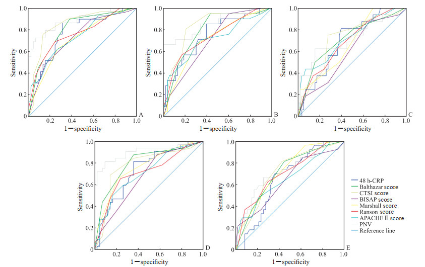
2.5 PNV与临床评分系统、影像学评分、实验室指标预测临床结局的比较ROC曲线比较PNV与APACHE Ⅱ评分、改良Marshall评分、Ranson评分、BISAP评分、Balthazar评分、CTSI评分及48 h-CRP对急性胰腺炎患者临床结局的预测价值,结果显示,PVN预测感染、并发症、MOF、入住ICU、器官功能不全的AUC值分别为0.73(95% CI0.60~0.85)、0.72(95% CI 0.57~0.88)、0.86(95% CI 0.76~0.97)、0.90(95% CI 0.82~0.98)、0.88(95% CI 0.80~0.97),灵敏度分别为0.72、0.67、0.67、0.63、0.81,特异度分别为0.99、0.98、0.75、0.81、0.91。与各评分系统及48 h-CRP相比,PNV是预测急性胰腺炎患者器官功能不全、MOF、入住ICU、感染等的最佳参数。APACHE Ⅱ评分对需要手术或介入干预的并发症有较好的预测价值(AUC值为0.73)。见图 2、表 1。

|
图 2 PNV、48 h-CRP及6种评分系统预测急性胰腺炎患者临床结局的ROC曲线 Fig 2 ROC curves of PNV, 48 h-CRP, and 6 scoring systems for predicting clinical outcomes of patients with acute pancreatitis A: Organ dysfunction; B: Multiple organ failure; C: Complication; D: Intensive care unit admission; E: Infection. PNV: Peripancreatic necrosis volume; CRP: C reactive protein; ROC: Receiver operating characteristic; CTSI: Computed tomography severity index; BISAP: Bedside index for severity of acute pancreatitis; APACHEⅡ: Acute physiological and chronic health evaluation Ⅱ. |
|
|
表 1 PNV、48 h-CRP及6种评分系统预测急性胰腺炎患者临床结局的AUC (95% CI) Tab 1 AUC (95% CI) of PNV, 48 h-CRP, and 6 scoring systems in predicting clinical outcomes of patients with acute pancreatitis |
3 讨论
急性胰腺炎是急诊科常见的疾病,也是导致器官功能障碍和病死率较高的疾病之一[13]。不同严重程度的急性胰腺炎病死率有明显差异,早期积极有效治疗可明显改善预后。鉴于此,早期预测重症急性胰腺炎至关重要。目前临床上评估急性胰腺炎病情严重程度的评分系统有APACHEⅡ、BISAP、Ranson、改良Marshall评分等,但效果均不理想[6, 14]。CRP是一个重要的炎症指标,发病后24~48 h升高使其在早期评估方面价值有限[15]。CT检查为当前简单、有效的检查方法,不仅扫描速度快、空间分辨率高,还能够判断疾病的严重程度和类型,并进行定性和定量分析[16]。有研究认为CTSI评分不能反映全身炎症反应状况,对器官衰竭、死亡的预测价值较低[17-18]。
胰腺局部坏死程度增加、周围渗出扩大可加重对患者全身各器官系统的影响,就诊时胰腺及其周围组织出现坏死迹象提示患者可能会发展为重症急性胰腺炎[19-20]。本研究结果显示,与常用的影像学评分CTSI或Balthazar评分相比,PNV为预测急性胰腺炎患者器官功能不全、MOF、入住ICU、感染等的最佳参数。此外,与其他临床评分系统或48 h-CRP相比,PNV预测器官功能不全、MOF、入住ICU、感染等预后指标的效能也更高。这表明PNV在预测急性胰腺炎严重程度方面是可靠和准确的。PNV也与住院时间呈正相关(相关系数为0.462,P<0.05)。Çakar等[21]研究表明,PNV可有效预测器官衰竭及感染,BISAP、Ranson、改良Marshall评分与患者器官功能不全及MOF具有明显的相关性,但没有将PNV的预测效能与经典的临床系统评分如APACHE Ⅱ评分或Ranson评分进行比较。
APACHE Ⅱ评分作为一个非特异性病情严重程度评价指标,被多项指南推荐为疾病严重程度评分指标,在预测急性胰腺炎患者预后中具有一定的临床参考价值,其预测灵敏度高但特异度较差。本研究结果显示APACHE Ⅱ评分对需要手术或介入干预的并发症有较好的预测价值。但PNV预测器官功能不全、MOF、入住ICU、感染等方面优于APACHE Ⅱ评分及其他方法。
PNV越大的患者发生器官功能不全及入住ICU、需要介入和手术干预的可能更大,住院时间也更长,故其预后较差。PNV作为影响患者预后的独立危险因素,其诊断效能高于Ranson评分及BISAPI评分。计算机可以简单、快速地量化胰腺坏死性炎症导致组织坏死的体积。因此,PNV是一个可以在急性胰腺炎CT检查中常规和前瞻性计算的参数。虽然腹部CT检查存在一定的辐射(剂量大约在8~20 mSv),但通过CT检查能在疾病早期对患者进行诊断和评估以便及早采取治疗措施,可以避免患者出现严重的并发症。
综上所述,PNV与急性胰腺炎患者病情严重程度相关,可对急性胰腺炎患者预后进行有效评估,具有较高的预测价值。本研究也存在一些局限性。首先,本研究为回顾性研究,PNV对重症急性胰腺炎的预测价值有待更大样本、多中心的前瞻性研究进一步检验。其次,未纳入未行胰腺增强CT检查的患者进行分析,减少了样本量,增加了研究的局限性。另一方面,虽然ROC曲线显示PNV的预测效能更高,但因较严重并发症病例数较少,可能存在选择偏倚,影响研究结果的可靠性。
| [1] |
刘建, 李非. 急性胰腺炎患者的诊治及预后[J]. 中华肝胆外科杂志, 2016, 22(10): 714-718. DOI:10.3760/cma.j.issn.1007-8118.2016.10.018 |
| [2] |
蔡兆辉, 左爽, 李海山, 等. BISAP和CTSI评分变化用于判断急性胰腺炎患者病情严重程度的临床价值[J]. 解放军预防医学杂志, 2019, 37(2): 90-92. DOI:10.13704/j.cnki.jyyx.2019.02.033 |
| [3] |
李应. 重症急性胰腺炎与CT严重度指数的关系研究[J]. 影像研究与医学应用, 2017, 1(13): 67-68. DOI:10.3969/j.issn.2096-3807.2017.13.040 |
| [4] |
MEYRIGNAC O, LAGARDE S, BOURNET B, et al. Acute pancreatitis: extrapancreatic necrosis volume as early predictor of severity[J]. Radiology, 2015, 276(1): 119-128. DOI:10.1148/radiol.15141494 |
| [5] |
PAMIES-GUILABERT J, DEL VAL ANTOÑANA A, COLLADO J J, et al. Pancreatic necrosis volume-a new imaging biomarker of acute pancreatitis severity[J]. Eur J Radiol, 2020, 130: 109193. DOI:10.1016/j.ejrad.2020.109193 |
| [6] |
BANKS P A, BOLLEN T L, DERVENIS C, et al. Classification of acute pancreatitis-2012:revision of the Atlanta classification and definitions by international consensus[J]. Gut, 2013, 62(1): 102-111. DOI:10.1136/gutjnl-2012-302779 |
| [7] |
ALBERTI P, PANDO E, MATA R, et al. Evaluation of the modified computed tomography severity index (MCTSI) and computed tomography severity index (CTSI) in predicting severity and clinical outcomes in acute pancreatitis[J]. J Dig Dis, 2021, 22(1): 41-48. DOI:10.1111/1751-2980.12961 |
| [8] |
KNAUS W A, DRAPER E A, WAGNER D P, et al. APACHE Ⅱ: a severity of disease classification system[J]. Crit Care Med, 1985, 13(10): 818-829. DOI:10.1097/00003246-198510000-00009 |
| [9] |
RANSON J H, RIFKIND K M, ROSES D F, et al. Objective early identification of severe acute pancreatitis[J]. Am J Gastroenterol, 1974, 61(6): 443-451. |
| [10] |
SINGH V K, BOLLEN T L, WU B U, et al. An assessment of the severity of interstitial pancreatitis[J]. Clin Gastroenterol Hepatol, 2011, 9(12): 1098-1103. DOI:10.1016/j.cgh.2011.08.026 |
| [11] |
宋彬, 徐隽. 急性胰腺炎的CT评价[J]. 中国普外基础与临床杂志, 2003, 10(6): 623-626. |
| [12] |
CHANDRA S, MURALI A, BANSAL R, et al. The bedside index for severity in acute pancreatitis: a systematic review of prospective studies to determine predictive performance[J]. J Community Hosp Intern Med Perspect, 2017, 7(4): 208-213. DOI:10.1080/20009666.2017.1361292 |
| [13] |
SELLERS Z M, BARAKAT M T, ABU-EL-HAIJA M. A practical approach to management of acute pancreatitis: similarities and dissimilarities of disease in children and adults[J]. J Clin Med, 2021, 10(12): 2545. DOI:10.3390/jcm10122545 |
| [14] |
BOLLEN T L, SINGH V K, MAURER R, et al. A comparative evaluation of radiologic and clinical scoring systems in the early prediction of severity in acute pancreatitis[J]. Am J Gastroenterol, 2012, 107(4): 612-619. DOI:10.1038/ajg.2011.438 |
| [15] |
任丽楠, 郭晓钟, 李宏宇, 等. C-反应蛋白在重症急性胰腺炎评估与转归中的临床意义[J]. 临床肝胆病杂志, 2015, 31(5): 691-693. DOI:10.3969/j.issn.1001-5256.2015.05.014 |
| [16] |
EVR MLER, ÇAKMAKÇI M, KARA BRAH MO LU A, et al. The prognostic value of fat necrosis deposits on CT imaging in acute pancreatitis[J]. Turk J Med Sci, 2021, 51(2): 749-756. DOI:10.3906/sag-1910-31 |
| [17] |
YADAV J, YADAV S K, KUMAR S, et al. Predicting morbidity and mortality in acute pancreatitis in an Indian population: a comparative study of the BISAP score, Ranson's score and CT severity index[J]. Gastroenterol Rep (Oxf), 2016, 4(3): 216-220. DOI:10.1093/gastro/gov009 |
| [18] |
CASAS J D, DÍAZ R, VALDERAS G, et al. Prognostic value of CT in the early assessment of patients with acute pancreatitis[J]. AJR Am J Roentgenol, 2004, 182(3): 569-574. DOI:10.2214/ajr.182.3.1820569 |
| [19] |
急性胰腺炎协作组. 中国6 223例急性胰腺炎病因及病死率分析[J]. 胰腺病学, 2006, 6(6): 321-325. |
| [20] |
于齐宏, 杜奕奇, 李兆申. 急性胰腺炎的治疗[J]. 中华胰腺病杂志, 2013, 13(1): 55-60. DOI:10.3760/cma.j.issn.1674-1935.2013.01.018 |
| [21] |
ÇAKAR, KEVEN A, ESERO LU E, et al. Role of extrapancreatic necrosis volume in determining early prognosis in patients with acute pancreatitis[J]. Abdom Radiol (NY), 2020, 45(5): 1507-1516. DOI:10.1007/s00261-019-02188-9 |
 2023, Vol. 44


