青少年特发性脊柱侧凸(adolescent idiopathic scoliosis,AIS)是一种三维畸形[1-3],特别是水平面椎体位置和旋转异常。目前已提出多种椎体水平面旋转的评价方法,其中Nash-Moe法和Perdriolle法最常用。近年一项新的骨骼成像技术——EOS全身骨骼成像系统投入临床使用[4]。对于AIS患者,该成像系统的优势是通过对EOS系统摄片得到的正侧位图像进行三维重建,准确地获得脊柱侧凸畸形的三维影像学参数,如冠状面三维Cobb角、矢状面三维胸椎后凸角(thoracic kyphosis,TK)和水平面的椎体旋转角度。鲍虹达等[5]指出,在控制冠状面畸形的条件下,矢状面TK较小的AIS患者主胸弯水平面椎体旋转角度显著大于TK正常的AIS患者。Chiari畸形患者通常伴有脊柱畸形,该类患者中矢状面TK与主胸弯水平面椎体旋转角度的关系目前仍不清楚。本研究选择冠状面形态匹配(主胸弯弯型相似且Cobb角大小相似)而矢状面TK存在较大差异的AIS患者和Chiari畸形合并脊柱侧凸患者作为研究对象,探讨Chiari畸形合并脊柱侧凸患者TK与椎体旋转的相关性。
1 资料和方法 1.1 研究对象回顾性选择2017年7月至2019年7月于南京大学医学院附属鼓楼医院进行EOS脊柱影像摄片的Chiari畸形伴脊柱侧凸患者30例和AIS患者34例。纳入标准:(1)MRI检查确诊Chiari畸形或AIS;(2)冠状面主胸弯脊柱侧凸Cobb角为20°~90°。排除标准:(1)伴先天性椎体分节不良、双下肢不等长或髋关节疾病,或既往有脊柱骨折病史等患者;(2)TK>20°的AIS患者,TK<30°的Chiari畸形伴脊柱侧凸患者;(3)影像学资料不完整的患者。本研究通过南京大学医学院附属鼓楼医院伦理委员会审批。
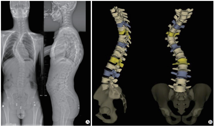
1.2 影像学测量获取入组患者的EOS影像摄片图像,由1名接受过EOS技术培训的脊柱外科住院医师对图像进行三维重建。三维重建方法:勾勒并调整脊柱弯曲曲线,按顺序调整各椎体在侧位X线片上的位置、高度及在正位X线片上的位置和椎体旋转方向,然后调整T1~L5椎体的上下终板、前后缘及椎弓根位置,最后由软件自动生成脊柱三维图像(图 1)。

|
图 1 1例Chiari畸形伴脊柱侧凸患者的EOS全脊柱正侧位X线片(A)及EOS三维重建图像(B) Fig 1 Anteroposterior and lateral radiographs of the full spine (A) and EOS three-dimensional reconstruction images (B) of a patient with Chiari malformation and scoliosis |
完成三维重建后用sterEOS软件自动测量患者的冠状面主胸弯Cobb角、次胸弯Cobb角、顶椎节段、椎体旋转角(左旋为正)等冠状面参数和T1~T12 TK、T4~T12 TK等矢状面参数,然后计算主胸弯平均椎体旋转角度(mean vertebral rotation,MTR),MTR计算方法为主弯胸区各椎体旋转角度之和除以椎体个数。
1.3 研究分组将入组患者分为AIS组和Chiari畸形伴脊柱侧凸组,根据冠状面形态相同或相似即主胸弯弯型和Cobb角大小相同或相似而矢状面TK不同将两组患者进行配对,以冠状面主胸弯Cobb角及顶椎节段为主要配对指标,配对原则为患者Cobb角相差≤5°、顶椎节段相差≤1个椎体。匹配的每对患者仅TK差异显著,AIS组患者TK≤20°,Chiari畸形伴脊柱侧凸组患者TK≥30°(图 2~4)。

|
图 2 1组配对的Chiari畸形伴脊柱侧凸患者和AIS患者的Cobb角和TK Fig 2 2 Cobb angle and TK in a matching group of a patient with Chiari malformation and scoliosis and a patient with AIS A: Male, 14 years old, Chiari malformation with scoliosis, coronal Cobb angle of 60° and TK (T4-T12) of 37° on the main curvature; B: Female, 12 years old, AIS, coronal Cobb angle of 65° and TK (T4-T12) of 10°. AIS: Adolescent idiopathic scoliosis; TK: Thoracic kyphosis |

|
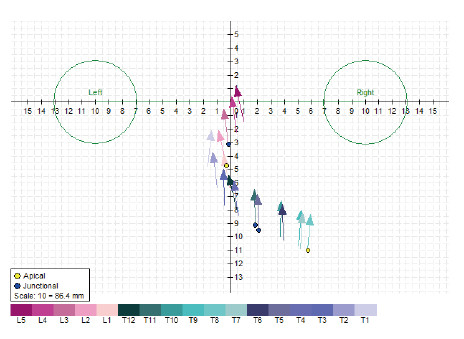
图 3 sterEOS软件给出的Chiari畸形伴脊柱侧凸患者椎体旋转向量图 Fig 3 Vertebral body rotation vector diagram of a patient with Chiari malformation and scoliosis given by sterEOS software |

|
图 4 sterEOS软件给出的AIS患者椎体旋转向量图 Fig 4 Vertebral body rotation vector diagram of a patient with AIS given by sterEOS software AIS: Adolescent idiopathic scoliosis |
1.4 统计学处理
应用SPSS 20.0软件进行数据分析。计量资料以x±s表示,组间比较采用配对t检验;计数资料以例数表示,组间比较采用χ2检验。采用Pearson相关分析研究AIS及Chiari畸形伴脊柱侧凸患者TK和主弯区MTR的相关性。检验水准(α)为0.05。
2 结果共24例(12对)患者入组,AIS组男4例、女8例,Chiari畸形伴脊柱侧凸组男5例、女7例,两组间性别构成差异无统计学意义(χ2=0.00,P>0.05)。AIS组和Chiari畸形伴脊柱侧凸组患者年龄分别为12~18(15.25±2.00)岁、11~18(14.42±2.43)岁,两组患者年龄差异无统计学意义(t=0.96,P=0.36)。AIS组和Chiari畸形伴脊柱侧凸组患者的冠状面主胸弯Cobb角分别为(58.13±11.45)°和(55.35±12.35)°,两组差异无统计学意义(t=2.20,P>0.05)。AIS组TK为(13.89±6.35)°,小于Chiari畸形伴脊柱侧凸组[(37.11±9.40)°],两组差异有统计学意义(t=-6.38,P<0.01)。利用三维重建图像测得AIS组和Chiari畸形伴脊柱侧凸组患者的MTR(绝对值)分别为(24.62±2.78)°和(21.53±1.66)°,差异有统计学意义(t=3.94,P=0.002)。Pearson相关分析显示,AIS组和Chiari畸形伴脊柱侧凸组患者TK均与MTR呈负相关(r=-0.667、-0.645,P=0.018、0.024)。
3 讨论脊柱侧凸作为一种三维畸形已得到广泛认同。秦乐等[6]认为脊柱侧凸顶椎水平面旋转角度与冠状面Cobb角之间存在良好的正相关性。南京大学医学院附属鼓楼医院的一项研究发现,对于AIS患者,其TK减小与主弯区椎体的水平面旋转相关[5],这一观点可指导相应的手术策略。关于AIS患者TK减小的原因目前尚存在争议。根据Hueter-Volkmann定律,由于椎体受力不对称,椎体前柱应力减小而后柱应力增大,可导致前柱过度生长从而出现TK减小[7]。Newton等[8]发现这一矢状面异常应力状态可能由椎体旋转引起。这也解释了本研究中AIS组椎体旋转较大而Chiari畸形伴脊柱侧凸组椎体旋转较小的现象。
Abelin-Genevois等[9]研究认为AIS患者二维侧视图像是由三维畸形的投影产生,因此传统的二维矢状面评估缺乏准确性[10],尤其椎体间旋转较大时。Newton等[8]借助EOS成像系统对AIS患者的脊椎进行三维重建,并通过软件模拟去旋转,结果显示排除旋转影响后的真实TK(三维TK)比二维矢状面测量的TK更小。而Burkus等[11]通过对中欧人群的对照研究发现,AIS患者TK明显减小,TK减小幅度与Cobb角直接相关。盆腔入射角的变化很小,但也与胸椎的变化相关,Sullivan等[12]发现去旋转后的三维胸椎后凸与冠状面主胸弯Cobb角相关。这一结果提示在冠状面畸形存在的情况下,TK和主弯区椎体旋转互相影响,这解释了本研究中在相同冠状面形态下Chiari畸形伴脊柱侧凸组主弯区旋转较AIS组小的现象。
由于EOS成像系统在我国应用时间较短,加上其本身存在的费用昂贵、人员培训要求高等局限性[13],致使本研究样本量小。此外,本研究中Chiari畸形伴脊柱侧凸组与AIS组之间因发病机制、伴随并发症及就诊年龄等原因也使本研究入组的样本较少。本研究主要探讨了主胸弯区椎体旋转与TK之间的关系,Schlösser等[14]研究表明,即使在早期阶段,胸椎AIS与腰椎脊柱侧凸的矢状位轮廓也有差异,因此关于次弯区等其他区域椎体旋转与后凸角的关系仍有待研究。对于椎体间旋转与矢状面TK之间的关系在实际临床中的应用及随访也有待研究。
综上所述,在特定冠状面形态的条件下,TK较大的Chirai畸形伴脊柱侧凸患者椎体水平面旋转较小,即较小的椎体水平面旋转可引起二维平面较大的后凸,提示脊柱外科医师在制定手术策略时在关注冠状面形态的同时也须关注矢状面的形态及椎体间的旋转。
| [1] |
LENKE L G, BETZ R R, HARMS J, BRIDWELL K H, CLEMENTS D H, LOWE T G, et al. Adolescent idiopathic scoliosis:a new classification to determine extent of spinal arthrodesis[J]. J Bone Joint Surg Am, 200(83): 1169-1181. |
| [2] |
QIU G, ZHANG J, WANG Y, XU H, ZHANG J, WENG X, et al. A new operative classification of idiopathic scoliosis:a Peking Union Medical College method[J]. Spine (Phila Pa 1976), 2005, 30: 1419-1426. DOI:10.1097/01.brs.0000166531.52232.0c |
| [3] |
邱勇, 朱丽华, 宋知非, 骆东山. 脊柱侧凸的临床病因学分类研究[J]. 中华骨科杂志, 2000, 20: 265-268. DOI:10.3760/j.issn:0253-2352.2000.05.002 |
| [4] |
HUMBERT L, DE GUISE J A, AUBERT B, GODBOUT B, SKALLI W. 3D reconstruction of the spine from biplanar X-rays using parametric models based on transversal and longitudinal inferences[J]. Med Eng Phys, 2009, 31: 681-687. DOI:10.1016/j.medengphy.2009.01.003 |
| [5] |
鲍虹达, 舒诗斌, 施健, 刘树楠, 孙明辉, 胡安宁, 等. 青少年特发性脊柱侧凸患者中同样的冠状面弯型不代表同样的三维畸形:基于EOS三维影像系统的配对研究[J]. 中华医学杂志, 2018, 98: 1691-1696. DOI:10.3760/cma.j.issn.0376-2491.2018.21.014 |
| [6] |
秦乐, 严福华, 杜联军, 师小凤. 基于EOSTM的青少年特发性脊柱侧凸椎体轴面旋转分析[J]. 中国骨与关节杂志, 2016, 5: 572-576. DOI:10.3969/j.issn.2095-252X.2016.08.004 |
| [7] |
STOKES I A. Analysis and simulation of progressive adolescent scoliosis by biomechanical growth modulation[J]. Eur Spine J, 2007, 16: 1621-1628. DOI:10.1007/s00586-007-0442-7 |
| [8] |
NEWTON P O, FUJIMORI T, DOAN J, REIGHARD F G, BASTROM T P, MISAGHI A. Defining the "three-dimensional sagittal plane" in thoracic adolescent idiopathic scoliosis[J]. J Bone Joint Surg Am, 2015, 97: 1694-1701. DOI:10.2106/JBJS.O.00148 |
| [9] |
ABELIN-GENEVOIS K, SASSI D, VERDUN S, ROUSSOULY P. Sagittal classification in adolescent idiopathic scoliosis:original description and therapeutic implications[J]. Eur Spine J, 2018, 27: 2192-2202. DOI:10.1007/s00586-018-5613-1 |
| [10] |
DEACON P, DICKSON R A. Vertebral shape in the median sagittal plane in idiopathic thoracic scoliosis. A study of true lateral radiographs in 150 patients[J]. Orthopedics, 1987, 10: 893-895. |
| [11] |
BURKUS M, SCHLÉGL Á T, O'SULLIVAN I, MÁRKUS I, VERMES C, TUNYOGI-CSAPÓ M. Sagittal plane assessment of spino-pelvic complex in a Central European population with adolescent idiopathic scoliosis: a case control study[J/OL]. Scoliosis Spinal Disord, 2018, 13: 10. doi: 10.1186/s13013-018-0156-0.
|
| [12] |
SULLIVAN T B, REIGHARD F G, OSBORN E J, PARVARESH K C, NEWTON P O. Thoracic idiopathic scoliosis severity is highly correlated with 3D measures of thoracic kyphosis[J/OL]. J Bone Joint Surg Am, 2017, 99: e54. doi: 10.2106/JBJS.16.01324.
|
| [13] |
师小凤, 严福华. EOSTM在青少年脊柱侧凸中的应用[J]. 国际医学放射学杂志, 2017, 40: 313-316. |
| [14] |
SCHLÖSSER T P, SHAH S A, REICHARD S J, ROGERS K, VINCKEN K L, CASTELEIN R M. Differences in early sagittal plane alignment between thoracic and lumbar adolescent idiopathic scoliosis[J]. Spine J, 2014, 14: 282-290. DOI:10.1016/j.spinee.2013.08.059 |
 2020, Vol. 41


