髌骨是人体最大的籽骨,其骨折发生率占全身骨折的1%[1]。髌骨下极粉碎性骨折常因错位明显造成伸膝装置损伤,应早期手术,将骨折解剖复位并行坚强内固定,以便进行早期功能锻炼。临床上治疗髌骨下极骨折多采用髌骨下极切除法[2-3]。但髌骨下极切除可导致髌骨下移,使髌股关节出现错位现象,容易造成创伤性髌股关节炎。有学者使用带线锚钉固定的方法治疗髌骨下极粉碎性骨折,不仅保留了远端碎骨块,维持了髌骨的正常高度,而且患者可以早期接受功能锻炼,获得良好的功能预后[4-5]。但是也有锚钉脱出骨质导致固定失败的报道[6]。我院通过改良锚钉置入角度和应用双滑轮技术治疗髌骨下级粉碎性骨折,取得了良好效果,现报告如下。
1 资料和方法 1.1 病例资料纳入2017年1月至2018年4月于我院接受双滑轮技术治疗髌骨下极粉碎性骨折的患者。纳入标准:(1)年龄18~80岁;(2)闭合性粉碎性髌骨下极骨折;(3)手术时间为伤后7 d以内;(4)手术均由本文第一作者完成;(5)随访至少12个月。排除标准:(1)严重骨质疏松;(2)合并同侧胫骨近端骨折、股骨远端骨折;(3)既往有膝关节手术史;(4)既往有代谢性疾病及精神病病史。
1.2 器械与材料采用3.5 mm Twinfix带线锚钉(美国施乐辉公司),每枚锚钉带2条缝线和4枚针。1.5 mm钢缆购自常州鼎健医疗器械有限公司。
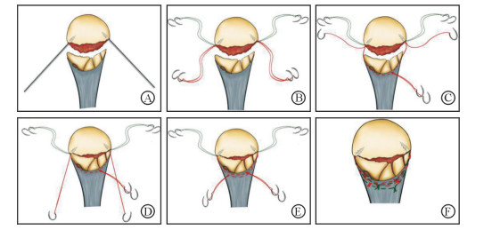
1.3 手术方法麻醉成功后,常规消毒,铺无菌巾。取膝前正中纵行切口,充分暴露断裂的髌骨、髌韧带和两侧支持带;暴露骨折端,将髌骨骨折近端清理干净。双滑轮技术手术步骤如下:(1)在近端骨折块的两下角,斜向内上45°打入2枚3.5 mm锚钉(图 1A、1B),注意锚钉位置尽可能靠近两侧骨皮质,锚钉尾部应埋入骨质。(2)取2枚锚钉的同色(红线)缝线分别从两侧紧贴髌骨下极骨折块下缘穿过髌韧带,在髌韧带中间同一点穿出并打结(图 1C)。(3)同时拉2根缝线的另一端(图 1D),以2个锚钉尾孔为滑轮,髌韧带内缝线将髌骨下极骨折块和软组织托举复位。(4)将红线另一端再次重复第3步操作穿入髌韧带内,然后收紧打结固定(图 1E)。(5)1枚锚钉一般带2根线,使用绿线重复第2~4步操作,完成锚钉双滑轮固定(图 1F)。(6)使用1.5 mm钢缆“8”字张力带固定以保护锚钉。术中屈曲膝关节至120°,检查骨折的稳定性,再予以修补两侧支持带、逐层缝合皮肤。

|
图 1 髌骨下极粉碎性骨折双滑轮技术手术步骤 Fig 1 Steps for the Double-Pulley technique for fixation of comminuted fractures of distal patellar pole A: Place two suture anchors of size 3.5 mm into the medial and lateral edges of the proximal fractured patella with an 45° angle oblique upward; B: Each anchor is loaded with 2 Ultrabraid sutures; C: Thread one suture limb from each anchor through half of the junction of patellar tendon and distal fractured bone from each side and tie them with knot; D: Perform the Double-Pulley procedure by pulling the remaining suture limbs of each anchor, and anatomic reduction is obtained; E: Tie the remaining suture limbs after threading them through patellar tendon; F: Repeat the same Double-Pulley procedure to another two sutures from each anchor |
1.4 术后康复训练与恢复活动时间
关节活动:术后第1天即服用止痛药物并开始被动膝关节弯曲训练和股四头肌等长收缩训练,住院期间由持续被动运动仪辅助膝关节活动,出院后由家属手法辅助膝关节伸屈活动,患者在直腿支具保护下主动直腿抬高训练,术后6周内每天增加膝关节屈曲角度直至120°。术后6周至3个月主动弯曲膝关节,配合下蹲锻炼,逐渐将膝关节屈曲至与健侧相同的角度。支具保护和负重时间:术后6周内佩戴直腿膝关节护具下地完全负重。术后6周后无需佩戴支具完全负重。恢复正常活动和运动时间:术后3个月后可正常生活和从事非体力劳动,术后半年可参加体育活动。
1.5 临床评估依据Bostman髌骨骨折功能评分[7]评价术后膝关节功能。评价标准:优,30~28分;良,27~20分;差,<20分。临床评估指标包括术后Bostman髌骨骨折功能评分优良率、随访时X线片检查结果和膝关节活动度。
1.6 统计学处理应用SPSS 12.0软件进行统计学分析。计量资料以x±s表示,计数资料以例数表示。
2 结果共纳入患者10例,均为新鲜骨折,左膝4例、右膝6例。男6例,女4例;年龄为22~58岁,平均(36.4±11.4)岁。伤后至手术时间1~5 d,平均(2.8±1.1)d。致伤原因:交通事故伤6例,摔伤3例,运动创伤1例。
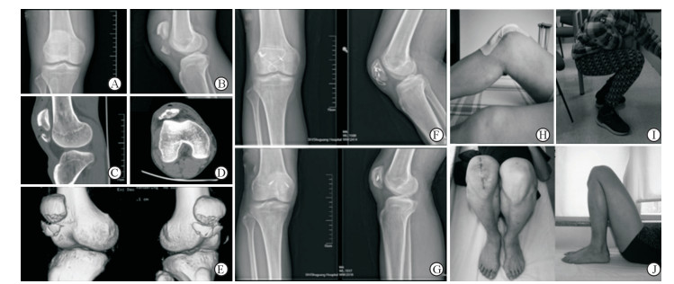
10例患者均获得随访,随访时间为16~24个月。所有患者术后切口均愈合良好。10例患者术后6个月Bostman髌骨骨折功能评分为(28.5±1.8)分,优9例、良1例,优良率为100%。术后6个月X线片检查示髌骨骨性愈合,未发现锚钉脱落、断裂及骨折不愈合等情况。术后膝关节稳定,术后2周膝关节屈曲角度为80.2°±8.4°,术后6周为117.8°±7.2°,术后6个月为132.0°±12.3°。股四头肌肌力均为5级,未发生膝前疼痛及其他并发症。典型病例影像学资料及患膝屈曲角度见图 2。

|
图 2 1例髌骨下极粉碎性骨折患者患膝影像资料及屈曲角度 Fig 2 Radiographs and flexion degrees of injured knee of a patient with distal pole patellar fractures A 38-year-old male patient sustained an injury to the right knee in a traffic accident. A: Anteroposterior X-ray before surgery; B: Lateral X-ray before surgery; C: Sagittal CT before surgery showed displacement of distal fragments; D, E: Cross section CT (D) and 3D-CT (E) showed comminuted distal fracture; F: Bone healing was confirmed by radiographs three months after surgery; G: No sign of patellofemoral joint degeneration was found by X-ray after cable removed at one year; H-J: Photographs of 2 weeks, 6 weeks and 6 months after surgery, respectively. CT: Computed tomography |
3 讨论
克氏针及张力带钢丝内固定是治疗髌骨骨折的首选,但在髌骨下极骨折中,由于骨折远端骨量少,克氏针难以在骨折远端找到固定点,因此不适用于髌骨下极骨折,而张力带即使在粉碎性髌骨骨折的治疗中也有很重要的作用[8-9]。我们使用带线锚钉在完整的骨折近端找到固定点并使用双滑轮技术把骨折远端全部包绕固定,同时使用钢缆“8”字张力带作为对带线锚钉的保护和辅助,前方张力带不但提供了屈膝时的张力带效应使骨折端加压,同时也可以限制屈膝时骨折远端向后的移动和前方骨折块的张开,为早期功能锻炼提供了保护。
使用带线锚钉的双滑轮技术可以为肌腱或韧带的修复提供较大的作用区域和稳定的固定效果,被广泛用于全身各处韧带或肌腱损伤的治疗,如骨性Bankart损伤[10]、肩袖损伤[11]、肩关节不稳的Remplissage修补[12]、SLAP损伤[13]和肱骨大结节的撕脱骨折[14]等。而髌骨下极骨折可以被视为一种髌韧带止点的损伤,也可以使用双滑轮技术来固定。我们在治疗髌骨下极骨折时使用的双滑轮技术有以下优点:(1)在髌骨远端与髌韧带交界处有4根线贯穿,对髌骨下极形成由线组成的吊床样弧形托举,将大部分骨折块包括其中,并对骨折端产生均匀而强大的压力,使早期功能锻炼成为可能。(2)螺钉的置入方向是斜向内上45°,这样可以使锚钉尾部所受拉力与锚钉长轴成90°角,锚钉可以获得更强的抗拔出力。Kadar等[6]报道的使用带线锚钉治疗的27例髌骨下极粉碎性骨折病例中,3例(11.11%)出现了早期内植物失败。Aktay和Kowaleski[15]的生物力学实验结果显示锚钉尾部受缝线拉力与锚钉长轴成90°比0°有更大的抗拔出力。这可能解释了Kadar等[6]的病例中出现早期锚钉松动的原因,在他们的病例中锚钉尾部受缝线拉力与锚钉长轴成0°。(3)由于滑轮(锚钉尾孔)的存在,4根线中两两可以平衡张力,避免了因松紧的差异使张力集中在某一根较紧的线上。Douglass等[16]发现,锚钉固定失败的最常见形式即锚钉拔出。Kadar等[6]报道的3例锚钉失败病例中都只有1枚锚钉拔出。这一现象也被Robb等[17]注意到,他们发现在这种每枚锚钉单独打结固定的系统中,总是有1枚锚钉比另1枚锚钉先失败。这可能与每枚锚钉缝线在打结时的收紧程度有关,收得最紧的线结在屈膝时首先被拉紧而使这枚锚钉承担了大部分张力。而双滑轮技术可以使2枚锚钉的受力均衡,避免这一现象的发生。
双滑轮技术中锚钉的固定依赖于近端骨折块的完整性,因此该技术对于近端骨折块也粉碎性骨折的病例并不适用,对于严重骨质疏松的患者也应谨慎使用。
本研究结果表明,双滑轮技术结合“8”字张力带治疗髌骨下极粉碎性骨折复位效果好,固定可靠,患者可早期开始功能锻炼。但其临床疗效还需开展更多病例、更长期临床随访的研究进一步验证。
| [1] |
BOSTRÖM A. Fracture of the patella. A study of 422 patellar fractures[J]. Acta Orthop Scand Suppl, 1972, 143: 1-80. |
| [2] |
ANDREWS J R, HUGHSTON J C. Treatment of patellar fractures by partial patellectomy[J]. South Med J, 1977, 70: 809-813, 817. DOI:10.1097/00007611-197707000-00014 |
| [3] |
BÖSTMAN O, KIVILUOTO O, NIRHAMO J. Comminuted displaced fractures of the patella[J]. Injury, 1981, 13: 196-202. DOI:10.1016/0020-1383(81)90238-2 |
| [4] |
ANAND A, KUMAR M, KODIKAL G. Role of suture anchors in management of fractures of inferior pole of patella[J]. Indian J Orthop, 2010, 44: 333-335. DOI:10.4103/0019-5413.65149 |
| [5] |
杨怀阔, 马占忠, 薛磊, 杨公博, 邓磊. 带线锚钉治疗髌骨粉碎性骨折13例[J]. 中国中医骨伤科杂志, 2018, 26: 53-55. |
| [6] |
KADAR A, SHERMAN H, DREXLER M, KATZ E, STEINBERG E L. Anchor suture fixation of distal pole fractures of patella:twenty seven cases and comparison to partial patellectomy[J]. Int Orthop, 2016, 40: 149-154. DOI:10.1007/s00264-015-2776-9 |
| [7] |
刘云鹏, 刘沂. 骨与关节损伤和疾病的诊断分类及功能评定标准[M]. 北京: 清华大学出版社, 2002: 223-224.
|
| [8] |
张如意, 唐佩福, 张立海, 陶笙, 梁向党, 郭义柱. 锚钉技术治疗髌骨下极撕脱骨折的疗效分析[J]. 中国矫形外科杂志, 2017, 25: 700-703. |
| [9] |
MELVIN J S, MEHTA S. Patellar fractures in adults[J]. J Am Acad Orthop Surg, 2011, 19: 198-207. DOI:10.5435/00124635-201104000-00004 |
| [10] |
ZHANG J, JIANG C. A new "double-pulley" dual-row technique for arthroscopic fixation of bony Bankart lesion[J]. Knee Surg Sports Traumatol Arthrosc, 2011, 19: 1558-1562. DOI:10.1007/s00167-010-1390-z |
| [11] |
ARRIGONI P, BRADY P C, BURKHART S S. The double-pulley technique for double-row rotator cuff repair[J/OL]. Arthroscopy, 2007, 23: 675.e1-e4. doi: 10.1016/j.arthro.2006.08.016.
|
| [12] |
KOO S S, BURKHART S S, OCHOA E. Arthroscopic double-pulley remplissage technique for engaging Hill-Sachs lesions in anterior shoulder instability repairs[J]. Arthroscopy, 2009, 25: 1343-1348. DOI:10.1016/j.arthro.2009.06.011 |
| [13] |
PARNES N, CIANI M, CARR B, CAREY P. The Double-Pulley anatomic technique for type Ⅱ slap lesion repair[J/OL]. Arthrosc Tech, 2015, 4: e545-e550. doi: 10.1016/j.eats.2015.05.009.
|
| [14] |
MURENA L, CANTON G, FALVO D A, GENOVESE E A, SURACE M F, CHERUBINO P. The "Double-Pulley" technique for arthroscopic fixation of partial articular-side bony avulsion of the supraspinatus tendon: a rare case of bony pasta lesion[J/OL]. Arthrosc Tech, 2012, 2: e9-e14. doi: 10.1016/j.eats.2012.09.002.
|
| [15] |
AKTAY S A, KOWALESKI M P. Analysis of suture anchor eyelet position on suture failure load[J]. Vet Surg, 2011, 40: 418-422. DOI:10.1111/j.1532-950X.2011.00834.x |
| [16] |
DOUGLASS N P, BEHN A W, SAFRAN M R. Cyclic and load to failure properties of all-suture anchors in synthetic acetabular and glenoid cancellous bone[J/OL]. Arthroscopy, 2017, 33: 977-985.e5. doi: 10.1016/j.arthro.2016.11.022.
|
| [17] |
ROBB J L, COOK J L, CARSON W. In vitro evaluation of screws and suture anchors in metaphyseal bone of the canine tibia[J]. Vet Surg, 2005, 34: 499-508. |
 2019, Vol. 40


