2. 海军总医院放射科, 北京 100048
2. Department of Radiology, Navy General Hospital, Beijing 100048, China
椎弓根螺钉在颈椎内固定中最坚强,1994年Abumi等[1]首次临床报道了后路C3~6椎弓根螺钉固定。颈椎前路椎弓根螺钉置入开始于2004年,于2008年首次报道[2]。早期颈椎前路椎弓根螺钉仅用于单侧,近年来颈椎前路椎弓根钉板系统可同时行双侧前路椎弓根螺钉置入[3], 颈椎前路椎弓根螺钉置钉不准可导致灾难性后果[4]。不少学者进行了解剖学研究,但多集中于测量椎弓根宽度、高度、进钉部位、进钉角度等[5-6]。有学者对颈椎椎弓根后入口皮质及椎弓根四壁皮质行厚度测量发现,后入口外壁及椎弓根外壁皮质菲薄,抵抗外力强度小,不宜作为进钉时的解剖学参考[7]。那么,下颈椎椎弓根前入口四壁皮质之间及椎弓根前半四壁皮质之间的厚度是否有差别?本研究对颈椎椎弓根前入口及前半四壁皮质厚度行影像学测量,为前路椎弓根钉置入时规避风险提供依据。
1 材料和方法 1.1 材料10例因意外急性颅脑伤死亡的健康男性,年龄23~40(30.0±4.4) 岁。死亡后2~6 h内取其下颈椎,剔除肌肉,保留骨、关节、韧带组织和关节囊,双层塑料袋密封。-80 ℃保存,检查前于室温自然复温。本研究获海军总医院医学伦理委员会批准。
1.2 影像学检查数字化X线摄影(digital radiography,DR)采用德国西门子公司MultixFD的DR系统,包括颈椎正、侧、双斜位。排除X线片显示颈椎椎弓根结构异常的标本。
对X线片无异常发现的标本行Lightspeed 256排螺旋CT(美国GE公司)扫描。标本取俯卧位(标本的周围软组织已经剔除,仰卧位摆放难以维持位置),以层厚5 mm自上而下行无间距连续容积扫描。扫描范围包含整个下颈椎(C3~7)。扫描条件:管电压120 kV,管电流150 mA,扫描周期1.2 s。以0.625 mm层间距通过内插法重建获得原始横断面影像。将原始数据输入Advantage Workstation上观察。原始图像显示下颈椎横断面满意(平行于椎体解剖横断面, 两侧椎弓根对称显露)的图像以Dicom格式保存;如扫描平面与椎体横断面不平行,则调整标本放置位置和角度,重新扫描,直至重建的原始横断面影像满意,再将原始图像以Dicom格式保存。
X线片与CT扫描同日完成,间隔时间小于2 h。影像学检查完毕后,标本再次于-80 ℃保存,供下一步研究。
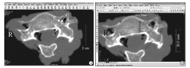
1.3 CT图像测量指标利用CT原始横断面图像完成的测量指标包括:下颈椎椎弓根前入口内皮质(medial cortex of anterior entrance,MAE)、外皮质(lateral cortex of anterior entrance,LAE)厚度,以及椎弓根前半内皮质(medial cortex of anterior half of pedicle,MPA)、外皮质(lateral cortex of anterior half of pedicle,LPA)厚度。方法为:在ImageViewer软件中打开源图像(图 1A),选择骨窗(窗位400 HU,窗宽2 000 HU),将拟测量的图像转换为JPG格式,然后作为光栅图像在AutoCAD 2007软件中打开,利用菜单栏中的“对齐标注”完成指标测量(图 1B)[8]。测量精确度为0.1 mm。

|
图 1 在原始CT横断面图像上测量MAE、LAE、MPA和LPA的厚度 Fig 1 Cortex thicknesses measurement of MAE, LAE, MPA and LPA on raw transaxial CT image A: Raw transaxial CT image opened at ImageViewer software; B: Thickness measurement based on raw CT image opened at AutoCAD 2007 software. a: Medial cortex of anterior entrance (MAE); b: Lateral cortex of anterior entrance (LAE); c: Medial cortex of anterior half of pedicle (MPA); d: Lateral cortex of anterior half of pedicle (LPA) |
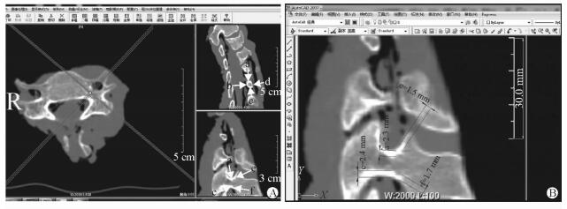
利用冠、矢状面重建图像完成的测量指标包括:下颈椎椎弓根前入口上皮质(superior cortex of anterior entrance,SAE)、下皮质(inferior cortex of anterior entrance,IAE)厚度,及椎弓根前半上皮质(superior cortex of anterior half of pedicle,SPA)、下皮质(inferior cortex of anterior half of pedicle,IPA)厚度。方法为:将源图像在ImageViewer软件中打开,行矢状面和冠状面重建,采用“交联技术”选择合适的重建平面,获得合适的冠、矢状面重建图像(图 2A),进行屏幕截图,作为新建“图层”粘贴至Adobe Photoshop CS4软件的新建空白文档上,利用该软件的“裁剪”选项保留拟行测量的图像部分,合并图层后另存为JPG格式。然后作为光栅图像在AutoCAD 2007软件中打开,利用“对齐标注”完成重建图像相应的指标测量(图 2B)。

|
图 2 在CT冠、矢状面重建图像上测量SPA、IPA、SAE和IAE的厚度 Fig 2 Cortex thicknesses measurement of SPA, IPA, SAE and IAE on reconstructed coronal and sagittal CT images A: Reconstructed sagittal and coronal CT images at ImageViewer interface; B: Thickness measurement based on reconstructed sagittal CT image opened at AutoCAD 2007 interface. a: Superior cortex of anterior half of pedicle (SPA); b: Medial cortex of anterior half of pedicle (MPA); c: Inferior cortex of anterior half of pedicle (IPA); d: Lateral cortex of anterior half of pedicle (LPA); e: Superior cortex of anterior entrance (SAE); f: Inferior cortex of anterior entrance (IAE) |
1.4 统计学处理
采用SPSS 18.0软件对数据进行统计学处理。测量数据用x±s表示。同一颈椎左、右侧同一测量指标的比较采用配对t检验,同一颈椎同侧不同测量指标的比较和不同颈椎序列同侧同一测量指标的比较均采用单因素随机区组方差分析。检验水准(α)为0.05。
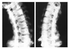
2 结果 2.1 通过X线片筛选合格颈椎标本10例颈椎标本X线片均未发现椎弓根异常或病变,全部纳入研究。斜位片上可清楚显示对侧椎弓根的矢状面和同侧椎弓根的横断面投影,椎弓根横断面投影可见椎弓根上、下、内壁均有明显的皮质骨结构,厚薄较一致,而外壁骨皮质菲薄,部分外壁皮质厚度接近0(图 3)。

|
图 3 颈椎斜位片示颈椎外侧壁菲薄 Fig 3 Oblique view of subaxial spine revealed thin lateral pedicle cortex |
2.2 下颈椎椎弓根前入口及前半四壁皮质厚度测量结果
椎弓根前入口及前半四壁皮质厚度测量结果见表 1。同一颈椎同一测量指标左右侧差异均无统计学意义(P>0.05)。对同一颈椎同侧的不同测量指标行单因素随机区组方差分析,差异有统计学意义(P<0.05);进一步行成对资料均数间两两比较,LAE与LPA的皮质厚度均薄于SAE、MAE、IAE、SPA、MPA和IPA,差异有统计学意义(P均<0.01),LAE与LPA间厚度差异无统计学意义(P>0.05);SAE、MAE、IAE、MPA与IPA间及IAE、SPA、MPA与IPA间差异均无统计学意义(P>0.05),MAE、SAE均小于SPA(P<0.05)。而C3、C7的IAE厚度均薄于C4、C5、C6(P<0.05),C3、C5、C7的LPA薄于同侧C4、C6(P<0.05,P<0.01)。其他同一颈椎同侧不同测量指标之间以及不同颈椎同侧同一测量指标之间的差异均无统计学意义。
|
|
表 1 下颈椎椎弓根前入口四壁及前半四壁皮质厚度 Tab 1 Cortex thickness measurement of anterior entrance and anterior half of pedicle |
3 讨论 3.1 后路颈椎椎弓根螺钉置钉偏差与椎弓根后入口及椎弓根四壁皮质厚度的相关性
早期认为颈椎椎弓根直径偏小,四周重要血管神经结构众多,颈椎椎弓根螺钉固定是禁忌证。C2后路椎弓根螺钉的应用于1964年首次报道,C2椎弓根被认为最宽、最安全;C7后路椎弓根螺钉于1989年首次报道,C7横突孔内无椎动脉通过,钉道偏外无血管损伤风险,相对安全;C3~6后路椎弓根螺钉固定由Abumi等[1]及Jeanneret等[9]于1994年先后报道,后者采用统计学测量的平均值决定进钉点及进钉方向,置钉失误率高,很快就被淘汰。Abumi等[10]发现颈椎椎弓根后入口处管径呈由宽变窄的漏斗状,发明了暴露椎弓根后入口的“漏斗(funnel)”技术,此外还有在透视下判定进钉点并沿椎弓根轴向进钉的研究[11]。实际应用后发现,置钉失误者中进钉点偏外或钉道偏外的概率高。而对后入口及后方椎弓根四壁皮质的观察测量发现,外壁皮质最为菲薄,如果在寻找入口及进钉过程中对四壁平均施力,外壁破溃及钉道偏外的概率增加[7]。
3.2 现有的前路颈椎椎弓根螺钉固定方法颈椎前路固定系统中,椎弓根螺钉最坚强[12]。Aramomi等[2]于2004年最早应用颈椎前路椎弓根螺钉置入并于2008年发表,与Abumi等[1]的方法相似,暴露椎弓根前入口,再行后续操作;但暴露时需注意哪些细节,作者并未深入讨论。此后Yukawa等[13]介绍了透视指引进钉法,Ikenaga等[14]在手术显微镜下寻找椎弓根前入口。复习有关文献发现置钉失误者中,偏外的概率最大[15]。
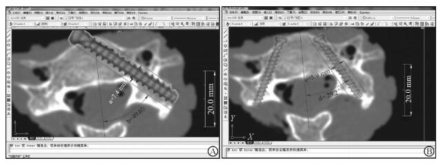
颈椎椎弓根呈由内前向后外约45°走向,椎弓根轴线与椎体前壁的交叉点靠近椎体中线或在对侧,顺轴线置钉只能为单侧[2, 4-5, 13-14]。但钉道角度等于椎弓根外倾角、钉道有效宽度等于椎弓根宽度时,椎弓根宽度可得到最大程度地利用(图 4A)。近年Zhang等[3]设计了前路椎弓根钉板系统,试图行双侧椎弓根螺钉固定,但此时钉道角度减小,钉道改变,钉道有效宽度(即可用椎弓根宽度)减小[16]。本研究实测发现,钉道角度可较椎弓根外倾角小20.4°,钉道有效宽度可较椎弓根宽度窄1.5 mm以上(图 4B)。前路椎弓根置钉时,如无钢板[3]或粗大钉尾[14]遮挡,可通过C形臂透视判断钉道准确性[4];在有遮挡而又无法凭肉眼直视[2]或手术显微镜[14]确定钉道准确性时,置钉过程的手感就显得极为重要[17]。手感显然需了解椎弓根前入口及椎弓根前部四壁的皮质强度,如强度和皮质厚度相平行,则皮质厚度可一定程度上反映其抵抗外力的强度。

|
图 4 颈椎前路双椎弓根螺钉固定时钉道有效宽度(ASTW)缩窄 Fig 4 Available screw trajectory width (ASTW) decreased while anterior cervical bi-pedicle screw insertion was done A: Only unilateral pedicle screw can be inserted when the screw’s trajectory coincided with pedicle axis, but insuring ASTW is equal to pedicle width (PW); B: Inserting pedicle screws at both sides make actual trajectory transverse angle (ATTA) smaller than pedicle transverse angle (PTA), and ASTW smaller than PW. a: PW/ASTW; b: PTA/ATTA; c: ASTW; d: ATTA |
3.3 同一下颈椎椎弓根前入口及前半四壁的皮质厚度特点
本研究通过CT影像学测量发现,同一颈椎LAE厚度较其他3壁菲薄,而其他3壁皮质厚度差异无统计学意义。如果皮质厚度与抵抗外力强度相平行,则LAE抵抗外力的强度弱于其他3壁。这样,在寻找颈椎弓根前入口时,应刻意避免在LAE附近查找,以免穿破LAE。
同样,同一颈椎LPA薄于其他3壁皮质,而SPA、MPA及IPA 3者差异无统计学意义。如果皮质厚度与抵抗外力强度平行,则LPA抵抗外力的强度小于其他3壁。提示在判定前入口正确的前提下进钉时,应避免向LPA施力,不宜以LPA作为参考。在判断钉道是否准确时,不宜像胸腰椎那样用探针探测四壁是否完整,而应注意避免探测LPA。在置入钉板系统或万向椎弓根钉无法透视判定钉道准确性时,上述参照尤为重要。
新近出现的前路双侧椎弓根螺钉钢板内固定系统[3]理论上虽然可达到双侧对称固定的效果,但不是沿椎弓根轴线进钉,而是要迁就钢板孔决定的进钉点,导致横断面上调整后的钉道角度减小,钉道有效宽度变小。而钢板的阻挡使得通过透视判定位置困难,需凭借手感实施进钉[16],此时更应注意以MAE为参照,逐步往外探查找到椎弓根入口;在后续进钉时,需以MPA为参照逐渐进钉,以免偏外穿破LPA进入横突孔内。
3.4 不同颈椎之间同一测量指标的差异不同颈椎之间同一测量指标的统计学结果显示,不同颈椎间MAE、SAE和LAE差异均无统计学意义,提示上述3壁在不同颈椎间抵抗外力的强度相似,其中LAE抵抗外力的强度最弱。C3、C7的IAE抵抗外力强度弱于C4~6。MPA、SPA和IPA在不同颈椎间差异也无统计学意义,可见其抵抗外力的强度也相似。LPA厚度虽在不同颈椎间有所不同,但其抵抗外力的强度均弱于其他3壁。临床实际操作更注重同一颈椎椎弓根不同侧壁抵抗外力强度的比较,而非不同颈椎的同一测量指标的比较,因此其临床意义有限。
| [1] | ABUMI K, ITON H, TANEICBI H, KANEDA K. Transpedicle screw fixation for traumatic lesion of the middle and lower cervical spine:description of the techniques and preliminary report[J]. J Spinal Disord, 1994, 7: 19–28. DOI: 10.1097/00002517-199407010-00003 |
| [2] | ARAMOMI M, MASAKI Y, KOSHIZUKA S, KADOTA R, OKAWA A, KODA M, et al. Anterior pedicle screw fixation for multilevel cervical corpectomy and spinal fusion[J]. Acta Neurochir (Wien), 2008, 150: 575–582. DOI: 10.1007/s00701-008-1574-1 |
| [3] | ZHANG Z, MU Z, ZHENG W. Anterior pedicle screw and plate fixation for cervical facet dislocation:case series and technical note[J]. Spine J, 2016, 16: 123–129. DOI: 10.1016/j.spinee.2015.09.040 |
| [4] | PATTON A G, MORRIS R P, KUO Y F, LINDSEY R W. Accuracy of fluoroscopy versus computer-assisted navigation for the placement of anterior cervical pedicle screws[J]. Spine (Phila Pa 1976), 2015, 40: E404–E410. DOI: 10.1097/BRS.dejydxxb-38-9-115200786 |
| [5] | KOKTEKIR E, TOKTAS Z O, SEKER A, AKAKIN A, KONYA D, KILIC T. Anterior transpedicular screw fixation of cervical spine:is it safe? Morphological feasibility, technical properties, and accuracy of manual insertion[J]. J Neurosurg Spine, 2015, 22: 596–604. DOI: 10.3171/2014.10.SPINE14669 |
| [6] | ZHAO L, HONG J, WANDTKE M E, XU R, MA W, JIANG W, et al. An RCT study on the feasibility of anterior transpedicular screw fixation in the cervicothoracic junction[J]. Eur Spine J, 2016, 25: 1716–1723. DOI: 10.1007/s00586-016-4470-z |
| [7] | 侯黎升, 阮狄克, 徐云可, 何勍, 林井付, 叶晓健, 等. 下颈椎椎弓根入口处皮质厚度的影像学测量[J]. 解剖与临床, 2008, 13: 8–12. DOI: 10.3969/j.issn.1671-7163.2008.01.002 |
| [8] | ZHANG J, LIN W, CHI Y, ZHENG N, XU Q, ZHANG G, et al. The error analysis of lobular and segmental division of right liver by volume measurement[J]. Clin Anat, 2017, 30: 585–590. DOI: 10.1002/ca.v30.5 |
| [9] | JEANNERET B, GEBHARD J S, MAGERL F. Transpedicular screw fixation of articular mass fracture-separation:results of an anatomical study and operative technique[J]. J Spinal Disord, 1994, 7: 222–229. DOI: 10.1097/00002517-199407030-00004 |
| [10] | ABUMI K. Cervical spondylotic myelopathy:posterior decompression and pedicle screw fixation[J]. Eur Spine J, 2015, 24(Suppl 2): 186–196. |
| [11] | NAKASHIMA H, YUKAWA Y, IMAGAMA S, KANEMURA T, KAMIYA M, YANASE M, et al. Complications of cervical pedicle screw fixation for nontraumatic lesions:a multicenter study of 84 patients[J]. J Neurosurg Spine, 2012, 16: 238–247. DOI: 10.3171/2011.11.SPINE11102 |
| [12] | KOLLER H, SCHMIDT R, MAYER M, HITZL W, ZENNER J, MIDDERHOFF S, et al. The stabilizing potential of anterior, posterior and combined techniques for the reconstruction of a 2-level cervical corpectomy model:biomechanical study and first results of ATPS prototyping[J]. Eur Spine J, 2010, 19: 2137–2148. DOI: 10.1007/s00586-010-1503-x |
| [13] | YUKAWA Y, KATO F, ITO K, NAKASHIMA H, MACHINO M. Anterior cervical pedicle screw and plate fixation using fluoroscope-assisted pedicle axis view imaging:a preliminary report of a new cervical reconstruction technique[J]. Eur Spine J, 2009, 18: 911–916. DOI: 10.1007/s00586-009-0949-1 |
| [14] | IKENAGA M, MUKAIDA M, NAGAHARA R, YASUNAGA T, UEDA Y, SOHMA Y. Anterior cervical reconstruction with pedicle screws after a 4-level corpectomy[J]. Spine (Phila Pa 1976), 2012, 37: E927–E930. DOI: 10.1097/BRS.0b013e31824ff7b2 |
| [15] | SCHAEFER C, BEGEMANN P, FUHRHOP I, SCHROEDER M, VIEZENS L, WIESNER L, et al. Percutaneous instrumentation of the cervical and cervico-thoracic spine using pedicle screws:preliminary clinical results and analysis of accuracy[J]. Eur Spine J, 2011, 20: 977–985. DOI: 10.1007/s00586-011-1775-9 |
| [16] | KOLLER H, HITZL W, ACOSTA F, TAUBER M, ZENNER J, RESCH H, et al. In vitro study of accuracy of cervical pedicle screw insertion using an electronic conductivity device (ATPS part Ⅲ)[J]. Eur Spine J, 2009, 18: 1300–1313. DOI: 10.1007/s00586-009-1054-1 |
| [17] | 贾卫斗, 王金, 王立全. 下颈椎椎弓根内固定术的个体化设计及临床应用探讨[J]. 中国矫形外科杂志, 2012, 20: 880–883. |
 2017, Vol. 38


