文章信息
- 原发性骨血管外皮细胞瘤的影像学表现
- Imaging Features of Primary Hemangiopericytoma of Bone
- 肿瘤防治研究, 2017, 44(4): 276-280
- Cancer Research on Prevention and Treatment, 2017, 44(4): 276-280
- http://www.zlfzyj.com/CN/10.3971/j.issn.1000-8578.2017.04.008
- 收稿日期: 2016-08-22
- 修回日期: 2016-10-10
血管外皮细胞瘤(Hemangiopericytoma)是一种罕见的发生于间叶组织的血管源性肿瘤,起源于毛细血管网状纤维鞘外的血管外皮细胞,可发生于全身各部位[1]。血管外皮细胞瘤主要侵犯软组织,发生于骨者较罕见[2]。目前,由于血管外皮细胞瘤发病率低,国内外报道跨越时间较长、随访时间较短,故对该肿瘤的认识仍不够全面[3]。笔者对收集本院5例经手术或穿刺后病理证实的原发性骨血管外皮细胞瘤的临床及影像学资料进行回顾性分析,以提高对该病影像改变的认识。
1 资料与方法一般资料:收集本院1993年4月至2016年6月间5例原发性骨血管外皮细胞瘤患者,男2例、女3例,患者年龄15~45岁,平均28.2岁。所有患者均表现为不同程度患处疼痛,3例伴有可触及的体表肿块。病程3月~10年,平均3.6年。
影像学检查:全部患者均行X线平片检查,3例行CT平扫,2例行MR平扫+增强扫描。(1)X线平片:采用常规X线、CR、DR多种机型。骨盆行X线正位片摄影,下肢长骨病变行常规正侧位摄影。(2)CT:采用GE公司生产的LightSpeed QX/i型8层螺旋CT机。采用各向同性的扫描参数,矩阵512×512,螺距1.0;层厚0.5 cm、120 kV、200~350 mA,标准算法及滤过算法重建。对扫描横断面图像进行冠状、矢状及横断面MPR骨窗和软组织窗重组。(3)MRI:使用Simens公司生产的Symphony Vision 1.5 T超导型MR扫描仪,采用SET1WI(TR 480~600 ms, TE15~18 ms);FSE T2WI(TR 3000~4000 ms, TE96~102 ms)及STIR(TR 3500 ms, TE 28 ms, TI 110 ms)序列。增强扫描:对比剂为Gd-DTPA,剂量0.1 ml/kg,注射速率2 ml/s,自肘静脉注药后20 s开始扫描。由2名高年资放射科医师阅片分析其影像表现。
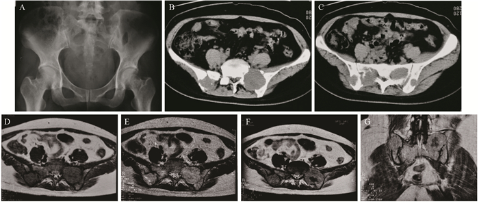
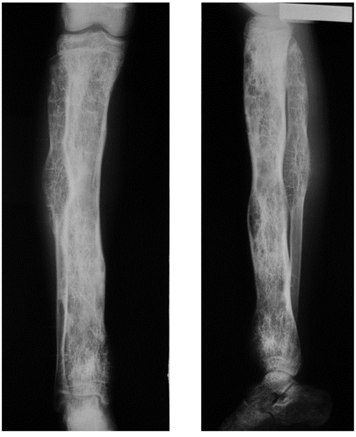
2 结果5例中,2例为多发病灶,X线平片显示为多发骨质破坏。其中1例为双侧骶骨翼病灶并跨越双侧骶髂关节累及双侧髂骨,病灶边缘不规则,周围见轻度骨质硬化,见图 1A;CT可见多发溶骨性骨质破坏,边界清晰,局部骨皮质破坏中断,骨质膨胀及软组织肿块不明显,无明显骨膜反应,见图 1B~1C;MRI扫描示肿瘤边界清晰,T1WI呈等低信号,见图 1D,T2WI呈中等或稍高信号,病灶信号不均匀,见图 1E,增强扫描可见肿瘤呈不均匀明显强化,其内囊变坏死区无强化见图 1F~IG。1例为胫骨全长及腓骨近中段病灶,表现为髓腔大范围斑片状高低混杂密度影,骨质出现不同程度的破坏,骨小梁增粗呈蜂窝、网格状改变,骨皮质可见轻度膨胀并变薄,见图 2。

|
| A: X-ray film of pelvis showed bilateral sacral wings lesions which crossed bilateral sacroiliac joints with bilateral ilia involvement; B, C: the lesions showed irregular edge with peripheral mild bone sclerosis. Axial CT images showed multiple osteolytic bone destruction in the sacrum and bilateral ilia. The boundary was clear, and local bone cortex was interrupted; D, E: the tumor showed uneven iso-or low signal intensity on T1-weighted MR image and iso-or slightly high signal intensity on T2-weighted MR image; F, G: the tumor was unevenly obvious enhancement with no enhanced cystic and necrotic areas on axial and coronal contrast-enhanced MR scan 图 1 骶骨及双侧髂骨血管外皮细胞瘤 Figure 1 Hemangiopericytoma of sacrum and bilateral ilium |

|
| Anteroposterior and lateral X-ray film of tibia and fibula showed the lesion involved the whole tibia and proximal and middle fibula. The medullary cavity showed large-scaled patchy mixed high and low density with different degree of bone destruction. The bone trabecular was honeycomb and latticed thickening, and the bone cortex was mild inflation and thinning 图 2 胫腓骨血管外皮细胞瘤 Figure 2 Hemangiopericytoma of tibia and fibula |
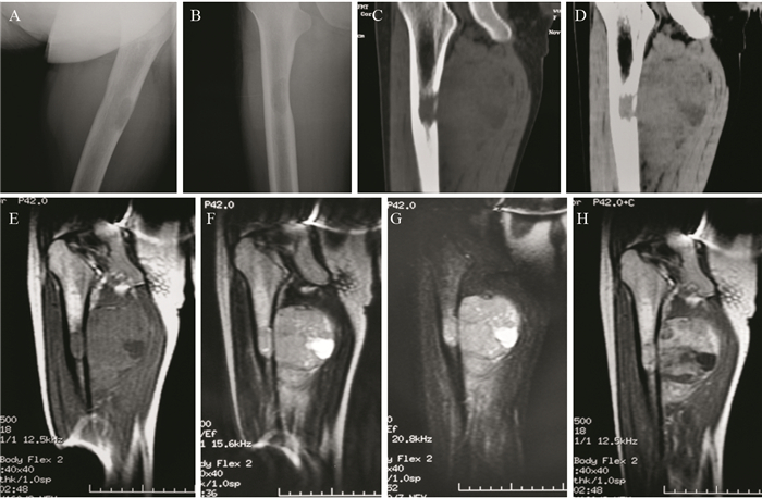
3例单发病灶均为长骨偏心性骨质破坏。其中1例腓骨远端病灶仅累及骨皮质,表现为骨皮质明显增厚硬化,表面形态不规则,可见串珠状骨质破坏,软组织肿块范围较小,其内可见少许钙化,见图 3A;CT可见局部骨皮质明显增厚硬化,未累及髓腔,病灶外侧骨皮质破裂,可见椭圆形软组织肿块影,其内可见散在钙化,见图 3B~3C。另2例病灶同时累及骨皮质和骨松质,呈局限性溶骨性骨质破坏,病灶内密度均匀,边界规则清晰,见图 4A~4B。CT可见其中1例股骨干病灶同时累及髓腔及骨皮质,可见骨皮质破坏中断,局部见多发骨质破坏后残存骨嵴,病灶内侧形成巨大软组织肿块,密度不均,其内可见更低密度坏死囊变区,边界欠清晰,见图 4C~4D。MRI显示肿瘤位于髓腔并破坏内侧骨皮质,T1WI呈等低信号,见图 4E,T2WI呈不均匀中等或稍高信号,见图 4F。内侧见巨大软组织肿块,MRI较平片和CT显示明显。软组织肿块在T1WI为等低信号,在T2WI为稍高或高信号,以STIR序列最明显,见图 4G,其内见T1WI更低和T2WI更高信号的囊变坏死区。肿块下方可见等长T1长T2信号软组织水肿灶,见图 4D~4F。增强扫描可见肿瘤及软组织肿块呈明显不均匀强化,其内囊变坏死区无强化,见图 4H。

|
| A: anteroposterior and lateral X-ray film of ankle showed the lesion only involved the bone cortex. The bone cortex was obvious thickening and hardening with irregular surface, and bead-like bone destruction. The range of surrounding soft tissue mass was smaller with a little calcification. B:the bone window of axial CT showed the lesion of distal fibula demonstrated obvious thickening and hardening of local bone cortex. C: the lateral cortical bone of lesion was ruptured. The soft tissue window of axial CT showed locally oval soft tissue mass with scattered calcification 图 3 腓骨远端血管外皮细胞瘤 Figure 3 Hemangiopericytoma of distal tibia |

|
| A, B: anteroposterior and lateral X-ray film of femur showed the lesion involved both the cortical bone and cancellous bone. It presented locally osteolytic bone destruction with uniform density, regular boundary, and large-scaled soft tissue mass around it; C: the bone window of CT showed the lesion of femur involved both the medullary cavity and bone cortex. The cortical bone was destroyed and interrupted with locally residual bone crest; D: the soft tissue window of CT showed a huge soft tissue mass located in the inner side of lesion. The mass was uneven density with lower density of necrosis and cystic change. The boundary was less clear; E: the tumor showed iso-or low signal intensity on T1-weighted MR image. The lesion located in the medullary cavity and destroyed medial bone cortex with locally iso-or low signal intensity soft tissue mass; F: the tumor showed unevenly slight high or high signal intensity on T2-weighted MR image and unevenly high signal intensity on short time inversion recovery image; G: the higher signal intensity inside the lesion was cystyic and necrotic areas, and soft tissue edema showed high signal intensity below the mass; H: the tumor and soft tissue mass showed unevenly obvious enhancement and no enhancement of the cystic and necrotic areas on contrast-enhanced MR scan 图 4 股骨干血管外皮细胞瘤 Figure 4 Hemangiopericytoma of femoral shaft |
血管外皮细胞瘤是由Stout和Murray于1942年首先发现的一种发生于血管外皮细胞的较罕见肿瘤。血管外皮细胞位于小血管周围,因此本病可发生于身体任何具有毛细血管的部位,以下肢、腹膜后、骨盆及颅内为多[1],原发于骨内组织者非常罕见[4]。本组病例均发生于下肢长骨及骨盆。血管外皮细胞瘤可发生于任何年龄,中青年多见,男女发病率无明显差别。本组病例发病年龄15~45岁,平均28.2岁,符合中青年好发及无性别差异的发病特征。
病理上肿瘤呈浸润性生长,表现为不同大小的实质性或海绵状病变。界限清或不清,质地软或硬,有的呈韧性。切面呈鱼肉样,灰红色,可见出血及坏死灶。大的肿瘤可具有透明样硬化灶。显微镜下见肿瘤由丰富的、分支状的薄壁新生血管组成,血管外皮细胞显著增生,呈肥硕梭形,在血管外排列成不规则的旋涡状,压迫血管致宫腔狭小[5]。胞核为圆形、卵圆形或梭形,排列从囊状到浓染。电子显微镜观察,这些肿瘤细胞显示具有周皮细胞的特征,这种细胞正常时环绕毛细血管或后毛细血管[6]。免疫组织化学染色可用于排除其他的诊断。约2/3的血管外皮细胞瘤患者CD34表达阳性[2],血管标记也有助于诊断。
临床表现多无特异性,其症状随肿瘤发生部位而异。一般有局部疼痛和可触及的体表肿块,多以无痛性肿块就诊。肿瘤发展速度不一,起初可无疼痛或肢体功能障碍,可能数年保持不变,也可能逐渐增大,但无炎性反应。本组病程最长者达10年之久,考虑与肿瘤恶性程度较低,生长缓慢有关。故此类患者一般被认为是良性肿瘤或其他病变,不为临床和患者重视。间变性和恶性者具有侵袭性生物学行为,容易复发,也可出现肺内转移[2,7-10]。本病的主要治疗措施是手术完整切除[11-13],力求全部切除肿瘤以降低复发风险,提高远期生存率[14]。然而,该肿瘤又存在易复发的特点,加之较大或特殊部位的肿瘤有时无法完全切除,需辅以放化疗[2,15],但也有学者认为常规放化疗无明显疗效[13]。
原发于骨的血管外皮细胞瘤的X线表现主要是非特异性溶骨性骨质破坏,可单发或多发。长骨病变好发于干骺端及骨干[16]。本组所有发生于下肢长骨病例病灶均位于干骺端及骨干,考虑与血管分布及密集程度相关。骨质破坏可为虫蚀状、斑片状或泡沫状,破坏区内可有大小不一的残留骨嵴,可出现病理性骨折。根据病变的良恶性程度不同,病灶边缘可整齐或不规则,边界可清晰或不清晰。良性或低度恶性病灶骨小梁可增粗呈蜂窝、网格状改变,皮质可见轻度膨胀,可似良性骨血管瘤,边缘可出现不同程度骨质硬化。本研究中1例发生于胫腓骨病例曾被误诊为骨血管瘤,可见骨血管瘤与良性或低度恶性血管外皮细胞瘤影像学表现有相似之处,鉴别困难。肿瘤侵及骨皮质致骨皮质变薄和轻度膨胀,虽然骨皮质可以膨胀变薄,但骨膜反应较少见。恶性程度高的病变突破骨皮质侵及软组织形成软组织肿块,偶有反应性皮质增生。本组所有病例均未见骨膜反应,与文献报道相符。
CT表现与X线平片所见类似,但可更清晰地显示骨质破坏和软组织肿块的部位以及病灶内的细节如残存骨嵴、钙化等,表现为溶骨性骨质破坏,可伴有或不伴有软组织肿块。病灶范围内有囊状改变、伴有周围反应性骨质硬化或骨质膨胀、钙化及病灶内有骨小梁代偿性增粗而形成的网格、皂泡、蜂窝状改变等提示病灶偏良性或低度恶性。本组1例位于腓骨远端病灶可见骨皮质明显增厚硬化伴散在钙化,病程达10年,提示其偏良性或低度恶性。病灶内散在钙化可能是血管源性肿瘤较具特异性的征象之一,但在骨血管外皮细胞瘤中出现未见相关文献报道且病例数少,具体临床价值有待积累更多病例进一步研究。而骨皮质破裂、软组织肿块形成及病灶边界模糊不清则提示病变恶性程度较高。因此上述具体征象能间接反映肿瘤的生物学行为,对鉴别良恶性很有帮助。
MRI的多参数多方位成像且软组织分辨率更高,能更准确地显示病变的范围、周围受侵情况及病变内部的结构。T1WI呈等低或混杂信号,T2WI呈中等或稍高信号,信号多不均匀,肿瘤内有出血、坏死时则伴有相应的信号改变,增强扫描呈明显不均匀强化,其内出血、坏死则不强化。其信号及强化特点可能为血管源性肿瘤的诊断提供线索,但各序列均无特异性,不能进一步定性诊断。有文献报道在MRI上由于放射状分布的低信号流空血管可出现特异性的车轮辐条状表现[16],但本组MR检查的2例均未见到,可能与病例数较少有关。
总之,原发性骨血管外皮细胞瘤多发生于中青年患者,骨盆和下肢长骨多见。影像学上以溶骨性骨质破坏为主,软组织肿块可有或无,无骨膜反应。影像学检查虽无明显特征性,但有助于了解病变的范围、治疗措施的制定和治疗效果的评价,最终确诊需依据病理学检查。
| [1] | Park BJ, Kim YI, Hong YK, et al. Clinical analysis of intracranial hemangiopericytoma[J]. J Korean Neurosurg Soc, 2013, 54(4): 309–16. DOI:10.3340/jkns.2013.54.4.309 |
| [2] | Ren K, Zhou X, Wu S, et al. Primary osseous hemangiopericytoma in the thoracic spine[J]. Clin Neuropathol, 2014, 33(5): 364–70. |
| [3] | Kim YJ, Park JH, Kim YI, et al. Treatment Strategy of Intracranial Hemangiopericytoma[J]. Brain Tumor Res Treat, 2015, 3(2): 68–74. DOI:10.14791/btrt.2015.3.2.68 |
| [4] | 陈迎春, 刘郑生. 骨原发血管外皮瘤 (附2例报告)[J]. 中国肿瘤临床与康复, 2002, 9(4): 97–8. [ Chen YC, Liu ZS. Hemangiopericytoma of bone[J]. Zhongguo Zhong Liu Lin Chuang Yu Kang Fu, 2002, 9(4): 97–8. ] |
| [5] | 陈克敏, 陆勇. 骨与关节影像学[M]. 上海: 上海科学技术出版社, 2015: 544.] [ Chen KM, Lu Y. Bone and joint imaging[M]. Shanghai: Shanghai Science and Technology Publishing House, 2015: 544. ] |
| [6] | Yu Y, Shi HY, Huang HF. Uterine perivascular epithelioid cell tumour[J]. J Obstet Gynaecol, 2014, 34(6): 519–22. DOI:10.3109/01443615.2014.914475 |
| [7] | 马春华, 张学斌, 姜镕, 等. 颅内间变性血管外皮瘤合并肺部多发性转移一例[J]. 中国现代神经疾病杂志, 2016, 16(2): 107–12. [ Ma CH, Zhang XB, Jiang R, et al. Intracranial anaplastic hemangiopericytoma with pulmonary metastases: one case report[J]. Zhongguo Xian Dai Shen Jing Ji Bing Za Zhi, 2016, 16(2): 107–12. ] |
| [8] | 于凤凯, 杨立臣, 苏炜, 等. 颅内血管外皮细胞瘤19例MRI分析[J]. 临床放射学杂志, 2014, 33(11): 1643–6. [ Yu FK, Yang LC, Su W, et al. MRI Manifestations of Intracranial Hemangiopericytoma: An Analysis of 19 Cases[J]. Lin Chuang Fang She Xue Za Zhi, 2014, 33(11): 1643–6. ] |
| [9] | 唐菲, 刘辉. 颅内血管周细胞瘤的MRI表现与病理对照分析[J]. 临床放射学杂志, 2014, 33(9): 1438–41. [ Tang F, Liu H. MRI manifestations of Intracranial Hemangiopericytoma: Comparison Study with Pathological Findings[J]. Lin Chuang Fang She Xue Za Zhi, 2014, 33(9): 1438–41. ] |
| [10] | Zhu H, Duran D, Hua L, et al. Prognostic Factors in Patients with Primary Hemangiopericytomas of the Central Nervous System: A Series of 103 Cases at a Single Institution[J]. World Neurosurg, 2016, 90: 414–9. DOI:10.1016/j.wneu.2016.02.103 |
| [11] | 古庆家, 徐刚, 何刚, 等. 鼻腔鼻窦血管外皮细胞瘤临床分析[J]. 中华耳鼻咽喉头颈外科杂志, 2014, 49(6): 452–6. [ Gu QJ, Xu G, He G, et al. Clinical analysis of sinonasal hemangiopericytoma[J]. Zhonghua Er Bi Yan Hou Tou Jing Wai Ke Za Zhi, 2014, 49(6): 452–6. ] |
| [12] | Liu HG, Yang AC, Chen N, et al. Hemangiopericytomas in the spine: clinical features, classification, treatment, and long-term follow-up in 26 patients[J]. Neurosurgery, 2013, 72(1): 16–24. DOI:10.1227/NEU.0b013e3182752f50 |
| [13] | 王康宁, 方强. 左肺下叶血管外皮细胞瘤伴淋巴结转移1例[J]. 中华胸心血管外科杂志, 2016, 32(2): 119–20. [ Wang KN, Fang Q. PEComa with lymph node metastasis in left lower lung:a case report[J]. Zhonghua Xionng Xin Xue Guan Wai Ke Za Zhi, 2016, 32(2): 119–20. ] |
| [14] | Shirzadi A, Drazin D, Gates M, et al. Surgical management of primary spinal hemangiopericytomas: an institutional case series and review of the literature[J]. Eur Spine J, 2013, Suppl 3: S450–9. |
| [15] | Melone AG, D'Elia A, Santoro F, et al. Intracranial hemangiopericytoma-our experience in 30 years: a series of 43 cases and review of the literature[J]. World Neurosurg, 2014, 81(3-4): 556–62. DOI:10.1016/j.wneu.2013.11.009 |
| [16] | 郭启勇. 实用放射学[M]. 第3版. 北京: 人民卫生出版社, 2007: 1185.] [ Guo QY. Practice of radiology[M]. 3rd ed. Beijing: People Health Publishing House, 2007: 1185. ] |
 2017, Vol. 44
