文章信息
- 张慧, 张馨月, 张含
- ZHANG Hui, ZHANG Xinyue, ZHANG Han
- 无赤光血管造影在眼底疾病检查中的图像特征研究
- Comparative study of image features of red-free angiography in the examination of fundus diseases
- 中国医科大学学报, 2023, 52(3): 247-252
- Journal of China Medical University, 2023, 52(3): 247-252
-
文章历史
- 收稿日期:2022-10-21
- 网络出版时间:2023-03-16 09:33:06
目前常用的眼底病变检查技术包括眼底荧光素血管造影(fluorescein fundus angiography,FFA)、吲哚菁绿血管造影(indocyanine green angiography,ICGA)、眼底自发荧光(fundus autofluorescence,FAF)以及光学相干断层成像(optical coherence tomography,OCT)等。其中,FFA检查主要用于诊断视网膜疾病,ICGA检查主要用于诊断脉络膜疾病[1],FAF检查用于观察眼底视网膜色素上皮层(retinal pigment epithelium,RPE)细胞中脂褐质代谢情况,OCT检查是利用光的干涉原理对视网膜各层进行解剖层面上的检查。无赤光血管造影(red-free angiography,RFA)作为一种新兴检查,相比于其他几种检查,临床上应用较少。无赤光眼底照相一般用于观察视网膜神经纤维层的缺损情况[1-2],也有学者[3]提出无赤光眼底照相能清晰反映RPE病变。RFA是在无赤光眼底照相的基础上,移除滤光片后,利用488 nm波长的激发光与染色剂荧光素钠结合进行检查[4]。本研究拟利用上述检查方法对主要眼底病变分别进行检查,比较RFA与其他检查方法的异同,探讨RFA检查在临床诊疗中的优势,为眼底病变针对性选择诊断技术提供依据。
1 材料与方法 1.1 研究对象选择2022年1月至8月期间于中国医科大学附属第一医院行RFA与FFA联合ICGA检查眼底疾病的患者29例(41眼)。纳入标准:接受RFA、FFA、ICGA、FAF、OCT等相关检查的眼底病变的所有患者。排除标准:屈光间质严重混浊,眼底检查图像质量不佳者;全身状态差或精神状态不佳,无法配合检查者。本研究已获得医院医学伦理委员会批准,所有患者均签署知情同意书。
1.2 检查方法采用共焦激光眼底血管造影仪(SPECTRALIS HRA 2,德国海德堡公司)完成RFA与FFA联合ICGA检查。检查前先将10%荧光素钠3 mL和含有吲哚菁绿25 mg的无菌注射液3 mL混合,在5 s内快速注入肘静脉进行试敏。本研究所有患者荧光素钠试敏结果均为阴性。将共焦激光眼底血管造影仪镜头聚焦眼底后,分别采集RFA及FFA的10 min内和ICGA的30 min内影像;在FFA和ICGA检查进行至造影中期5~7 min时,转换激光扫描模式为无赤光检查模式,同时其他参数不变,进行RFA影像采集;在检查进行至30 min时,采集ICGA晚期影像。吲哚菁绿通过主动转运方式聚集在RPE细胞,造影后期阶段更加突出[5],故本研究ICGA造影图像的时间选择为造影晚期30 min以上。检查结束后选择FFA、RFA 5~7 min内的影像及ICGA30 min的影像进行比较。
1.3 统计学分析采用Excel软件录入数据,采用SAS 9.4软件的McNemar’s检验进行分析。采用Kappa值评价各种检查方法结果的一致性,Kappa值越接近于1,检查结果越一致,越接近于0,检查结果越不一致。P < 0.05为差异有统计学意义。
2 结果 2.1 患者的基本情况本研究共纳入29例患者(41眼),平均年龄(43.2±14.24)岁,其中,男12例(20眼),女17例(21眼)。诊断主要包括葡萄膜炎(24.38%)、中心性浆液性脉络膜视网膜病变(24.38%)、黄斑出血(17.07%),以及其他10种眼底病变,见表 1。
| Diagnosis | n | Percentage(%) |
| Uveitis | 10 | 24.38 |
| Central serous chorioretinopathy | 10 | 24.38 |
| Macular hemorrhage | 7 | 17.07 |
| Punctate inner choroidopathy | 2 | 4.88 |
| Pathologic myopia | 2 | 4.88 |
| Retinitis pigmentosa | 2 | 4.88 |
| Multiple evanescent white dot syndrome | 2 | 4.88 |
| Acute regional occult outer retinopathy | 1 | 2.44 |
| Macular edema | 1 | 2.44 |
| Retinal capillary hemangioma | 1 | 2.44 |
| Retinal myelinated nerve fibers | 1 | 2.44 |
| Visual field defect | 1 | 2.44 |
| Multiple evanescent white dot syndrome combined with punctate inner choroidopathy | 1 | 2.44 |
| n,number of eyes. | ||
2.2 各检查方法对视网膜下积液诊断的差异分析
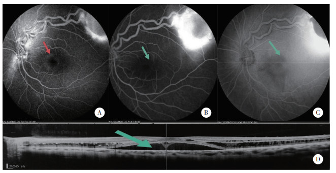
分别比较RFA与FFA、ICGA和OCT对视网膜下积液的诊断差异,结果显示,RFA诊断结果与ICGA和OCT完全一致(Kappa=1),均显著高于FFA(Ka-ppa=0.580,S = 9.000,P = 0.004)。见表 2。OCT检查结果显示,23眼存在视网膜下积液,与RFA检查结果完全一致。视网膜毛细血管瘤不同眼底检查影像结果如图 1所示,RFA可见黄斑区视网膜下液环状隆起病灶,但FFA相应位置未见荧光渗漏;ICGA晚期图像和OCT图像均可见明显异常。
| Method | Clear extent of subretinal fluid detachment | Unclear extent of subretinal fluid detachment | S | P | Kappa |
| RFA | 24 | 17 | - | - | - |
| FFA | 15 | 26 | 9.000 | 0.004 | 0.580 |
| ICGA | 24 | 17 | -* | -* | 1 |
| OCT | 23 | 18 | -* | -* | 1 |
| * The results of the RFA and OCT and ICGA groups were completely consistent for all subjects without specific statistics. | |||||

|
| A, RFA showed disc-shaped subretinal fluid detachment in the macular region, as shown by the red arrow; B, FFA showed no obvious fluorescence leakage in the macular region, as shown by green arrow; C, late phase of ICGA faintly showed annular leakage in the macular region, as shown by green arrow; D, OCT showed dark areas of subretinal fluid, as shown by green arrow. 图 1 临床诊断为视网膜毛细血管瘤患眼的眼底不同检查影像 Fig.1 Different fundus examination images of eyes clinically diagnosed with retinal capillary hemangioma |
2.3 各检查方法对黄斑区膜状高荧光的差异分析
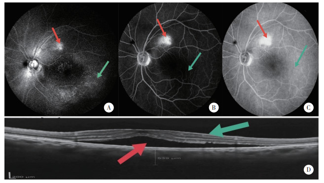
在1例中心性浆液性脉络膜视网膜病变患者接受FFA、ICGA、RFA检查过程中,RFA发现黄斑区膜状高荧光,而FFA及ICGA均未见此异常。该患者RFA检查可见黄斑区上方视网膜下液高荧光灶(图 2A红色箭头),黄斑区可见环状金箔样高反光灶(图 2A绿色箭头)。FFA检查虽然可见黄斑区上方视网膜下液高荧光灶(图 2B红色箭头),但黄斑区未见荧光渗漏(图 2B绿色箭头)。ICGA晚期检查可见黄斑区上方视网膜下液高荧光灶(图 2C红色箭头),黄斑区隐约可见环状暗区(图 2C绿色箭头)。OCT检查确认黄斑前存在条带样高反射和视网膜下液暗区(图 2D红、绿色箭头)。对于黄斑区膜样高荧光的观察,RFA检查与OCT图像判读一致,比FFA、ICGA晚期检查有优势。

|
| A, RFA showed subretinal fluid above the macular region, as shown by red arrow, a ringed gold foil-like hyperfluorescence focus, as shown by green arrow; B, FFA showed subretinal fluid above the macular region, as shown in red arrow, no fluorescence leakage was observed in the macular region, as shown in green arrow; C, late phase of ICGA showed subretinal fluid above the macular region, as shown in red arrow, annular dark areas are faintly visible in the macular region, as shown in green arrow; D, OCT showed premacular strip-like hyperreflex, as shown in green arrow, dark area of subretinal fluid was shown in red arrow. 图 2 临床诊断为中心性浆液性脉络膜视网膜病变患眼的眼底不同检查影像 Fig.2 Different fundus examination images of the eye clinically diagnosed with central serous chorioretinopathy |
2.4 各检查方法对视网膜点片状异常荧光的差异分析
分别比较RFA与FFA、ICGA和FAF对网膜点片状异常荧光的诊断差异,可见RFA发现网膜点片状异常荧光的能力高于其他3种检查,但无统计学差异,见表 3。
| Method | Clear patched abnormal fluorescence of retina | Unclear patched abnormal fluorescence of retina | S | P | Kappa |
| RFA | 9 | 32 | - | - | - |
| FFA | 7 | 34 | 2.000 | 0.500 | 0.845 |
| ICGA | 8 | 33 | 1.000 | 1.000 | 0.926 |
| FAF | 8 | 33 | 1.000 | 1.000 | 0.926 |
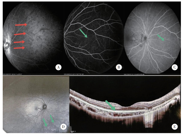
对于视网膜点片状异常荧光,RFA与其他3种检查不一致者为多发性一过性白点综合征和点状内层脉络膜病变。RFA可见鼻侧网膜点片状低荧光灶。FFA和ICGA晚期未见明显异常荧光。FAF检查可见周边视网膜边界不清斑点状高自发荧光。OCT检查可见椭圆体带结构不清。见图 3。

|
| A, RFA showed diffuse punctal hypofluorescence in the nasal retina, as shown in red arrow; B, FFA showed diffuse punctal hypofluorescence in the nasal retina, as shown in green arrow; C, late phase of ICGA showed no obvious abnormal fluorescence in the nasal retina, but only showed spot-like low fluorescence in the nasal macula, as shown in green arrow; E, OCT showed unclear structure of the ellipsoid band, as shown in green arrow. 图 3 临床诊断为多发性一过性白点综合征联合点状内层脉络膜病变患眼的眼底不同检查影像 Fig.3 Different fundus examination images of eyes clinically diagnosed with multiple evanescent white dot syndrome combined with punctate inner choroidopathy |
2.5 RFA与其他检查方法对视乳头周围多发性片状病灶的差异分析
结果显示,RFA检查与其他检查方法对于视乳头周围多发性片状病灶的影像一致。
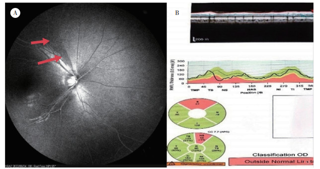
2.6 各检查方法对视网膜神经纤维层状态的差异分析RFA检查可清晰观察到视网膜神经纤维层状态,而FFA和ICGA晚期检查均无法观察到视网膜神经纤维层状态。本研究中,1例视野缺损患者在行FFA及ICGA晚期检查时,未见异常影像改变。但RFA检查发现视盘周围1处神经纤维的缺失改变(图 4A红色箭头)。经OCT检查确认为视盘上方视网膜神经纤维层厚度变薄(图 4B)。

|
| A, RFA showed fibrous strip of dark area above the optic papilla, as shown in red arrow; B, OCT of optic disc showed the thinning layer of retinal nerve fibers above the optic disc. 图 4 临床诊断为视野缺损患眼的眼底不同检查影像 Fig.4 Different fundus examination images of eyes clinically diagnosed with visual field defect |
3 讨论
无赤光眼底照相具有波长短的特点,有助于排除深层组织反光干扰,使眼底表层的变化在眼底图像上更加清晰[6]。而RFA是在去掉滤光片的情况下,在无赤光眼底照相短波长激光和荧光素双重作用下,结合眼底血红蛋白、视网膜色素基团,并因视网膜色素沉着和其他组织吸收参数不同[4]等形成高质量图像的一种检查技术。本研究中,在视网膜下积液的检查方面,RFA和ICGA晚期结果一致,比FFA检查更有优势,这是由于RFA检查的上述成像特点可以敏锐地捕捉到FFA检查所漏掉的信息所致。
对于黄斑区膜状高荧光的观察,RFA检查比FFA及ICGA晚期检查有优势。RFA在清晰显示视盘、筛板结构的同时,也可观察到视网膜神经纤维层特异性的染色情况,而FFA、ICGA检查中的激发光波长较长,穿透力较强,易受视网膜深层组织的干扰,故在此方面不如RFA成像效果好。本研究中仅有1例中心性浆液性脉络膜视网膜病变的患者在进行RFA检查时发现了黄斑区膜状高荧光,而FFA和ICGA均未发现。提示了RFA在临床检查工作中的重要性。
对于视乳头周围多病灶性点片状异常荧光图像,RFA检查和FFA及ICGA晚期检查在检出率上无明显差异。因此异常荧光现象多发生于点状内层脉络膜疾病,而该疾病以围绕视乳头出现多灶性黄白色、大小不等的点片状灰黄色病灶为典型眼底表现之一,病灶累及视网膜及脉络膜[7],因此这3种检查图像无差异。
本研究在临床诊断为葡萄膜炎、多发性一过性白点综合征、点状内层脉络膜病变、视网膜色素变性、急性局灶性隐匿性外层视网膜病变等9个病例中,发现RFA对于周边视网膜点片状异常荧光的检出率与其他检查方法有所差异。虽然这些疾病的病因不同,尤其对于多发性一过性白点综合征的病因比较有争议,有学者认为其是一种脉络膜疾病,可继发RPE和外层视网膜病变;也有学者认为其原发病灶是椭圆体带,而RPE和脉络膜是继发病变;甚至有学者认为脉络膜、RPE和外层视网膜均受到影响[8-10],但这几种疾病中RPE层均受累。而RFA在RPE层的观察中,对于周边视网膜点状异常荧光,比FFA及ICGA更有优势,RFA检查范围更广、更清晰。提示RFA的短波长激光虽然无法穿过RPE层,但其对于RPE层的病变检查也许有一定优势。
综上所述,本研究发现,RFA检查在眼底病变的检查方面,尤其是视网膜下积液、视网膜神经纤维层等视网膜浅层结构的观察方面,比FFA、ICGA等检查方法更有优势。而且,在检查时间方面,ICGA造影晚期图像需要等候30 min以上方能获得,而RFA具有耗时更短、更易操作、患者花费少、病灶影像对比度清晰等优点。故建议在今后的眼底血管造影检查中,应同时行RFA检查。然而需要注意的是,RFA需借助荧光素钠造影剂,与ICGA类似,属于有创检查,且RFA为短波长扫描激光,受屈光系统影响较大,穿透性差,可能影响图像质量。另外,本研究为回顾性分析,样本量较小,有待通过扩大样本量以及检查仪器和时间选择节点多样化等手段,进一步完善研究结果。
| [1] |
JI MJ, PARK JH, YOO C, et al. Comparison of the progression of localized retinal nerve fiber layer defects in red-free fundus photograph, en face structural image, and OCT angiography image[J]. J Glaucoma, 2020, 29(8): 698-703. DOI:10.1097/IJG.0000000000001528 |
| [2] |
LIM AB, PARK JH, JUNG JH, et al. Characteristics of diffuse retinal nerve fiber layer defects in red-free photographs as observed in optical coherence tomography en face images[J]. BMC Ophthalmol, 2020, 20(1): 16. DOI:10.1186/s12886-019-1302-z |
| [3] |
孙心铨, 刘晓玲. 眼底疾病影像学检查的合理应用及联合应用[J]. 中华眼视光学与视觉科学杂志, 2011, 13(3): 161-164. |
| [4] |
华瑞, 文峰. 图说眼底影像技术从多模影像到人工智能[M]. 北京: 人民卫生出版社, 2020: 53-61.
|
| [5] |
CHANG AA, ZHU MD, BILLSON F. The interaction of indocyanine green with human retinal pigment epithelium[J]. Invest Ophthalmol Vis Sci, 2005, 46(4): 1463-1467. DOI:10.1167/iovs.04-0825 |
| [6] |
李瑞峰. 眼底荧光血管造影及光学影像诊断[M]. 北京: 人民卫生出版社, 2010: 38.
|
| [7] |
张承芬. 眼底病学[M]. 2版. 北京: 人民卫生出版社, 2010.
|
| [8] |
MONFERRER ADSUARA C, REMOLÍ SARGUES L, MONTERO HERNÁNDEZ J, et al. Multimodal imaging in multiple evanescent white dot syndrome and new insights in pathogenesis[J]. J Fr Ophtalmol, 2021, 44(10): 1536-1544. DOI:10.1016/j.jfo.2021.05.011 |
| [9] |
ONG AY, BIRTEL J, CHARBEL ISSA P. Multiple evanescent white dot syndrome (MEWDS) associated with progression of lacquer cracks in high myopia[J]. Klin Monbl Augenheilkd, 2021, 238(10): 1098-1100. DOI:10.1055/a-1515-6065 |
| [10] |
JABBARPOOR BONYADI MH, HASSANPOUR K, SOHEILIAN M. Recurrent focal choroidal excavation following multiple evanescent white dot syndrome (MEWDS) associated with acute idiopathic blind spot enlargement[J]. Int Ophthalmol, 2018, 38(2): 815-821. DOI:10.1007/s10792-017-0511-9 |
 2023, Vol. 52


