扩展功能
文章信息
- 何英, 殷启凯, 付士红, 王环宇
- HE Ying, YIN Qi-kai, FU Shi-hong, WANG Huan-yu
- 库蚊黄病毒的病毒滴度测定方法建立
- Establishment of a method for determining the virus titer of the Culex flavivirus isolated in China
- 中国媒介生物学及控制杂志, 2021, 32(4): 412-414
- Chin J Vector Biol & Control, 2021, 32(4): 412-414
- 10.11853/j.issn.1003.8280.2021.04.005
-
文章历史
- 收稿日期: 2021-05-17
库蚊黄病毒(Culex flavivirus,CxFV)属于黄病毒科黄病毒属的蚊传虫媒病毒。2003年,日本学者首次在日本和印度尼西亚的淡色库蚊(Culex pipiens pallens)和三带喙库蚊(Cx. tritaeniorhynchus)中分离到CxFV毒株[1]。随后在北美洲的美国,中美洲的危地马拉、墨西哥、特立尼达和多巴哥,南美洲的阿根廷,非洲的乌干达相继分离到CxFV。目前分离的CxFV分为2个基因型别,日本、印度尼西亚、美国的毒株属于基因1型,非洲、危地马拉和墨西哥的毒株属于基因2型。我国于2006年从山东省东明市采集的淡色库蚊中分离到1株库蚊黄病毒(SDDM06-11)。基于E基因的系统进化分析显示,该毒株属于基因1型病毒,但不同于日本、印度尼西亚和美国的病毒株,在进化树中单独成支[2]。CxFV能够引起白纹伊蚊(Aedes albopictus)卵细胞(C6/36)病变,但在其他哺乳动物细胞系,如金黄地鼠肾细胞(BHK-21)、非洲绿猴肾细胞(Vero)均未发现细胞病变,接种乳鼠后不能引起乳鼠死亡。为进一步开展此类病毒系统生物学研究,首先要准确测定病毒滴度,完成病毒定量。病毒噬斑试验是经典的病毒定量方法,但是C6/36细胞属于半悬浮细胞,传统的琼脂糖培养基覆盖方法操作难度较大,因此,本研究尝试建立由甲基纤维素代替琼脂糖覆盖,结晶紫染色后,通过噬斑计算病毒滴度。
1 材料与方法 1.1 细胞C6/36细胞(白纹伊蚊卵细胞)由本实验室保存。
1.2 病毒CxFV(SDDM06-11株)由本实验室分离、鉴定并保存。
1.3 病毒滴度测定(1)将对数生长期C6/36细胞接种至6孔培养板中(300 μl/孔),置于28 ℃,5% CO2培养箱培养,细胞长至单层待用。(2)取出冻存的SDDM06-11株病毒悬液,做10倍递次稀释,使之成为10-1~10-11,共11个稀释度的病毒悬液。(3)每个病毒稀释度中吸取100 μl依次加入到6孔细胞培养板中,28 ℃,5% CO2孵箱孵育1 h。(4)加入1.2%的甲基纤维素4 ml/孔,置于28 ℃、5% CO2培养箱培养,观察。(5)培养4 d后,在显微镜下观察到明显的噬斑时,弃去培养基,用磷酸盐缓冲液(PBS)洗2遍后,加入染色液(结晶紫∶草酸混合液为1∶4),室温染色1 h后,用水轻缓冲洗掉染色液,计算每个稀释度的噬斑数。
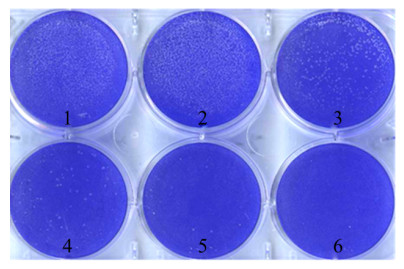
2 结果 2.1 噬斑试验使用甲基纤维素-结晶紫空斑方法观察CxFV(SDDM06-11株)发现,感染C6/36细胞后形成清晰的噬斑,大小为针尖样圆形。稀释度为10-3的CxFV形成的噬斑基本成片,不易于计算,稀释度为10-4~10-6形成的噬斑较好,利于计算(图 1)。

|
| 注:1~5表示病毒稀释度为10-1~10-5;6表示细胞对照。 图 1 SDDM06-11株感染C6/36细胞4 d后的噬斑(结晶紫染色) Figure 1 Plaque of C6/36 cells infected with SDDM06-11 strain for 4 days(crystal violet staining) |
| |
在10-4、10-5、10-6稀释度中分别有72、7和1个噬斑,10-7~10-11稀释度无噬斑。实验的阳性和阴性对照均成立,计算病毒的滴度为1×106.85 pfu/ml。
3 讨论通过对蚊虫的病原谱进行分析表明,蚊虫体内可以携带多种昆虫病毒,并且未表现出任何严格的地理限制[3]。其中,从库蚊属中鉴定出大量病毒,表明库蚊属蚊虫可能是具有广泛地理分布和高病毒丰度的各种病毒的主要宿主或载体[4]。CxFV在我国分布广泛,我国于2006年在山东省首次分离到1株此病毒,命名为SDDM06-11[2]。随后,在我国的辽宁省丹东地区[5]、甘肃省河西走廊地区[6]、台湾地区[7]、新疆维吾尔自治区艾比湖湿地[8]、陕西省、河南省相继分离到大量CxFV毒株或检测到病毒核酸序列[9]。
CxFV虽然对人类和其他脊椎动物致病性尚不清楚,但由于与其他具有明确致病性的黄病毒成员如登革病毒(Dengue virus,DENV)、西尼罗病毒(West Nile virus,WNV)、圣路易脑炎病毒(St Louis encephalitis virus,SLEV)、日本脑炎病毒(Japanese encephalitis virus,JEV)和黄热病毒(Yellow fever virus,YFV)等在生态学和遗传学上存在密切的关系[10],且近年来在辽宁省发现的朝阳病毒(Chaoyang virus)[11]以及在云南省分离的广平病毒(Quang Binh virus,QBV)[12],在遗传进化分析上也显示与CxFV亲缘关系较近,近年来受到人们越来越多的关注。
目前关于CxFV的研究主要以地区分布研究为主,对于进一步的病毒学研究尚待深入。病毒定量是对病毒进行深入研究的基础,测定病毒滴度经典的方式是噬斑检测(Plaque assay),以噬斑形成单位(pfu/ml)表示。CxFV只能引起C6/36细胞病变,且C6/36细胞属于半悬浮细胞(28 ℃培养)。常规的病毒空斑形成试验多采用琼脂糖作为培养基,中性红染色,由于琼脂糖对于温度控制要求较高,实验操作难度较大,并且中性红较易褪色,不适合长期保存。本文改用甲基纤维素法在6孔细胞培养板中首次完成我国新分离的CxFV SDDM06-11株病毒定量,结果清晰,易于计算,而且方法简便,便于操作,同时结果可以长期保存。在我国新分离的虫媒病毒中,还有病毒具有类似的情况,如:东南亚十二节段双链RNA病毒属(South-East Asian dodeca RNA virus,Seadornavirus)中的版纳病毒[13](Banna virus,BAV)、辽宁病毒[14](Liaoning virus,LNV)和Kadipiro病毒[15](Kadipiro virus,KDV),这3种病毒均只能引起C6/36细胞病变,因此,对于这些病毒的定量同样可以采用此方法完成。
对于虫媒病毒而言,对相关传播媒介以及病原体进行检测,对于预防和控制虫媒疾病十分重要[16]。我们已经建立了CxFV的实时荧光PCR(Real-time polymerase chain reaction,RT-PCR)又称实时定量PCR(qPCR)的检测方法[17],从而直接测定病毒母液中的病毒RNA拷贝数(vg/ml),减少接种细胞、病毒液稀释和感染等繁琐操作步骤。下一步,我们还将利用宏基因组测序技术对我国不同地区的蚊虫进行进一步调查,以得到更多CxFV的本底资料。
利益冲突 无
| [1] |
Hoshino K, Isawa H, Tsuda Y, et al. Genetic characterization of a new insect flavivirus isolated from Culex pipiens mosquito in Japan[J]. Virology, 2007, 359(2): 405-414. DOI:10.1016/j.virol.2006.09.039 |
| [2] |
Wang HY, Wang HY, Fu SH, et al. Isolation and identification of a distinct strain of Culex flavivirus from mosquitoes collected in Mainland China[J]. Virol J, 2012, 9(1): 73. DOI:10.1186/1743-422X-9-73 |
| [3] |
Du J, Li F, Han YL, et al. Characterization of viromes within mosquito species in China[J]. Sci China Life Sci, 2020, 63(7): 1089-1092. DOI:10.1007/s11427-019-1583-9 |
| [4] |
Shi M, Neville P, Nicholson J, et al. High-Resolution metatranscriptomics reveals the ecological dynamics of mosquito-associated RNA viruses in western Australia[J]. J Virol, 2017, 91(17): e00680-17. DOI:10.1128/JVI.00680-17 |
| [5] |
安淑一, 刘家松, 任毅, 等. 辽宁发现库蚊黄病毒[J]. 病毒学报, 2012, 28(5): 511-516. An SY, Liu JS, Ren Y, et al. Isolation of the Culex flavivirus from mosquitoes in Liaoning province, China[J]. Chin J Virol, 2012, 28(5): 511-516. |
| [6] |
查冰, 于德山, 付士红, 等. 甘肃省河西走廊地区2011年蚊虫种类及虫媒病毒调查研究[J]. 中国媒介生物学及控制杂志, 2012, 23(5): 424-427. Zha B, Yu DS, Fu SH, et al. Investigation of mosquitoes and arboviruses in Hexi Corridor of Gansu province, China in 2011[J]. Chin J Vector Biol Control, 2012, 23(5): 424-427. |
| [7] |
Chen YY, Lin JW, Fan YC, et al. First detection of the Africa/Caribbean/Latin American subtype of Culex flavivirus in Asian country, Taiwan[J]. Comp Immunol, Microbiol Infect Dis, 2013, 36(4): 387-396. DOI:10.1016/j.cimid.2013.02.001 |
| [8] |
刘然, 郑旸, 党荣理, 等. 利用深度测序技术发现新疆库蚊黄病毒[J]. 中华微生物学和免疫学杂志, 2014, 34(7): 513-516. Liu R, Zheng Y, Dang RL, et al. Identification of Culex flavivirus by deep sequencing approach in Xinjiang, China[J]. Chin J Microbiol Immunol, 2014, 34(7): 513-516. DOI:10.3760/cma.j.issn.0254-5101.2014.07.005 |
| [9] |
Liang WK, He XX, Liu GF, et al. Distribution and phylogenetic analysis of Culex flavivirus in mosquitoes in China[J]. Arch Virol, 2015, 160(9): 2259-2268. DOI:10.1007/s00705-015-2492-1 |
| [10] |
梁文凯, 荆春霞, 王环宇. 库蚊黄病毒的研究进展[J]. 中华实验和临床病毒学杂志, 2015, 29(2): 188-190. Liang WK, Jing CX, Wang HY. Research progress of Culex flavivirus[J]. Chin J Exp Clin Virol, 2015, 29(2): 188-190. DOI:10.3760/cma.j.issn.1003-9279.2015.02.032 |
| [11] |
王作虪, 安淑一, 王燕, 等. 朝阳病毒: 黄病毒新种-辽宁首次发现[J]. 中国公共卫生, 2009, 25(7): 769-772. Wang ZS, An SY, Wang Y, et al. A new virus of flavivirus: Chaoyang virus isolated in Liaoning province[J]. Chin J Public Health, 2009, 25(7): 769-772. DOI:10.3321/j.issn:1001-0580.2009.07.001 |
| [12] |
Zuo SQ, Zhao QM, Guo XF, et al. Detection of Quang Binh virus from mosquitoes in China[J]. Virus Res, 2014, 180: 31-38. DOI:10.1016/j.virusres.2013.12.00 |
| [13] |
Liu H, Li MH, Zhai YG, et al. Banna virus, China, 1987-2007[J]. Emerg Infect Dis, 2010, 16(3): 514-517. DOI:10.3201/eid1603.091160 |
| [14] |
Attoui H, Jaafar FM, Belhouchet M, et al. Liaoning virus, a new Chinese seadornavirus that replicates in transformed and embryonic mammalian cells[J]. J Gen Virol, 2006, 87(Pt 1): 199-208. DOI:10.1099/vir.0.81294-0 |
| [15] |
孙肖红, 孟维珊, 付士红, 等. 中国首次分离到Kadipiro病毒[J]. 病毒学报, 2009, 25(3): 173-177. Sun XH, Meng WS, Fu SH, et al. The first report of Kadipiro virus isolation in China[J]. Chin J Virol, 2009, 25(3): 173-177. DOI:10.3321/j.issn:1000-8721.2009.03.003 |
| [16] |
Liang GD, Li XL, Gao XY, et al. Arboviruses and their related infections in China: A comprehensive field and laboratory investigation over the last 3 decades[J]. Rev Med Virol, 2018, 28(1): e1959. DOI:10.1002/rmv.1959 |
| [17] |
Cao YX, He XX, Fu SH, et al. Real-time RT-PCR assay for the detection of Culex flavivirus[J]. Biomed Environ Sci, 2015, 28(12): 917-919. DOI:10.3967/bes2015.126 |
 2021, Vol. 32


