 2020, Vol. 31
扩展功能
文章信息
- 崔耀仁, 康东梅, 郑楠
- CUI Yao-ren, KANG Dong-mei, ZHENG Nan
- 3种蚤类幼虫触角与肛柱结构量度的测定和比较
- Measurement and comparison of the antennae and anal column structure of three species of flea larvae
- 中国媒介生物学及控制杂志, 2020, 31(5): 575-579
- Chin J Vector Biol & Control, 2020, 31(5): 575-579
- 10.11853/j.issn.1003.8280.2020.05.015
-
文章历史
- 收稿日期: 2020-04-02


蚤属于节肢动物门、昆虫纲、蚤目,是哺乳动物和鸟类的体外寄生虫。全世界共记录2 574种,我国已知有655种,约占世界蚤类的25%。蚤类分布广泛,种类多,成虫的分类研究较为深入。但是,蚤类幼虫的分类研究仍然是一个薄弱环节。自1872年Laboulbene描述了猫栉首蚤(Ctenocephalides felis)的幼虫形态开始,我国学者已描述国内蚤类幼虫形态近60多种(亚种),隶属于6科22属[1-15]。其研究主要从幼虫整体形态、幼虫破卵器形态、幼虫气门、幼虫刚毛形态及幼虫“肛背孔”“肛柱腹小孔”等几个方面的不同角度进行了形态研究和描述,但整体上缺少结合量化指标的相关研究。本文对居民区常见的印鼠客蚤(Xenopsylla cheopis)、缓慢细蚤(Leptopsylla segnis)和猫栉首蚤3种幼虫的触角和触角间距长度、肛柱和肛柱间距长度,分别进行了测量和量化分析,现将结果报告如下。
1 材料与方法 1.1 试虫来源印鼠客蚤:2003年引进于中国疾病预防控制中心鼠疫布氏菌病预防控制基地,该种群2002年采自吉林省四平市郊区的褐家鼠(Rattus norvegicus)体表,成蚤以小白鼠为供血动物饲养至今。猫栉首蚤:2004年采集自北京市昌平区的家猫和家犬体表,用家猫饲养1年,以小白鼠供血饲养至今。缓慢细蚤:2005年引进于云南省地方病研究所,成蚤以小白鼠为供血动物饲养至今。
1.2 饲养条件3种试虫饲养温度(24±1) ℃,相对湿度(80±10)%,在黑暗的条件下人工饲养。
1.3 饲养方法将印鼠客蚤、猫栉首蚤和缓慢细蚤分别养于小白鼠体,每日早晚各观察1次,雌虫和雄虫各占120和20匹。当产卵时,将卵分别收集至铺有湿滤纸的平皿中,待幼虫从卵中孵化后,饲以猪血粉和酵母粉,在(24±1) ℃,相对湿度(80±10)%的条件下培养。其中破卵器未消失前为1龄幼虫,破卵器消失,虫体内可见黑褐色物质的为2龄幼虫,虫体内黑褐色物质消失,虫体体积最大时为3龄幼虫。
1.4 幼虫触角及肛柱测量方法选择发育正常,虫体完整的幼虫,在其不排泄的情况下,将1~3龄幼虫分别移入75%乙醇溶液中处死,置于载玻片上,用体式解剖镜及显微成像系统进行拍照后测量。
1.4.1 幼虫触角测量方法触角长(CD):从触角基部到其顶端的距离,触角间距(AB):2个触角基部之间的距离,如图 1所示。k:触角长与触角间距的比值定义为k,即k=CD/AB。

|
| 注:AB.触角间距;CD.触角长 图 1 蚤类幼虫触角长与触角间距示意图 Figure 1 Schematic diagram of antenna length and antenna distance of flea larvae |
| |
肛柱长(GH):从肛柱基部到其顶端的距离,肛柱间距(EF):2个肛柱基部之间的距离,如图 2所示。r:肛柱长与肛柱间距的比值定义为r,即r=GH/EF。

|
| 注:GH.肛柱长;EF.肛柱间距。 图 2 蚤类幼虫肛柱长与肛柱间距示意图 Figure 2 Schematic diagram of anal column length and anal column spacing of flea larvae |
| |
对印鼠客蚤、猫栉首蚤、缓慢细蚤的触角长与触角间距的比值k及肛柱长与肛柱间距的比值r进行比较,利用SPSS 17.0软件,采用秩和检验进行统计学分析,P < 0.05为差异有统计学意义。
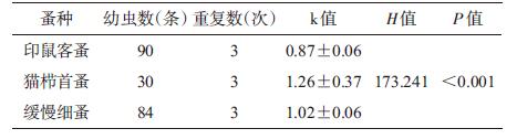
2 结果 2.1 3种幼虫触角长与触角间距的比较印鼠客蚤幼虫的触角从基部到端部形态变化均匀,细长如柱,触角的长度略小于触角间的距离(图 3),k=0.87±0.06;猫栉首蚤幼虫的触角形态与印鼠客蚤的幼虫触角形态近似,但其触角长略大于触角间距(图 4),k=1.26±0.37;缓慢细蚤的幼虫触角形态与前者的形态几乎相同,但其触角长几乎等于触角间距离(图 5),k=1.02±0.06。将3种蚤类幼虫的k值进行比较,差异有统计学意义(H=173.241,P < 0.001)。见表 1。

|
| 图 3 印鼠客蚤幼虫触角(×40) Figure 3 Larval antennae of Ctenocephalides felis (×30) |
| |

|
| 图 4 猫栉首蚤幼虫触角(×30) Figure 4 Schematic diagram of anal column length and anal column spacing of flea larvae |
| |

|
| 图 5 缓慢细蚤幼虫触角(×40) Figure 5 Larval antennae of Leptopsylla segnis (×40) |
| |
|
将3种幼虫触角长与触角间距的比值k进行两两比较,印鼠客蚤与猫栉首蚤(Z=4.239,P < 0.001)、印鼠客蚤与缓慢细蚤(Z=6.028,P < 0.001)、缓慢细蚤与猫栉首蚤(Z=4.793,P < 0.001),3种幼虫的触角长与触角间距的比值差异均有统计学意义。
2.2 3种幼虫肛柱长与肛柱间距的比较印鼠客蚤幼虫的肛柱长略大于肛柱间距离,其肛柱从基部到端部形态变化较缓(图 6),肛柱长与肛柱间距的比值r=1.31±0.13;猫栉首蚤幼虫的肛柱长约为肛柱间距的2倍多,其肛柱从基部逐渐变细,至端部为最细,形成较明显的基部粗、端部细的结构(图 7),肛柱长与肛柱间距的比值r=2.60±0.55;缓慢细蚤幼虫的肛柱基部与端部几乎等粗(图 8),且其肛柱长约是肛柱间距的4倍之多,r=4.82±1.47。将3种蚤类幼虫的r值进行比较,差异有统计学意义。见表 2。

|
| 图 6 印鼠客蚤幼虫肛柱(×80) Figure 6 Anal column of Xenopsylla cheopis larvae (×80) |
| |

|
| 图 7 猫栉首蚤幼虫肛柱(×60) Figure 7 Anal column of Ctenocephalides felis larvae (×60) |
| |

|
| 图 8 缓慢细蚤幼虫肛柱(×100) Figure 8 Anal column of Leptopsylla segnis larvae (×100) |
| |

|
将3种幼虫肛柱长与肛柱间距的比值r进行两两比较,印鼠客蚤与猫栉首蚤(Z=3.243,P < 0.001)、印鼠客蚤与缓慢细蚤(Z=3.072,P < 0.001)、缓慢细蚤与猫栉首蚤(Z=3.072,P < 0.001),3种幼虫的肛柱长与肛柱间距的比值差异均有统计学意义。
3 讨论我国蚤类幼虫研究始于1956年,至今已有60多年,报道的蚤类幼虫亦有60多种,但相对于成虫的研究,略显不足[16]。纵观蚤类幼虫研究,其重点多在形态的阐述,如幼虫破卵器形态[5]、体节毛序、大颚齿数、肛梳、肛柱及支柱毛等[17],且多以蚤类幼虫侧面标本或背腹标本为研究对象,少有使用新鲜幼虫的报道。本文以新鲜幼虫为研究对象,将其置于体式解剖镜下拍摄照片,放大若干倍,对其进行形态观察,因同一幼虫,其各个部位放大倍数相同,故其触角长与触角间距的比值,肛柱长与肛柱间距的比值为常数。经过观察发现:印鼠客蚤、猫栉首蚤、缓慢细蚤的触角形态几乎相同,但触角长与触角间距的比值差异较大,其中印鼠客蚤的触角长 < 触角间距,缓慢细蚤的触角长和触角间距大致相等,猫栉首蚤的触角长 > 触角间距。通过测量、比较触角长与触角间距的数值关系,对3种蚤类幼虫的鉴定具有一定的应用价值和意义。另外,通过比较3种幼虫的肛柱形态,发现其差异较大:印鼠客蚤幼虫的肛柱基部到端部近同粗,较纤细;猫栉首蚤幼虫肛柱端部细基部粗;缓慢细蚤幼虫肛柱端部至基部近等粗,但整体粗壮,明显比印鼠客蚤幼虫肛柱粗。3种蚤类幼虫肛柱长均 > 肛柱间距,其中缓慢细蚤差距最大,肛柱长约为肛柱间距的4倍之多,猫栉首蚤次之,也有2倍多,印鼠客蚤最小也在1倍左右。由此可见,根据该3种幼虫肛柱长与肛柱间距比值的对照,亦能做出简单的物种判断。
随着科学技术的不断进步,显微成像等先进的观察技术越来越多地应用到蚤类形态特征的分类研究中,通过形态结构的量化和比较,对蚤类幼虫形态的研究提供了新的技术手段,可为蚤类幼虫的分类鉴定和制定有效的蚤类防治措施提供科学依据。
志谢 中国疾病预防控制中心传染病预防控制所媒介生物控制室孟凤霞研究员对本研究大力支持,特此志谢| [1] |
王敦清. 几种常见蚤类幼虫形态的比较研究[J]. 昆虫学报, 1956, 6(3): 311-321, 378. Wang DQ. Comparative morphology of some common flea larvae (Siphonaptera)[J]. Acta Entomol Sin, 1956, 6(3): 311-321, 378. |
| [2] |
柳支英, 高钜镇, 吴厚永, 等. 黄鼠(Citellus dauricus Brandt)体外寄生的松江黄鼠蚤(Ceratophyllus tesquorum sungaris Jordan)的季节消长调查[J]. 人民军医, 1960(2): 33-40. Liu ZY, Gao JZ, Wu HY, et al. A study of the seasonal fluctuation of the suslik flea (Ceratophyllus tesquorum sungaris Jordan)[J]. People's Military Surgeon, 1960(2): 33-40. |
| [3] |
虞以新. 麻雀脊蚤幼虫形态的研究[J]. 动物学杂志, 1957, 1(2): 119-120. Yu YX. Study on larval morphology of sparrows' fleas[J]. Chin J Zool, 1957, 1(2): 119-120. |
| [4] |
孙昌秀. 草原黄鼠蚤的季节消长[J]. 寄生虫学报, 1965, 2(3): 310. Sun CX. The season of prairie fleas waxes and wanes[J]. J Para Med Ento, 1965, 2(3): 310. |
| [5] |
叶瑞玉, 于心, 王敦清. 我国若干蚤类幼虫形态的比较[J]. 昆虫学报, 1982, 25(2): 209-216. Ye RY, Yu X, Wang DQ. Larvae known in China, in addition with four new descriptions and studies on their spiracles[J]. Acta Entomol Sin, 1982, 25(2): 209-216. |
| [6] |
叶瑞玉, 于心. 新蚤属一新种及其幼虫形态的记述(蚤目:多毛蚤科)[J]. 昆虫学报, 1993, 36(3): 371-374. Ye RY, Yu X. On a new species of Neopsylla and its larva morphology (Siphonaptera:Hystrichopsyllidae)[J]. Acta Entomol Sin, 1993, 36(3): 371-374. |
| [7] |
费荣中, 徐宝娟, 石杲, 等. 方形黄鼠蚤松江亚种幼虫形态及与同属蚤幼虫的比较[J]. 昆虫学报, 1986, 29(1): 81-84. Fei RZ, Xu BJ, Shi G, et al. Morphological observations of the larvae of Citellophilus tesquorum sungaris and their comparison with larvae of two other forms of Citellophilus[J]. Acta Entomol Sin, 1986, 29(1): 81-84. |
| [8] |
王志军, 李明立. 无棘额蚤的养殖及其一龄幼虫破卵器形态[J]. 地方病通报, 1990, 5(1): 34-37. Wang ZJ, Li ML. Laboratorial breeding of Frontopsylla aspiriformis and morphologic observation on the egg buster of the first instar larvae[J]. Endem Dis Bull, 1990, 5(1): 34-37. |
| [9] |
肖柏林. 红羊新蚤的幼虫形态及与二齿新蚤幼虫的比较[J]. 昆虫学报, 1993, 36(1): 67-70. Xiao BL. Morphological observation of the larvae of Neopsylla hongyangensis and their comparison with larvae of N. bidentatiformis[J]. Acta Entomol Sin, 1993, 36(1): 67-70. |
| [10] |
肖柏林. 曲扎角叶蚤幼虫的形态描述[J]. 昆虫学报, 1995, 38(2): 188-190. Xiao BL. Description of the larval of morphology of Ceratophyllus chutsaensis (Siphonaptera:Ceratophyllidae)[J]. Acta Entomol Sin, 1995, 38(2): 188-190. |
| [11] |
Qi YM, He JH. Morphological description on the larva of Neopsylla specials specialis[J]. Entomol Sin, 1997, 4(1): 59-66. |
| [12] |
王键, 阴利群, 石杲. 蚤类幼虫肛柱腹小孔的形态及其分类意义[J]. 中国媒介生物学及控制杂志, 2000, 11(6): 466. Wang J, Yin LQ, Shi G. Morphology and taxonomic significance of antral lacunae of fleas larvae[J]. Chin J Vector Biol Control, 2000, 11(6): 466. DOI:10.3969/j.issn.1003-4692.2000.06.020 |
| [13] |
鲁亮, 吴厚永. 窄板额蚤华北亚种幼虫形态研究(蚤目:细蚤科)[J]. 昆虫学报, 2002, 45(3): 380-383. Lu L, Wu HY. The larval morphology of Frontopsylla nakagawai borealosinica (Siphonaptera:Leptopsyllidae)[J]. Acta Entomol Sin, 2002, 45(3): 380-383. DOI:10.3321/j.issn:0454-6296.2002.03.018 |
| [14] |
刘井元, 马立名, 周毅德. 吴氏角叶蚤幼虫形态记述及其与同属另外3种的比较[J]. 昆虫学报, 2003, 46(1): 85-89. Liu JY, Ma LM, Zhou YD. Description of larval morphology of fleas and comparison with other three species of the same genus[J]. J Ento, 2003, 46(1): 85-89. |
| [15] |
刘艳华, 温海军, 石杲. 内蒙古蚤类研究概述[J]. 医学动物防制, 2013, 29(1): 42-43. Liu YH, Wen HJ, Shi G. Introduction to research on fleas in Inner Mongolia[J]. J Med Pest Control, 2013, 29(1): 42-43. DOI:10.7629/yxdwfz201301013 |
| [16] |
王海波, 石杲. 中国蚤类幼虫分类研究概述[J]. 医学动物防制, 2003, 19(9): 533-534. H B, Shi G. An overview of the classification of flea larvae in China[J]. J Med Pest Control, 2003, 19(9): 533-534. DOI:10.3969/j.issn.1003-6245.2003.09.010 |
| [17] |
李景原, 石杲, 王建国, 等. 弯鬃蝠蚤的幼期及蚤类幼虫刚毛形态的观察[J]. 昆虫知识, 1988, 25(1): 35-36. Li JY, Shi G, Wang JG, et al. Observation on the juvenile stage and the morphology of the bristles of fleas[J]. Entomol Knowl, 1988, 25(1): 35-36. |