2. 广东省阳江市人民医院超声医学科, 广东 阳江 529500;
3. 广东省阳江市人民医院心内一科, 广东 阳江 529500
2. Department of Ultrasound, Yangjiang Municipal People’s Hospital, Yangjiang 529500 China;
3. The First Department of Cardiology, Yangjiang Municipal People’s Hospital, Yangjiang 529500 China
甲状腺结节是一种发生于甲状腺内的临床常见症状,其影像学特征与周围甲状腺实质不同[1]。由于诊断方法差异,一般人群甲状腺结节患病率为2%~65%[2];且由于人口老龄化、肥胖、工作生活压力、熬夜等多种因素影响,近年来甲状腺结节患病率成上升趋势[3]。近期一项系统综述和meta分析结果显示,全球甲状腺结节总体患病率为24.83%(95%可信区间:21.44%~28.55%),且患病率与高龄、女性、肥胖高度相关[4]。我国甲状腺结节总体患病率为38.0%(95%可信区间:37.0%~39.1%),且女性甲状腺结节患病率(44.7%)及年龄标化患病率(45.2%)均显著高于男性(分别为29.9%和31.2%);甲状腺结节患病率在18~25岁呈下降趋势,而在25岁后随年龄增加而上升;多因素logistic回归分析结果显示,年龄、性别、体质指数、血压、尿酸、空腹血糖、甘油三酯、高密度脂蛋白、低密度脂蛋白与甲状腺结节患病率显著相关[5]。
目前,甲状腺细针抽吸活检(fine needle aspiration biopsy,FNAB)是评估甲状腺结节性质最具诊断价值的方法[6-7]。而甲状腺细胞病理学分离依据Bethesda报告系统,其中第Ⅲ类为意义不明确的细胞非典型病变(atypia of undetermined significance,AUS),该病变恶性率高达10%~30%[8] ,随访、重复穿刺及外科手术是治疗AUS的主要措施[9]。凭借无创、无辐射、操作简易、成本-效益高等优势,超声检查为目前用于甲状腺结节筛查的首选方法[10-11];但甲状腺超声在AUS临床决策中的作用尚无定论,尤其是高分类甲状腺影像报告和数据系统(thyroid imaging reporting and data system,TI-RADS)的AUS甲状腺结节[12]。本研究旨在评价多模态超声成像用于TI-RADS 3~5类AUS甲状腺结节的诊断价值。
1 对象与方法 1.1 研究对象收集2019年1月—2022年4月88例甲状腺结节患者、90个经细胞病理学检查的AUS甲状腺结节为研究对象。纳入标准:① 经Bethesda报告系统诊断为AUS的甲状腺结节;② 包括超声和病理结果在内的患者病历资料完整;③ 甲状腺结节均成功获得超声弹性成像和超微血管成像;④ 所有甲状腺结节在超声检查前未进行射频消融术。本研究获得南方医科大学附属广东省人民医院医学伦理委员会批准,患者均知情同意,全部操作均符合相关伦理规范。
1.2 甲状腺超声检查采用TOSHIBA APLIO 500彩色超声诊断仪进行甲状腺超声检查,线阵探头频率5 ~14 MHz。所有患者均在FNAB检查前行甲状腺常规超声、超声弹性成像、超微血管成像检查。常规超声应用美国放射学会(American College of Radiology,ACR)推荐的TI-RADS系统对每个甲状腺结节成分、回声、形状、边缘和钙化等5个特征进行超声评估,每个特征均有相应分值,依据分值总和分为TI-RADS 1~5类[13]。参照如下标准进行甲状腺结节超声弹性成像评分:0分,结节内以囊性成分为主,呈红蓝/蓝绿红相间;1分,结节及其周围组织呈均匀绿色;2分,结节蓝绿相间,以绿色为主;3分,结节蓝绿相间,以蓝色为主;4分,蓝色完全覆盖结节[14]。甲状腺结节超微血管成像选择灰阶模式[15],采用半定量方法对结节内血流分布模式进行分型:Ⅰ型,甲状腺结节内无血流或少量血流,可见1 ~ 2个点状或短棒状血流信号;Ⅱ型,结节内可见 ≥ 3个血流信号,以周边血流为主;Ⅲ型,结节内可见 ≥ 3个血流信号,以中央血流为主;Ⅳ型,结节周边及中央均存在 ≥ 2个血流信号的混合型[16]。
1.3 甲状腺FNAB检查对甲状腺结节FNAB检查,对切除的甲状腺结节及颈部转移淋巴结标本进行细胞病理学检查,以甲状腺结节细胞病理结果为金标准评估常规超声、超声弹性成像、超微血管成像和多模态超声成像的诊断价值。
1.4 统计方法应用多因素logistic回归模型构建AUS甲状腺结节多模态超声成像诊断预测模型,绘制受试者工作特征(receiver operating characteristic,ROC)曲线,计算曲线下面积(area under curve,AUC)。采用SPSS 25.0软件进行统计学分析,P < 0.05为差异有统计学意义,组间率的比较采用χ2检验和Fisher精确概率法,AUC值比较采用Z检验。
2 结 果 2.1 甲状腺结节细胞病理学检查结果经细胞病理学检查,90个AUS甲状腺结节中,69个恶性结节包括65个甲状腺乳头状癌、3个甲状腺滤泡癌、1个甲状腺髓样癌,21个良性结节包括17个结节性甲状腺肿、1个甲状腺腺瘤和3个桥本氏甲状腺炎。
2.2 良恶性甲状腺结节临床特征比较良恶性甲状腺结节性别构成、年龄构成、位置差异均无统计学意义(P均 > 0.05),但恶性结节中 ≤ 1 cm的甲状腺结节比例显著高于良性结节(χ² = 9.610,P = 0.002)(表1)。
|
|
表 1 良恶性甲状腺结节人口学和临床特征比较 Table 1 Comparison of demographic and clinical features between benign and malignant thyroid nodules |
恶性甲状腺结节中,结节呈低回声/极低回声比例、边界模糊结节比例、结节纵横比 > 1比例及结节呈微小钙化/无钙化特征比例显著多于良性结节(P均 < 0.05)(表2),超声弹性成像评分为 ≥ 3分及超微血管成像分型为III型血流模式提示恶性结节可能性更高(P 均 < 0.001)(图1)。
|
|
表 2 良恶性甲状腺结节常规超声、超微血管成像和超声弹性成像特征比较 Table 2 Comparison of features derived from conventional ultrasonography, ultrasound elastography, and superb microvascular imaging between benign and malignant thyroid nodules |

|
图 1 甲状腺乳头状癌病例甲状腺结节超声图像 Figure 1 Ultrasound images of thyroid nodules in a patient with papillary thyroid carcinoma 注:A 常规超声图像示甲状腺右侧叶一大小约8 mm实性低回声结节,边界模糊,周边弧形钙化,纵横比 < 1,TI-RADS 4类;B 甲状腺结节超声弹性成像评分为3分;C 甲状腺结节超微血管成像示III型血流模式。 |
多因素logistic回归分析结果显示,甲状腺结节大小、回声、边界、纵横比及超微血管成像分型均与甲状腺结节良恶性无统计学意义(P均 > 0.05),而微小钙化/无钙化和超声弹性成像评分 ≥ 3分是AUS甲状腺恶性结节的独立危险因素(P均 < 0.05)(表3)。
|
|
表 3 超声图像特征与AUS甲状腺结节良恶性关联的多因素logistic回归分析 Table 3 Multivariable logistic regression analysis of the relationship between ultrasonographic features and benign/malignant potential of AUS thyroid nodules |
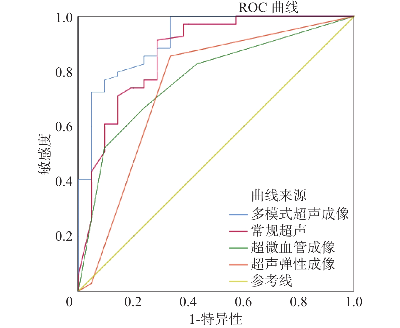
以甲状腺结节FNAB细胞病理学检查结果为金标准,结果常规超声诊断AUS甲状腺结节良恶性的敏感度、特异度、准确度、假阳性率、假阴性率分别为91.30%、71.40%、62.70%、28.60%和8.70%,超声弹性成像分别为85.50%、66.70%、52.20%、33.30%和14.50%,超微血管成像分别为66.70%、76.20%、42.90%、23.80%和33.30%,多模态超声成像分别为75.20%、92.50%、67.70%、24.80%和7.50%。常规超声、超声弹性成像、超微血管成像、多模态超声成像诊断AUS甲状腺结节良恶性的AUC值分别为0.866、0.745、0.774和0.918(图2),表明多模态超声成像诊断AUS甲状腺结节良恶性效能显著高于其他3种方法。

|
图 2 4种超声方法诊断AUS甲状腺结节良恶性的ROC曲线 Figure 2 Receiver operating characteristic curves for distinguishing between benign and malignant AUS thyroid nodules using four ultrasound methods |
在我国31个省(直辖市、自治区)开展的流行病学调查结果显示,我国甲状腺疾病总体患病率为50.96%[17]。既往研究表明,年龄是甲状腺癌预后的独立危险因素;多数甲状腺癌预后良好,但甲状腺癌复发转移风险与年龄呈线性相关[18]。此外,生存率随年龄增长而下降,血管侵袭和早期转移在老年患者中更为常见[19]。因此,早期、精准诊断甲状腺结节对于改善疾病预后、提高生存率具有重要意义。本研究针对TI-RADS 3~5类AUS甲状腺结节,该类结节较1~2类结节良恶性更难以诊断,探索单个及多模态超声成像对AUS甲状腺结节诊断能力对甲状腺癌临床诊疗有重大意义。既往较少有同时比较常规超声、超声弹性成像、超微血管成像及多模态超声成像诊断TI-RADS 3~5类AUS甲状腺结节的报道[20-21]。这类结节良恶性特征往往并不明显,常规超声较其他超声技术筛查灵敏度高,但其假阳性率高,并不能较高诊断良恶性。多模态超声成像将超声弹性成像、超微血管成像与常规超声技术相结合,各技术得以互补后综合评估AUS甲状腺结节,可提高其诊断准确率。
本研究结果显示,以甲状腺结节FNAB细胞病理学检查结果为金标准,多模态超声成像诊断TI-RADS 3~5类AUS甲状腺结节良恶性的准确度和AUC值显著均高于常规超声、超声弹性成像和超微血管成像,而假阳性率显著低于其他3种超声诊断技术,提示多模态超声成像较单一超声成像技术对诊断TI-RADS 3~5类AUS甲状腺结节良恶性准确性更高。与常规超声筛查相比,多模态超声成像增加了甲状腺结节检查成本,优先获得特异度较高的检查结果。超微血管成像可弥补超声弹性成像不能评估甲状腺结节种血流分布的缺点;对于一些较小的恶性甲状腺结节,超微血管成像的血流分布模式仅为I或II型,假阴性结果较常见,而超声弹性成像可通过评分获得结节“硬度”信息,从而实现准确诊断。
本研究采用超声弹性成像评估TI-RADS 3~5类AUS甲状腺结节,以3分作为诊断临界值,即 ≥ 3分为恶性、< 3为良性。结果显示,超声弹性成像诊断AUS甲状腺结节的敏感度为85.50%,与既往研究结果一致[22-24]。本研究结果显示,超声弹性成像诊断AUS甲状腺结节良恶性的灵敏度较常规超声低,且其假阳性率较高,分析其原因可能部分结节表现为内部粗大钙化或周边弧形钙化导致弹性评分升高,另外一些结节因长期纤维化也会导致弹性评分升高。
本研究结果显示,超微血管成像评估AUS甲状腺结节良恶性的敏感度为66.70%,较既往报道低[25-26],这可能与纳入的参数较少有关。超微血管成像较超声弹性成像诊断甲状腺结节良恶性假阳性率较低,分析其原因可能是环形和弧形钙化存在导致评估不准确,而超微血管成像可以准确评估结节内血流分布模式,从而避免粗大钙化的影响。
本研究也存在一定局限性。如入组样本量较少,结节病理类型相对简单等。在今后的研究中,将收集更多数据,以进一步证实研究的可行性。
本研究结果表明,多模态超声成像可提高常规超声、超声弹性成像、超声血管成像用于诊断TI-RADS 3~5类AUS甲状腺结节良恶性的诊断效能,有助于弥补常规超声在评估AUS甲状腺结节方面的不足,有助于临床医生对AUS甲状腺结节的恶性风险分层和管理。
| [1] |
Burman KD, Wartofsky L. Thyroid nodules[J]. N Engl J Med, 2015, 373(24): 2347-2356. DOI:10.1056/NEJMcp1415786 |
| [2] |
Dean DS, Gharib H. Epidemiology of thyroid nodules[J]. Best Pract Res Clin Endocrinol Metab, 2008, 22(6): 901-911. DOI:10.1016/j.beem.2008.09.019 |
| [3] |
Uppal N, Collins R, James B. Thyroid nodules: Global, economic, and personal burdens[J]. Front Endocrinol (Lausanne), 2023, 14: 1113977. DOI:10.3389/fendo.2023.1113977 |
| [4] |
Mu CY, Ming X, Tian Y, et al. Mapping global epidemiology of thyroid nodules among general population: a systematic review and meta-analysis[J]. Front Oncol, 2022, 12: 1029926. DOI:10.3389/fonc.2022.1029926 |
| [5] |
Li YH, Jin C, Li J, et al. Prevalence of thyroid nodules in China: a health examination cohort-based study[J]. Front Endocrinol (Lausanne), 2021, 12: 676144. DOI:10.3389/fendo.2021.676144 |
| [6] |
Belfiore A, La Rosa GL. Fine-needle aspiration biopsy of the thyroid[J]. Endocrinol Metab Clin North Am, 2001, 30(2): 361-400. DOI:10.1016/s0889-8529(05)70191-2 |
| [7] |
周乐, 付吉涛, 孙辉. 超声引导下细针穿刺技术在甲状腺外科中的应用进展[J]. 中华医学超声杂志(电子版), 2021, 18(9): 895-897. Zhou L, Fu JT, Sun H. Application and expansion of ultrasonic-guided fine needle puncture technology in thyroid surgery[J]. Chin J Med Ultrasound Electron Ed, 2021, 18(9): 895-897. DOI:10.3877/cma.j.issn.1672-6448.2021.09.015 |
| [8] |
Cibas ES, Ali SZ. The 2017 Bethesda system for reporting thyroid cytopathology[J]. Thyroid, 2017, 27(11): 1341-1346. DOI:10.1089/thy.2017.0500 |
| [9] |
Erivwo P, Ghosh C. Atypia of undetermined significance in thyroid fine-needle aspirations: follow-up and outcome experience in Newfoundland, Canada[J]. Acta Cytol, 2018, 62(2): 85-92. DOI:10.1159/000486779 |
| [10] |
Boers T, Braak SJ, Rikken NET, et al. Ultrasound imaging in thyroid nodule diagnosis, therapy, and follow-up: current status and future trends[J]. J Clin Ultrasound, 2023, 51(6): 1087-1100. DOI:10.1002/jcu.23430 |
| [11] |
王萱, 芮忠颖, 郑薇, 等. 超声新技术诊断甲状腺结节的应用进展[J]. 国际放射医学核医学杂志, 2021, 45(7): 455-460. Wang X, Rui ZY, Zheng W, et al. Application of new ultrasonic technology in the diagnosis of thyroid nodule[J]. Int J Radiat Med Nucl Med, 2021, 45(7): 455-460. DOI:10.3760/cma.j.cn121381-202103018-00071 |
| [12] |
Abelardo AD, Sotalbo KCJ. Clinical management of thyroid aspirates diagnosed as atypia of undetermined significance in the Philippines[J]. Gland Surg, 2020, 9(5): 1788-1796. DOI:10.21037/gs-20-426 |
| [13] |
Hoang JK, Middleton WD, Tessler FN. Update on ACR TI-RADS: Successes, challenges, and future directions, from the AJR special series on radiology reporting and data systems[J]. AJR Am J Roentgenol, 2021, 216(3): 570-578. DOI:10.2214/AJR.20.24608 |
| [14] |
吴琼, 王燕, 李艺, 等. 超声引导下细针穿刺活检和实时弹性成像联合应用诊断可疑甲状腺结节的价值[J]. 中国介入影像与治疗学, 2015, 12(1): 43-46. Wu Q, Wang Y, Li Y, et al. Value of ultrasound-guided fine-needle aspiration combined with real-time ultrasound elastography in diagnosis of suspicious thyroid nodules[J]. Chin J Interv Imag Ther, 2015, 12(1): 43-46. DOI:10.13929/j.1672-8475.2015.01.011 |
| [15] |
杨艳, 胡金花, 夏群, 等. 超微血管成像技术与彩色多普勒血流成像分别联合甲状腺影像报告与数据系统在甲状腺结节鉴别诊断中的应用[J]. 中国医药导报, 2023, 20(7): 165-168. Yang Y, Hu JH, Xia Q, et al. Application of superb microvascular imaging and color Doppler flow imaging combined with thyroid image report and data system in the differential diagnosis of thyroid nodule[J]. China Med Herald, 2023, 20(7): 165-168. DOI:10.20047/j.issn1673-7210.2023.07.37 |
| [16] |
Lan Y, Li N, Song Q, et al. Correlation and agreement between superb micro-vascular imaging and contrast-enhanced ultrasound for assessing radiofrequency ablation treatment of thyroid nodules: a preliminary study[J]. BMC Med Imaging, 2021, 21(1): 175. DOI:10.1186/s12880-021-00697-y |
| [17] |
Li YZ, Teng D, Ba JM, et al. Efficacy and safety of long-term universal salt iodization on thyroid disorders: epidemiological evidence from 31 provinces of mainland China[J]. Thyroid, 2020, 30(4): 568-579. DOI:10.1089/thy.2019.0067 |
| [18] |
韩郁壬, 李利梅, 王睿. 甲状腺癌临床病理特点与其预后影响因素分析[J]. 实用癌症杂志, 2022, 37(6): 1000-1002. Han YR, Li LM, Wang R. Clinical characteristics and prognostic factors of thyroid cancer patients[J]. Pract J Cancer, 2022, 37(6): 1000-1002. DOI:10.3969/j.issn.1001-5930.2022.06.037 |
| [19] |
门伯媛, 高海燕, 侯铁军, 等. 甲状腺癌生存率及其影响因素[J]. 西安医科大学学报, 2001, 22(3): 267-269. Men BY, Gao HY, Hou TJ, et al. Survival rate and its influential factors of the thyroid cancer[J]. J Xi’an Med Univ, 2001, 22(3): 267-269. DOI:10.3969/j.issn.1671-8259.2001.03.026 |
| [20] |
李朝喜, 温德惠, 刘伟亮, 等. SMART 3D-SMI在甲状腺TI-RADS 4类结节良恶性鉴别诊断中的应用[J]. 影像科学与光化学, 2022, 40(3): 510-514. Li CX, Wen DH, Liu WL, et al. Application of SMART 3D-SMI in the differential diagnosis of benign and malignant in TI-RADS 4 thyroid nodules[J]. Imag Sci Photochem, 2022, 40(3): 510-514. DOI:10.7517/issn.1674-0475.211204 |
| [21] |
钟文乐, 张碧宏, 赖胜坤, 等. 多模式超声成像联合应用在甲状腺实性低回声结节 良恶性诊断中的价值[J]. 临床医学工程, 2019, 26(12): 1609-1610. Zhong WL, Zhang BH, Lai SK, et al. The value of combined application of multi-mode ultrasound imaging in the diagnosis of benign and malignant thyroid solid hypoechoic nodules[J]. Clin Med Eng, 2019, 26(12): 1609-1610. DOI:10.3969/j.issn.1674-4659.2019.12.1609 |
| [22] |
Magri F, Chytiris S, Zerbini F, et al. Maximal stiffness evaluation by real-time ultrasound elastography, an improved tool for the differential diagnosis of thyroid nodules[J]. Endocr Pract, 2015, 21(5): 474-481. DOI:10.4158/EP14504.OR |
| [23] |
裴书芳, 丛淑珍, 钱隽, 等. 甲状腺良恶性结节的弹性成像和常规超声特征及联合诊断效能[J]. 中国医学影像技术, 2015, 31(5): 725-728. Pei SF, Cong SZ, Qian J, et al. Elastic and conventional ultrasonic characteristics of benign and malignant thyroid nodules and efficacy of combined diagnosis[J]. Chin J Med Imag Technol, 2015, 31(5): 725-728. DOI:10.13929/j.1003-3289.2015.05.022 |
| [24] |
王燕, 金佳美, 陈林, 等. 超声联合硬度评分系统诊断TI-RADS 4类甲状腺结节[J]. 中国医学影像技术, 2016, 32(7): 1039-1042. Wang Y, Jin JM, Chen L, et al. United stiffness score system for diagnosis of TI-RADS 4 classification thyroid nodules[J]. Chin J Med Imag Technol, 2016, 32(7): 1039-1042. DOI:10.13929/j.1003-3289.2016.07.013 |
| [25] |
陶玲玲, 詹维伟, 樊金芳, 等. 超微血管成像结合TI-RADS鉴别诊断甲状腺良恶性结节[J]. 中国医学影像技术, 2020, 36(5): 671-674. Tao LL, Zhan WW, Fan JF, et al. Superb micro-vascular imaging combined with TI-RADS in differential diagnosis of benign and malignant thyroid nodules[J]. Chin J Med Imag Technol, 2020, 36(5): 671-674. DOI:10.13929/j.issn.1003-3289.2020.05.007 |
| [26] |
杨光旭, 万静, 吴作辉, 等. 超微血管成像评估甲状腺影像报告和数据系统(TI-RADS)4级甲状腺结节血流特征[J]. 中国医学影像技术, 2021, 37(6): 867-870. Yang GX, Wan J, Wu ZH, et al. Superb micro-vascular imaging in evaluation on flow characteristics of thyroid imaging reporting and data system(TI-RADS) grade 4 thyroid nodules[J]. Chin J Med Imag Technol, 2021, 37(6): 867-870. DOI:10.13929/j.issn.1003-3289.2021.06.017 |


