2. 湖南省株洲市中心医院肿瘤内科,湖南 株洲 412007
2. Department of Oncology, Zhuzhou Central Hospital, Zhuzhou 412007 China
子宫内膜癌是一种源自子宫内膜的常见女性恶性肿瘤,也是发达国家最常见的妇科肿瘤[1]。2018年,全球累计有38万子宫内膜癌新发病例,约有9万人死于子宫内膜癌[2]。基于全球疾病负担(Global Burden of Disease)数据分析显示,全球子宫内膜癌发病率呈上升趋势,1990—2019年全球子宫内膜癌年龄标化发病率每年增长0.69%(95%可信区间为0.57%~0.81%);虽然30年间全球子宫内膜癌死亡率整体呈下降趋势,但在91个国家和地区子宫内膜癌死亡率仍然上升[3]。子宫内膜癌不断上升的发病率和患病率给全球社会、经济、医疗资源造成了沉重负担[4]。在我国,子宫内膜癌发病率亦呈逐年上升趋势,成为仅次于宫颈癌的我国女性生殖系统第二大恶性肿瘤,2000—2011年子宫内膜癌年龄标化发病率年度变化百分比为3.7%;2015年,我国共有6万余例子宫内膜癌新发病例,2.2万人死于子宫内膜癌[5]。
早期和精准诊断对于优化子宫内膜癌治疗方案、改善预后具有重要意义[6]。术前影像学检查有助于了解子宫内膜癌局部病灶、是否发生远端转移,从而指导、优化治疗方案[7]。虽然国际妇产科联合会(The International Federation of Gynecology and Obstetrics,IFGO)外科分级指南已用于子宫内膜癌分级,但经阴道超声、CT、磁共振成像(MRI)、PET-CT、PET-MRI等影像学技术已用于术前子宫内膜癌分级工具;此外,这些影像学技术还在疗效评估、早期复发监测等方面发挥了重要作用[6]。本研究旨在分析经阴道彩色多普勒超声用于术前鉴别子宫内膜癌病理分级的价值。
1 对象与方法 1.1 对象回顾性分析2017年1月—2020年8月收治于株洲市中心医院妇产科的68例子宫内膜癌患者病例资料,所有68例患者均经组织病理学确诊。本研究方案经株洲市中心医院伦理审查委员会批准,所有研究对象均知情并签署知情同意书。
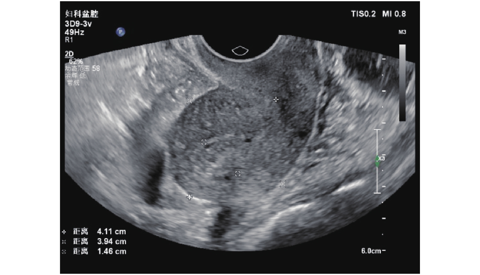
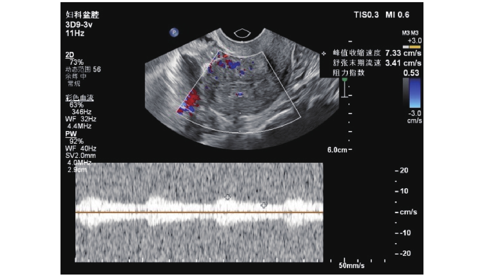
1.2 超声检查应用PHILIPS Affiniti70型彩色多普勒超声诊断仪仔细检测矢状面和横切面子宫体和子宫内膜,阴道探头频率7.5 MHz。测量子宫前后壁上子宫内膜和子宫肌层交接处的子宫内膜厚度、血流情况(图1、2),观察子宫肌层浸润及子宫平滑肌瘤、子宫内膜异位等发生情况。在子宫颈内口水平采用彩色多普勒超声诊断仪观察左、右侧子宫动脉血流,同时测定左、右侧子宫动脉血流阻力指数。全部超声检查均由同一位高年资超声科医师完成,且对受检对象临床资料不知情。

|
图 1 子宫内膜癌患者子宫内膜增厚,回声不均 Figure 1 Endometrial thickening and uneven echo in patients with endometrial cancer |

|
图 2 子宫内膜癌患者血流阻力指数 Figure 2 Blood flow resistance index in patients with endometrial cancer |
采取经腹子宫全切除术和双侧输卵管、卵巢切除术,术中收集腹膜细胞学标本,术后3 d内进行病变组织病理分级的超声评估。2~3级子宫内膜癌患者、子宫乳头状浆液性腺癌患者或子宫内膜癌侵犯深肌层患者给予系统性盆腔和腹主动脉旁淋巴结清扫术。术中将子宫解剖分成2部分,以观察子宫内膜癌侵犯子宫肌层情况。全部患者根据子宫肌层浸润程度分成两组:≤ 50%组和 > 50%组。根据WHO分类标准对子宫内膜癌进行组织学分类 [8]。以组织学评价结果为金标准,分析彩色多普勒超声检测子宫肌层浸润的准确度、灵敏度、特异度、阳性预测值和阴性预测值。
1.3 统计分析采用Microsoft Excel 2010软件建立数据库,应用SPSS 22.0软件进行统计学分析。计量资料采用均数 ± 标准差描述,组间差异的统计学比较采用Student’s t检验;计数资料采用百分比和率描述,组间差异的统计学比较采用χ2检验;多组间比较采用方差分析。检验水准α = 0.05。
2 结 果 2.1 病例概况68例子宫内膜癌患者中,年龄51~67岁,平均(56.6 ± 7.8)岁;绝经58例,占85.3%;经阴道彩色多普勒超声测定子宫内膜厚度7~19 mm,平均(27.1 ± 14.9) mm。根据组织学类型,其中腺癌48例,占70.6%;乳头状浆液性腺癌和腺鳞癌各9例,均占13.2%;透明细胞癌2例,占3.0%;根据WHO分级标准,1级26例,2级33例,3级9例(表1)。
|
|
表 1 研究对象肿瘤分级及子宫肌层浸润情况 Table 1 Grading and myometrial invasion of the study subjects |
经阴道彩色多普勒超声检测子宫肌层浸润的灵敏度为70.0%、特异度为67.9%、阳性预测值为75.7%、阴性预测值为61.3%、准确度为69.1%(表2)。进一步分析显示,子宫内膜癌侵犯子宫肌层 ≤ 50%的子宫内膜癌患者左、右侧子宫动脉阻力指数均显著高于子宫肌层浸润 > 50%的患者(均 P < 0.01);肿瘤分级越低,其阻力指数越高(均 P < 0.05)( 表3、4)。
|
|
表 2 经阴道彩色多普勒超声检测子宫肌层浸润效果 Table 2 Efficacy of transvaginal color Doppler ultrasonography for detection of myometrial invasion |
|
|
表 3 子宫动脉阻力指数与子宫内膜浸润的关联 Table 3 Correlation between resistance index of uterine artery and myometrial invasion |
|
|
表 4 子宫动脉阻力指数与子宫内膜癌分级的关联 Table 4 Correlation between resistance index of uterine artery and grading of endometrial cancer |
彩色多普勒超声利用人体组织与超声探头间距离相关声波或超声波的频率变化,常用于探查生殖系统肿块;而彩色多普勒超声还可以测定血流阻力指数,常用于筛查妇科和产科病变[9]。既往国内外已对经阴道彩色多普勒超声用于子宫内膜癌的诊断价值进行评价。杨冬[10]研究发现,经阴道彩色多普勒超声术前诊断子宫内膜癌与术后病理诊断结果符合率为96.7%,认为其对子宫内膜癌病理分级和恶性淋巴结肿大有一定的术前评估意义。李旭霞等[11]以手术及病理结果作为金标准,发现经阴道彩色多普勒超声用于子宫内膜病变的诊断符合率为95.87%。
此外,经阴道彩色多普勒超声还被用于子宫内膜癌浸润深度和分期评估。刘爱民等[12]报道经阴道彩色多普勒超声检查与手术病理T分期的结果符合率为73.2%。谭莉等[13]报道,经阴道彩色多普勒超声检测子宫内膜癌深肌层浸润的准确度、灵敏度、特异度、阳性预测值和阴性预测值分别为81.9%、60.0%、85.4%、40.0%和93.0%。张金玲等[14]研究发现,经阴道彩色多普勒超声检测子宫内膜癌肌层浸润程度与病理结果符合率为93.2%,诊断子宫内膜癌分期和病理分期结果符合率为65.2%,诊断子宫内膜癌分期与血流分级呈显著正相关(r = 0.794,P < 0.05)。王莉 [15]研究发现,经阴道彩色多普勒超声对子宫内膜癌的分期结果与病理分期结果显著相关(r = 0.893,P < 0.01),且与病理分期结果高度一致( Kappa值 = 0.673,P < 0.01)。刘晓芳等 [16]报道,根据2009版FIGO分期标准,经阴道彩色多普勒用于评估Ia、Ib及Ⅱ期子宫内膜癌分期的准确度分别为80%、83.33%及100%。陆娟仪等[17]亦报道,,经阴道彩色多普勒预测的子宫内膜癌分期与血流分级呈显著正相关(r = 0.638,P = 0.008),与FIGO分期结果亦呈显著正相关(r = 0.887,P = 0.003),且与FIGO分期结果较为一致(Kappa值 = 0.607,P = 0.010)。
本研究纳入的68例子宫内膜癌患者中,绝经女性占85.3%,与子宫内膜癌高发于绝经女性的流行病学数据一致[1-2, 18]。本研究结果发现,经阴道彩色多普勒超声检测子宫肌层浸润的准确度为69.1%,与既往土耳其学者报道结果类似[19]、但低于国内的相关报道[13-14]。本研究结果还发现,子宫内膜癌侵犯子宫肌层 ≤ 50%的子宫内膜癌患者左、右侧子宫动脉阻力指数均显著高于子宫肌层浸润 > 50%的患者(均 P < 0.01),肿瘤分级越低、其阻力指数越高(均 P < 0.05),与既往报道结果一致 [19]。
本研究结果表明,经阴道彩色多普勒超声检测子宫内膜癌患者子宫肌层浸润的准确度较高,可作为一种无创工具用于评估子宫内膜癌患者子宫肌层浸润程度和术前病理分级。建议将经阴道彩色多普勒超声检测纳入子宫内膜癌术前常规评估的一部分,用于指导患者个体化治疗方案的制定。
| [1] |
Morice P, Leary A, Creutzberg C, et al. Endometrial cancer[J]. Lancet, 2016, 387(10023): 1094-1108. DOI:10.1016/S0140-6736(15)00130-0 |
| [2] |
Lu KH, Broaddus RR. Endometrial cancer[J]. N Engl J Med, 2020, 383(21): 2053-2064. DOI:10.1056/NEJMra1514010 |
| [3] |
Gu B, Shang X, Yan M, et al. Variations in incidence and mortality rates of endometrial cancer at the global, regional, and national levels, 1990-2019[J]. Gynecol Oncol, 2021, 161(2): 573-580. DOI:10.1016/j.ygyno.2021.01.036 |
| [4] |
Zhang S, Gong TT, Liu FH, et al. Global, regional, and national burden of endometrial cancer, 1990-2017: results from the global burden of disease study, 2017[J]. Front Oncol, 2019, 9: 1440. DOI:10.3389/fonc.2019.01440 |
| [5] |
Jiang X, Tang H, Chen T. Epidemiology of gynecologic cancers in China[J]. J Gynecol Oncol, 2018, 29(1): e7. DOI:10.3802/jgo.2018.29.e7 |
| [6] |
Faria SC, Devine CE, Rao B, et al. Imaging and staging of endometrial cancer[J]. Semin Ultrasound CT MR, 2019, 40(4): 287-294. DOI:10.1053/j.sult.2019.04.001 |
| [7] |
Haldorsen IS, Salvesen HB. What is the best preoperative imaging for endometrial cancer[J]. Curr Oncol Rep, 2016, 18(4): 25. DOI:10.1007/s11912-016-0506-0 |
| [8] |
Petru E, Lück HJ, Stuart G, et al. Gynecologic Cancer Intergroup (GCIG) proposals for changes of the current FIGO staging system[J]. Eur J Obstet Gynecol Reprod Biol, 2009, 143(2): 69-74. DOI:10.1016/j.ejogrb.2008.12.015 |
| [9] |
Fleischer AC, Andreotti RF. Color Doppler sonography in obstetrics and gynecology[J]. Expert Rev Med Devices, 2005, 2(5): 605-611. DOI:10.1586/17434440.2.5.605 |
| [10] |
杨冬. 经阴道彩色多普勒超声对子宫内膜癌诊断价值研究[J]. 河北医药, 2012, 34(13): 1966-1967. Yang D. Study on the diagnostic value of transvaginal color Doppler ultrasonography for endometrial cancer[J]. Hebei Med J, 2012, 34(13): 1966-1967. DOI:10.3969/j.issn.1002-7386.2012.13.021 |
| [11] |
李旭霞, 张宏, 王保健. 经阴道彩色多普勒超声诊断子宫内膜病变[J]. 中国介入影像与治疗学, 2009, 6(6): 520-522. Li XX, Zhang H, Wang BJ. Transvaginal color Doppler ultrasound in identifying endometrium abnormality[J]. Chin J Interv Imaging Ther, 2009, 6(6): 520-522. |
| [12] |
刘爱民, 马志伟, 杨军. 经阴道彩色多普勒超声对临床I期子宫内膜癌浸润深度与手术病理T分期的对照研究[J]. 世界最新医学信息文摘, 2015, 15(A4): 28-29. Liu AM, Ma ZW, Yang J. Comparative study on invasion depth of clinical stage I endometrial carcinoma and T staging of surgical pathology by transvaginal color Doppler ultrasound[J]. World Latest Med Inf, 2015, 15(A4): 28-29. |
| [13] |
谭莉, 姜玉新, 潘凌亚, 等. 经阴道超声对术前诊断子宫内膜癌肌层浸润程度的价值[J]. 中华超声影像学杂志, 2002, 11(6): 345-347. Tan L, Jiang YX, Pan LY, et al. The value of transvaginal sonography in preoperative assessment of the myometrial invasion in endometrial carcinoma[J]. Chin J Ultrason, 2002, 11(6): 345-347. DOI:10.3760/j.issn:1004-4477.2002.06.007 |
| [14] |
张金玲, 付瑞, 李豆豆, 等. 经阴道彩色多普勒超声检测子宫内膜癌及对肌层浸润程度和血流分级的诊断分析[J]. 实用癌症杂志, 2021, 36(7): 1140-1143. Zhang JL, Fu R, Li DD, et al. Transvaginal color Doppler ultrasound detection of endometrial cancer and diagnosis and analysis of myometrial infiltration and flow classification[J]. Pract J Cancer, 2021, 36(7): 1140-1143. DOI:10.3969/j.issn.1001-5930.2021.07.024 |
| [15] |
王莉. 经阴道彩色多普勒超声扫查子宫内膜癌声像图特征及其在病理分期中的应用价值[J]. 临床超声医学杂志, 2018, 20(2): 98-101. Wang L. Value of transvaginal color Doppler ultrasound in diagnosis of endometrial carcinoma and its pathological staging[J]. J Clin Ultrasound Med, 2018, 20(2): 98-101. DOI:10.16245/j.cnki.issn1008-6978.2018.02.009 |
| [16] |
刘晓芳, 戴常平, 王红英. 经阴道彩色多普勒超声在子宫内膜癌新分期中的价值研究[J]. 肿瘤影像学, 2016, 25(2): 174-178. Liu XF, Dai CP, Wang HY. Value of transvaginal color Doppler sonography in new preoperative staging of endometrial carcinoma[J]. Oncoradiology, 2016, 25(2): 174-178. |
| [17] |
陆娟仪, 韩虹, 吴慧, 等. 子宫内膜癌阴道超声图像的特征及其对病理分期的意义[J]. 中国妇幼保健, 2020, 35(3): 562-564. Lu JY, Han H, Wu H, et al. Transvaginal color Doppler sonographic features of endometrial carcinoma and its significance in pathologic staging[J]. Matern Child Heal Care China, 2020, 35(3): 562-564. DOI:10.19829/j.zgfybj.issn.1001-4411.2020.03.057 |
| [18] |
张伊彤, 白田妹. 子宫内膜恶变危险因素分析[J]. 临床军医杂志, 2021, 49(6): 690-691. Zhang YT, Bai TM. Analysis of risk factors for endometrial malignancies[J]. Clin J Med Off, 2021, 49(6): 690-691. |
| [19] |
Kanat-Pektas M, Gungor T, Mollamahmutoglu L. The evaluation of endometrial tumors by transvaginal and Doppler ultrasonography[J]. Arch Gynecol Obstet, 2008, 277(6): 495-499. DOI:10.1007/s00404-007-0517-z |


