食管癌是常见的恶性肿瘤,放射治疗是食管癌治疗主要手段[1-2]。随着放疗技术的不断发展,动态调强放疗现已成为上段食管癌放疗的主流技术[3-4],动态调强放疗技术在实际治疗中对叶片运动同步性要求高,子野较多,故患者在放射治疗前需对动态调强放疗计划进行剂量验证[5-7]。上段食管癌靶区前端有大气管空腔,由于剂量的二次建成效应可能导致空腔后方靶区的剂量计算不准确,并且常规的剂量验证方式也是在均匀模体上进行,不能真实地反映出患者体内空腔计算不确定性所带来的剂量验证结果[8-9]。为此探究我院Monaco TPS中Monte Carlo和Pencil Beam 2种算法在不同空腔厚度模体剂量计算的准确性,以及上述2种算法在不同空腔厚度模体对上段食管癌剂量验证的影响。
1 材料与方法 1.1 病例选取及一般资料在本科室2018年下半年接受固定野(0°、50°、150°、210°、300°)动态调强放疗的上段食管癌患者中,采用单纯随机抽样方法选取20例,其中男14例、女6例,年龄43~72岁;年龄中位数59岁。根据第8版AJCC食管癌分期标准,其中ⅠB期4、ⅡA期8例、ⅡB期6例、ⅢB期2例。参考ICRU83号报告勾画危及器官,同时勾画原发肿瘤区(GTV)、转移淋巴结肿瘤区(GTVnd)、亚临床病灶及高危淋巴引流区(CTV),根据上述靶区外扩5 mm得到相应计划靶区(PGTV、PGTVnd、PTV),并分别给予对应靶区60、60 Gy和51 Gy的处方剂量,分30次完成治疗。
1.2 设备与材料配有Monaco5.11 TPS的瑞典医科达Versa HD型医用直线加速器;美国Sun Nuclear 的MapCHeck半导体探测器阵列;PTW的RW3固体水(带有电离室插口)、UNIDOSE剂量仪和0.6CC的指型电离室(30013);自制标准厚度(5,10 mm和20 mm)小木块若干。
1.3 测量方法将带有指型电离室插口的固体水和MapCheck平板分别与标准厚度的小木块组成不同空腔厚度的模体,空腔厚度分别为0、5、10 mm和20 mm。将这些模体放在CT模拟定位机(PHILIPS Brilliance)上进行扫描,共获得8套CT图像,并将获取的图像传至Monaco TPS,在Monaco TPS中分别对含有指型电离室插口的4套空腔模体添加以电离室插口为中心、机架角度0°、能量为6 MV和100 MU的4个照射方野(15 cm × 15 cm、10 cm × 10 cm、5 cm × 5 cm和3 cm × 3 cm),并用Monaco TPS中自带的Monte Carlo和Pencil Beam 2种算法分别以3 mm计算网格、每计划1%不确定度计算电离室空腔的剂量值。将含有指型电离室插口的4套空腔模体分别放置加速器治疗床上,摆位条件、机架角度、射野能量和大小与Monaco TPS中计算时相同,并使用UNIDOSE剂量仪在加速器上进行测量,测量剖面图如图1,每射野测量3次取平均值,结果以均匀模体中的测量值进行归一,对比不同空腔厚度模体中计算值与测量值的相对偏差,公式如下:
相对偏差 = (计算值 − 测量值)/测量值。

|
图 1 不同空腔模体的绝对剂量测量剖面图 Figure 1 The cross-section of different Cavity thickness phantoms for absolute dose measurement |
在接受固定野动态调强的上段食管癌患者中采用单纯随机抽样方法选取20例纳入研究对象,在Monaco TPS中对20例患者的放疗计划新建QA计划时,将射野归零并分别放在MapCheck和固体水共同组成的不同空腔厚度模体上采用Monte Carlo和Pencil Beam 2种算法进行剂量计算,计算条件为3 mm计算网格、每计划1%不确定度。将剂量计算结果传至MapCheck自带软件,在软件中将剂量计算结果与在加速器同等测量条件下的输出剂量结果进行γ分析比较,以3 mm/3%为标准[10],分析20例患者放疗计划的2种算法在不同空腔厚度模体的剂量验证γ通过率,验证测量剖面图如图2。

|
图 2 不同空腔模体的上段食管癌剂量验证测量剖面图 Figure 2 The cross-section phantoms with different cavity thickness for dose verification of upper esophageal cancer |
数据采用IBM SPSS22.0进行配对样本t检验,结果以
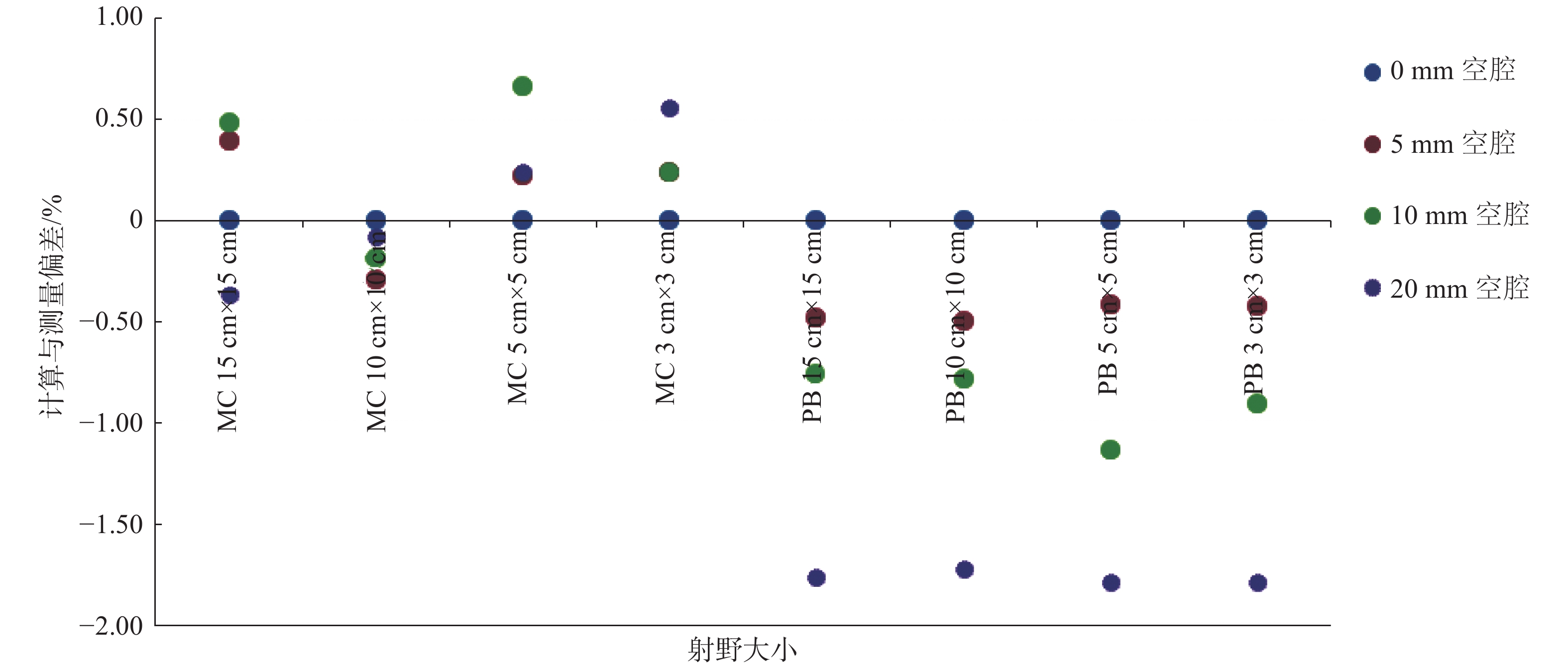
在不同空腔厚度模体中,Monaco TPS的Monte Carlo算法和Pencil Beam算法的计算值与实际测量值的相对偏差如图3。Monte Carlo算法的计算值与实际测量值差别较小,最大偏差发生在5 cm × 5 cm照射野、10 mm厚的空腔模体且最大偏差是0.66%;Pencil Beam算法的计算值均低于实际测量值且随着空腔厚度增加偏差增大,最大偏差发生在3 cm × 3 cm照射野、20 mm厚的空腔模体且最大偏差是−1.8%,其中未排除由摆位带来的随机误差[11]。

|
图 3 不同空腔厚度模体中TPS 2种算法计算值与实际测量值的偏差 Figure 3 Deviation between two algorithms of TPS calculated value and actual measured value in phantoms with cavity thickness |
20例上段食管癌放疗计划在MapCheck和固体水组成不同空腔厚度模体上共进行160次剂量验证,使用MapCheck自带的γ分析软件得到剂量验证γ通过率结果320个,不同空腔厚度模体的剂量验证平均γ通过率结果(%,
|
|
表 1 20例上段食管癌放疗计划在不同空腔厚度模体中剂量验证平均γ通过率(
|
由表1可以看出Monte Carlo算法和Pencil Beam算法在均匀模体(0 mm空腔模体)的3 mm 3% RD(相对剂量)、AD(绝对剂量)的γ通过率均大于97%,都能满足临床要求;Monte Carlo算法在不同空腔厚度模体的剂量验证平均γ通过率差别很小,Pencil Beam算法在不同空腔厚度模体中的剂量验证平均γ通过率存在较大差别,相比于Pencil Beam算法,Monte Carlo算法在不同空腔厚度模体的剂量验证平均γ通过率普遍偏高,详细差异需要进一步统计学分析。
在相同比对标准下,2种算法在不同空腔模体的剂量验证γ通过率配对t检验如表2。可以看出Monte Carlo算法在不同空腔厚度模体剂量验证γ通过率(RD)的配对t检验结果均P > 0.05,无统计学意义;不同空腔厚度模体剂量验证通过率(AD)中(0 mm,10 mm)、(5 mm,10 mm)和(10 mm,20 mm)组的配对 t检验P < 0.05,有统计学意义,且最大偏差为1.06%(0 mm,10 mm),其余组 P > 0.05,无统计学意义,说明应用不同空腔厚度模体做剂量验证,剂量通过率之间无临床差异;Pencil Beam算法中RD(0 mm,20 mm)和(5 mm,20 mm)组配对 t检验结果P < 0.05,最大偏差为0.58%,其余组 P > 0.05,无统计学意义。AD组 P < 0.05,最大偏差为2.78%。
|
|
表 2 20例上段食管癌放疗计划在不同空腔厚度模体中剂量验证γ通过率的配对t检验 Table 2 Paired t-test of dose verification gamma pass rate of 20 cases of upper esophageal cancer radiotherapy plan in different Cavity thickness phantoms |
在相同空腔厚度模体和比对标准下2种算法的剂量验证γ通过率配对t检验结果如表3所示。从表3中可以看出,相同空腔厚度和相同比对标准下两种算法的剂量验证γ通过率配对t检验结果P< 0.05,有统计学意义,Monte Carlo算法比Pencil Beam算法的剂量验证γ通过率高,RD和AD组中最大偏差均发生在20 mm厚度空腔模体,且分别为2.49%和4.14%(20 mm)。
|
|
表 3 20例上段食管癌放疗计划两种算法在相同空腔厚度模体中剂量验证γ通过率的配对t检验 Table 3 Paired t-test of dose verification gamma pass rate of 20 cases of upper esophageal cancer radiotherapy plan in identical Cavity thickness phantoms |
目前,用二维电离室矩阵进行调强验证在许多医院得到了广泛的应用[12]。大多数都是基于均匀模体来进行测量[13],该方法忽略了空腔带来的剂量计算不确定性。本课题组在自制的空腔模体上进行剂量验证,该方法可以更真实地反映计划系统对于射线穿过空腔后剂量计算的准确性。在本研究中,Monaco TPS中的Monte Carlo算法在3 mm计算网格在不同空腔厚度模体中的计算值与实际测量值最大偏差是0.66%(10 mm厚的空腔模体,5 cm × 5 cm射野),其中未排除由摆位带来的随机误差。Monte Carlo算法用3 mm计算网格对射线穿过空腔后的剂量计算结果准确,能满足临床对剂量计算准确度的要求[14];Monaco TPS中的Pencil Beam算法在3 mm计算网格在不同空腔厚度模体中的计算值均低于实际测量值,随着空腔厚度增加偏差增大,且最大偏差是−1.80%(20 mm厚的空腔模体,3 cm × 3 cm射野),在实际临床中会低估空腔边缘靶区或正常组织的受量,考虑Pencil Beam算法在非均匀组织计算中未考虑计算点周围散射线的影响,现已不能作为放疗剂量的精确计算,尤其是含有大量空腔的组织。因此在Monaco TPS中,在靶区周边含有大量空腔时不推荐使用Pencil Beam算法进行计算。
Monte Carlo算法在均匀模体(0 mm空腔模体)剂量验证的3 mm/3%RD、AD的平均γ通过率为(99.72 ± 0.38)%、(98.98 ± 0.83)%,Pencil Beam算法在均匀模体剂量验证的3 mm/3%RD、AD的平均通过率为(97.53 ± 1.43)%、(97.02 ± 1.29)%,2种算法的剂量验证的3 mm/3%RD、AD的平均γ通过率值都大于97%,均能满足临床要求,且普遍高于相关研究者结论[15],考虑主要是本研究课题平板探测器点数为445个,采集剂量信息低于上诉研究结论中所使用的1177个平板探测器平板,故此得到相对更高的剂量验证γ通过率。在含有空腔的不均匀模体上进行剂量验证的结果暂无报道,Monte Carlo算法计算的20例上段食管癌放疗计划在不同空腔厚度模体的剂量验证平均γ通过率差别很小,相同比对标准中的配对t检验结果中,(0、10)和(5、10)mm组的3 mm/3%AD通过率P < 0.05,有统计学意义,最大偏差发生在(0、10)mm组且数值为1.1%。其余组无统计学意义,说明20例上段食管癌放疗计划在不同空腔模体上进行剂量验证通过率无差异;Pencil Beam算法在不同空腔厚度模体的剂量验证平均γ通过率随着空腔厚度的增大而减小。Pencil Beam算法中RD(0、20)和(5、20)mm组配对 t检验结果P < 0.05,最大偏差为0.58%,其余组 P > 0.05,无统计学意义。AD组 P < 0.05,最大偏差为2.78%;Pencil Beam算法在不同空腔厚度模体的剂量验证对相对γ通过率影响很小,对绝对剂量γ通过率影响较大,进一步证明了Pencil Beam算法在非均匀组织计算中,尤其是含有大量空腔的组织时剂量计算的局限性 [16]。在相同空腔厚度模体、相同比对标准下,Monte Carlo算法的剂量验证平均γ通过率都高于Pencil Beam算法,说明Monte Carlo算法在剂量计算准确度上具有优势。
总之,Monaco TPS中Monte Carlo算法3 mm计算网格在不同空腔厚度模体中计算准确,与Pencil Beam算法相比,Monte Carlo算法在固定野动态调强的上段食管癌的剂量验证中可以得到更高的γ通过率,Monte Carlo算法在不同空腔厚度模体间的剂量验证γ通过率无临床差异且均能满足临床要求。在非均匀组织计算中,尤其是含有大量空腔的组织时,不推荐使用Pencil Beam算法进行剂量计算。
| [1] |
石婷婷, 韩济华, 张艳, 等. 食管癌固定野调强和旋转容积调强计划的剂量学比较[J]. 中国辐射卫生, 2020, 29(1): 89-92. Shi TT, Han JH, Zhang Y, et al. Dosimetry comparison of the 5-field IMRT and VMAT planning for esophageal carcinoma[J]. Chin J Radiol Health, 2020, 29(1): 89-92. DOI:10.13491/j.issn.1004-714X.2020.01.021 |
| [2] |
何栋成, 张晓晔, 张艳, 等. ArcCheck剂量验证角度分析食管上段癌调强放疗技术[J]. 中国辐射卫生, 2019, 28(2): 206-208, 213. He DC, Zhang XY, Zhang Y, et al. The analysis of the radiotherapy techniques for the treatment of upper esophageal cancer form the dose verification by ArcCheck[J]. Chin J Radiol Health, 2019, 28(2): 206-208, 213. DOI:10.13491/j.issn.1004-714x.2019.02.025 |
| [3] |
洪君, 单鹄声, 周玉凤. 医用直线加速器早晚绝对剂量输出稳定性对比分析[J]. 医学研究生学报, 2018, 31(5): 512-515. Hong J, Shan HS, Zhou YF. Comparative analysis on morning and evening absolute dose output stability of medical linear accelerator[J]. J Med Postgraduates, 2018, 31(5): 512-515. DOI:10.16571/j.cnki.1008-8199.2018.05.014 |
| [4] |
刘博宇, 杨筑春, 卜明伟, 等. 正向射野在颈及胸上段食管癌调强放疗中的应用[J]. 中华肿瘤防治杂志, 2017, 24(18): 1301-1304. Liu BY, Yang ZC, Bu MW, et al. Application of positive fields in IMRT treatment of cervical and upper esophageal cancer[J]. Chin J Cancer Prev Treat, 2017, 24(18): 1301-1304. DOI:10.16073/j.cnki.cjcpt.2017.18.008 |
| [5] |
Zhen HM, Nelms BE, Tome WA. Moving from gamma passing rates to patient DVH-based QA metrics in pretreatment dose QA[J]. Med Phys, 2011, 38(10): 5477-5489. DOI:10.1118/1.3633904 |
| [6] |
Olch AJ. Evaluation of the accuracy of 3DVH software estimates of dose to virtual ion chamber and film in composite IMRT QA[J]. Med Phys, 2012, 39(1): 81-86. DOI:10.1118/1.3666771 |
| [7] |
Yan GH, Lu B, Kozelka J, et al. Calibration of a novel four-dimensional diode array[J]. Med Phys, 2010, 37(1): 108-115. DOI:10.1118/1.3266769 |
| [8] |
Kadoya N, Saito M, Ogasawara M, et al. Evaluation of patient DVH-based QA metrics for prostate VMAT: correlation between accuracy of estimated 3D patient dose and magnitude of MLC misalignment[J]. J Appl Clin Med Phys, 2015, 16(3): 5251. DOI:10.1120/jacmp.v16i3.5251 |
| [9] |
唐斌, 康盛伟, 王先良, 等. 基于直线加速器虚拟源模型的蒙特卡洛剂量计算及在IMRT独立验算中的初步应用[J]. 中华放射肿瘤学杂志, 2016, 25(4): 372-375. Tang B, Kang SW, Wang XL, et al. Monte Carlo dose calculation based on the virtual source model with linear accelerator and its preliminary application in independent dose calculation for IMRT plans[J]. Chin J Radiat Oncil, 2016, 25(4): 372-375. DOI:10.3760/cma.j.issn.1004-4221.2016.04.014 |
| [10] |
郭冉, 张艺宝, 李明辉, 等. 调强计划验证通过率的影响因素分析[J]. 中国医学物理学杂志, 2018, 35(10): 1117-1121. Guo R, Zhang YB, Li MH, et al. Factors affecting the passing rate of intensity-modulated radiotherapy plans[J]. Chin J Med Phys, 2018, 35(10): 1117-1121. |
| [11] |
洪君, 单鹄声, 周玉凤. 全身集成定位架对放疗靶区吸收剂量的影响[J]. 中国医学物理学杂志, 2018, 35(7): 785-789. Hong J, Shan HS, Zhou YF. Effects of systemic integrated orientation frame on absorbed dose of target areas in radiotherapy[J]. Chin J Med Phys, 2018, 35(7): 785-789. DOI:10.3969/j.issn.1005-202X.2018.07.009 |
| [12] |
Low DA, Moran JM, Dempsey JF, et al. Dosimetry tools and techniques for IMRT[J]. Med Phys, 2011, 38(3): 1313-1338. DOI:10.1118/1.3514120 |
| [13] |
吴仕章, 陈进琥, 李振江, 等. IMRT计划剂量验证924例结果分析[J]. 中华肿瘤防治杂志, 2018, 25(18): 1323-1327. Wu SZ, Chen JH, Li ZJ, et al. Analysis of dose verification results for 924 IMRT plans[J]. Chin J Cancer Prev Treat, 2018, 25(18): 1323-1327. DOI:10.16073/j.cnki.cjcpt.2018.18.009 |
| [14] |
张帆, 伍海彪, 肖爱农, 等. 医用加速器射线参数对百分深度剂量影响的蒙特卡罗模拟[J]. 中华放射医学与防护杂志, 2018, 38(2): 145-149. Zhang F, Wu HB, Xiao AN, et al. Monte Carlo-based simulation of influence of linear accelerator beam parameter on percentage depth dose[J]. Chin J Radiol Med Prot, 2018, 38(2): 145-149. DOI:10.3760/cma.j.issn.0254-5098.2018.02.013 |
| [15] |
高璇, 李克新, 鞠永健, 等. 前列腺癌患者调强放疗中的摆位误差及其对剂量分布影响分析[J]. 中国辐射卫生, 2018, 27(3): 228-230, 251. Gao X, Li KX, Ju YJ, et al. The study about the setup error and its effect on dose distribution in IMRT for prostate cancer patients[J]. Chin J Radiol Health, 2018, 27(3): 228-230, 251. DOI:10.13491/j.issn.1004-714x.2018.03.010 |
| [16] |
贺瑶瑶, 荆亮, 黄晓铧, 等. 基于个性化组织等效体模的肺癌三维适形放疗计划算法比较[J]. 中国医学物理学杂志, 2019, 36(7): 755-759. He YY, Jing L, Huang XH, et al. Comparison of three-dimensional conformal radiotherapy planning algorithms for lung cancer based on personalized tissue-equivalent phantom[J]. Chin J Med Phys, 2019, 36(7): 755-759. DOI:10.3969/j.issn.1005-202X.2019.07.003 |


