2. 中国农业大学动物医学院,北京 100193;
3. 芭比堂爱心动物医院,宁波 330212
2. College of Veterinary Medicine, China Agricultural University, Beijing 100193, China;
3. Puppytown Love Animal Hospital, Ningbo 330212, China
桡尺骨骨折(radius and ulna fractures,RUF)是玩具犬常见的骨科疾病。治疗玩具犬RUF的技术包括铸件、外固定支架和骨板内固定等。与大型犬相比,玩具犬桡骨髓内血液供应差且骨膜软组织覆盖有限,使得玩具犬RUF发生不愈合或延迟愈合的概率更高[7]。尽管过去的研究报道了玩具犬RUF的并发症及不愈合的发生率高[8],但近期的研究结果表明当选择合适的骨板治疗这些骨折时并不会导致较高的并发症发生率。
目前国内关于玩具犬RUF治疗的报道仅见零星几例,尚未有对多个病例的回顾性分析。本文将报道使用普通圆洞骨板和兽医可剪裁骨板(veterinary cuttable plate,VCP)治疗玩具犬RUF的有效性及临床效果,报道相关并发症,以期为兽医临床中玩具犬RUF的诊断与治疗、手术方案的制定与实施提供借鉴。
1 材料与方法 1.1 病例来源病例来自于中国农业大学动物医院开放式复位和骨板内固定治疗RUF的玩具犬共63只。病例入选条件:体重不超过7 kg;回访时间大于12个月;病例信息记录完整;无可能影响骨折愈合的疾病(如糖尿病、库兴氏综合征等)。记录动物的相关信息,包括就诊日期、病史、品种、性别、体重、年龄、患肢和骨折时间等。拍摄患肢正侧位的X线片。通过X线片,记录骨折类型,测量远端骨段长度、近端骨段长度,并计算远端骨段与桡骨长度的比值(distal fragment length/radius length,DL/RL)。手术后第4、8周时分别拍摄X线片评估骨折的愈合情况。
1.2 骨板使用VCP、VCP加宽及圆洞骨板。所有骨板均为直骨板,材质为316LVM不锈钢,宽度、厚度详见表 1。
|
|
表 1 骨板的宽度及厚度 Table 1 Width and tickness of bone plate |
根据桡骨骨折的具体位置及手术医生的倾向选择头外侧或头内侧手术通路[11],暴露桡骨断端后确认骨折线,将骨折复位后安置骨板及合适长度的骨螺钉。检查复位良好、稳固后,使用0.9%生理盐水冲洗术部,用可吸收缝线常规缝合手术切口。较活泼或双侧RUF的患犬术后包扎罗伯特-琼斯绷带。罗伯特-琼斯绷带的拆除时机根据动物的实际情况而定。术中每90 min静脉注射氨苄西林钠50 mg·kg-1,术后1周给予阿莫西林克拉维酸钾(25 mg·kg-1,每天2次,口服)和非甾体抗炎药。
手术后3~5 d复查伤口,10~14 d拆线。拆线前犬一直佩戴伊丽莎白圈。
1.4 并发症及手术效果评估通过病历记录和回访,获得并发症和术后患肢功能情况。并发症的评估标准见表 2。患肢功能的评估标准见表 3。
2 结果 2.1 骨折犬基本情况62只(98.4%)犬为单侧骨折,1只(1.6%)犬为双侧骨折。品种:玩具贵宾犬(n=56,88.9%)、博美犬(n=6,9.5%)、约克夏犬(n=1,1.6%)。雄性犬30只(47.6%),雌性犬33只(52.4%)(包括1只已绝育犬)。患犬的体重及年龄的中位数分别是2.8 kg(1~5.8 kg)和11月龄(4~108月龄)。根据美国动物医院协会标准[15],将患犬按年龄进行划分,幼年犬(≤10月)共30只(47.6%),青年犬(>10月且≤2岁)共21只(33.3%),中年犬(>2岁且≤7岁)共11只(17.4%),老年犬(>7岁)共1只(1.6%)。年龄段性别分布见图 1。

|
图 1 患犬的年龄段性别分布图 Fig. 1 Age and sex distribution of affected dogs |
64例中61例(95.3%)为闭合性骨折,3例(4.7%)为开放性骨折。所有骨折中40例(62.5%)为横骨折,23例(35.9%)为斜骨折,1例(1.6%)为粉碎性骨折。33例(51.6%)为左侧桡尺骨骨折,31例(48.4%)为右侧桡尺骨骨折。远端骨段的平均长度为20.6 mm,平均远端骨段桡骨长度比为0.26(范围:0.09~0.70)。59例(92.2%)骨折线位于桡骨远端1/3以下,40例(62.5%)位于1/4以下。
2.3 所用骨板骨折修复所使用的骨板根据动物的体型、骨骼大小、骨折线以及医生的倾向而定,所使用的骨板见表 4。
|
|
表 4 骨板的使用情况 Table 4 Bone plates used in affected dogs |
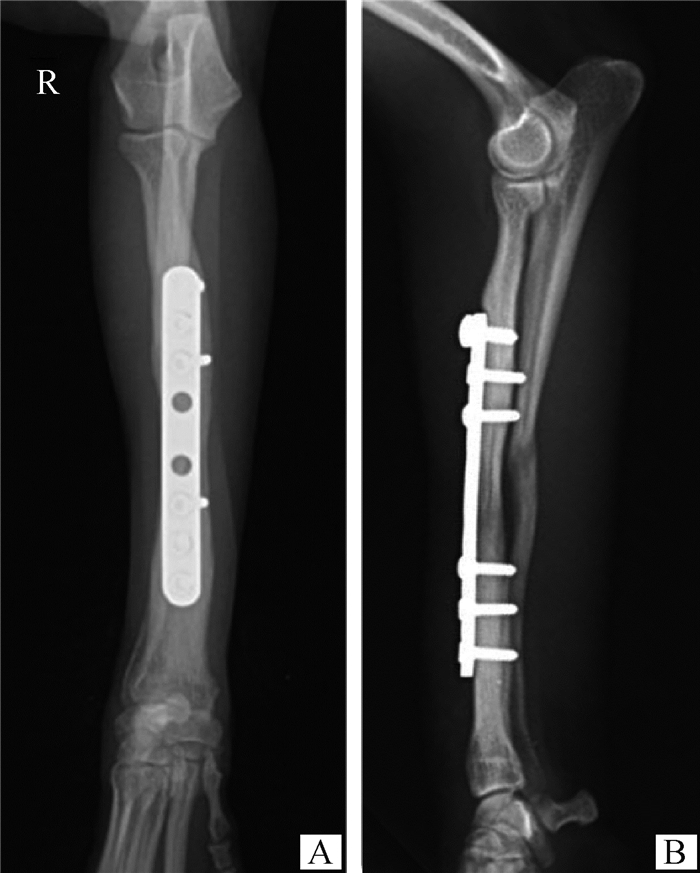
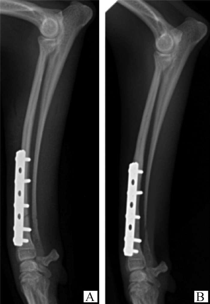
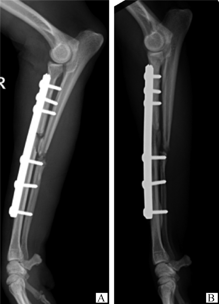
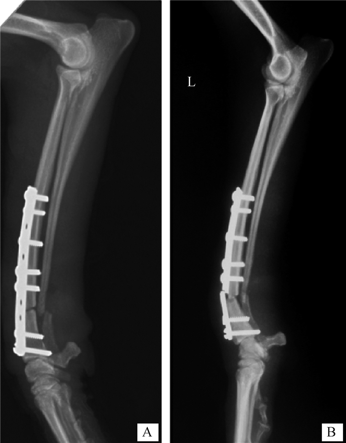
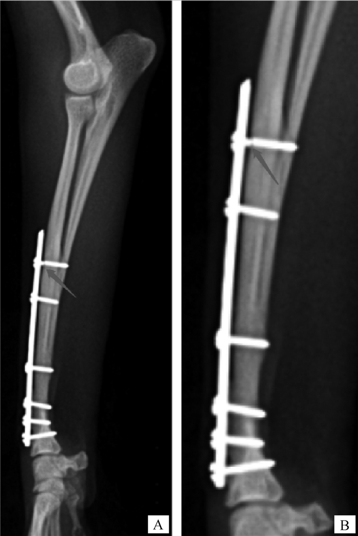
17例(27%)患犬术后发生轻微并发症(图 2~5),详见表 5;2例(3.2%)患犬术后发生严重并发症(图 6、图 7),详见表 6。患犬术后患肢活动情况见表 7。
|
术后4个月的X线片,A、B分别为前后位及内外位X线片,可见到桡骨中段骨密度降低 Radiographs at 4 months postoperatively, A and B are Anteroposterior and lateral radiograph, respectively, which demonstrate decreased bone mineral density at midshaft of radius 图 2 骨质减少(48号犬) Fig. 2 Osteopenia (Dog No.48) |
|
52号患犬的X线片。A、B均为内外位X线片。A为术后即刻的X线片,B为术后2个月时的X线片。B图可见尺骨骨干变细,远端发生吸收 A and B are lateral radiographs. A is the immediate postoperative radiograph and B is the postoperative radiograph at 2 months. Ulnar resorption is seen in B 图 3 尺骨吸收(52号犬) Fig. 3 Ulnar resorption (Dog No.52) |
|
A、B均为内外位X线片。A为手术后即刻的X线片,B为手术后74 d的X线片,可见到尺骨萎缩性不愈合 A and B are lateral radiographs. A is the immediately postoperative radiograph and B is the radiograph at 74 days postoperatively which shows atrophic nonunion of the ulna 图 4 尺骨不愈合(63号犬) Fig. 4 Ulnar nonunion (Dog No.63) |

|
A、B分别为前后位及内外位X线片,C为B图的局部细节放大图。X线片可见:第3、4螺钉(红色箭头)周围低密度带,骨折线清晰,周围骨痂过度生长,软组织肿胀 A and B are anteroposterior and lateral radiographs, respectively, and C is a magnified view of B.Radiographs showed: hypodense bands around the 3rd and 4th screws (red arrows) with clear fracture lines, overgrowth of the surrounding callus, and soft tissue swelling 图 5 感染(49号犬) Fig. 5 Infection (Dog No.49) |
|
|
表 5 轻微并发症 Table 5 Minor complication |
|
A、B均为内外位X线片。38号患犬,年龄4岁,体重4.7 kg,使用2.0 mm 10孔VCP。A为术后即刻的X线片,B为术后16 d的X线片,可见到骨板断裂 A and B are anteroposterior radiographs. Dog No.38, 4 years old, weighing 4.7 kg, used a 2.0 mm 10 holes VCP. A is the immediately postoperative radiograph, and B is radiograph at 16 days postoperatively in which bone plate fracture was visible 图 6 植入物断裂(38号犬) Fig. 6 Bone plate fracture (Dog No.38) |
|
A为内外位X线片,B为A图的局部细节放大图。54号患犬,年龄12月龄,体重1.1 kg,使用1.0 mm 10孔VCP。X线片可见:近端第1颗螺钉在螺钉头与螺纹交界处断裂(红色箭头所指) A is an anteroposterior radiograph and B is a magnified view of A. Dog No.54, 12 months old, weighing 1.1 kg, used a 1.0 mm 10 holes VCP. Radiographs showed: 1 st screw fractured at the junction of screw head and thread (red arrow) 图 7 螺钉断裂的X线片(54号犬) Fig. 7 Bone screw fracture (Dog No.54) |
|
|
表 6 严重并发症 Table 6 Major complication |
|
|
表 7 患肢跛行评分 Table 7 Lameness score |
骨质减少的4例,2例患肢功能正常,1例跛行评分为1分,1例评为2分(药物治疗有效);3例尺骨吸收的病例患肢功能均正常;患肢外旋的2例无X线片的异常,但与健侧肢体对比发现存在患肢外旋;63号患犬(3岁,4 kg),因被自行车撞击发生粉碎性RUF,使用1.5 mm 11孔VCP治疗,在术后74 d发现尺骨萎缩性不愈合;感染患犬在术后4 d发现患肢温度高,肿胀明显,第4周时患肢仍肿胀明显,X线片(图 5)发现第3、4颗螺钉周围轻微低密度带,周围骨痂过度生长,穿刺肿胀处需氧培养确认为革兰阳性菌,药敏试验显示,该菌对阿莫西林克拉维酸钾敏感,最终通过口服阿莫西林克拉维酸钾(25 mg·kg-1,每日2次,1个月)控制住了感染;骨板断裂的病例(4岁4.7 kg)最初使用2.0 mm 10孔VCP修复骨折,16 d后骨板在远端第3、4孔间发生断裂,后改用2.0 mm 10孔有桥VCP修复,并进行松质骨移植,最终成功愈合;螺钉断裂的病例(12月龄1.1 kg)使用1.0 mm 10孔VCP,术后17个月因跛行就诊,发现近端第1颗螺钉断裂。
3 讨论本研究结果表明,使用普通骨板(圆洞骨板或兽医可剪裁骨板)开放式复位和内固定治疗玩具犬RUF具有良好的临床效果,不会导致高的并发症发生率。
玩具贵宾犬(n=56)在本研究中的占比为88.9%,与加拿大[4]、美国[16]、日本[3]等地报道的动物品种存在一定差异,可能与玩具贵宾犬在北京地区的饲养数量较大或地区间品种喜好差异相关。尽管这几个研究报道的品种存在差异,但都属于玩具型品种,与中大型犬相比,玩具犬更易发生RUF,可能是玩具犬与中大型犬桡尺骨横截面特性的差异导致[17-19]。此外,本研究中的患犬以青幼年犬(80.9%)为主,与其他研究报道的结果相似[2-3, 10, 17, 20],Aikawa等[2]认为这种年龄差异可能与青幼年犬桡尺骨相比老年犬更脆弱有关,但尚无研究支持该观点。性别方面,本研究中雄性(n=30)与雌性(n=33)占比基本相当,但仅1只绝育犬,绝育率低于现有文献报道的39.76%,此差异可能是因为北京地区犬绝育率低、犬绝育后活动量下降因而受伤概率降低[22],但具体原因仍待进一步研究。
本研究中92.2%的患犬桡骨骨折位置位于远端1/3以下,62.5%位于远端1/4以下,这与之前的3个研究[2-4]结果相似,说明玩具犬RUF时远端骨折(尤其远端1/4以下)的概率更高。Brianza等[23]的研究指出玩具犬桡尺骨的相对面积惯性矩显著低于中大型犬种,使其在承受同比例的负荷时更容易承受较高的应力,可能是造成玩具犬RUF时骨折线常位于远端的原因之一。
轻微并发症是不需要手术治疗或不需要医疗干预的并发症,在本研究中,发生率最高的轻微并发症是皮肤相关并发症(n=6,9.4%),与de Arburn等[4]及Aikawa等[3]研究中,报道的轻微并发症结果相符,这可能与玩具犬前臂的软组织覆盖较少相关[7]。
骨质减少(n=4,6.4%)的发生率,低于其他研究报道的7.1%~20%。在Muroi等[1]的研究中,236例玩具犬RUF骨板内固定术后桡骨在1~12月内均发生了骨质减少,与本研究、Kang等[21]和Ramirez等[10]的结果差异较大。造成较大差异的可能原因包括:①Muroi等对术后X线片的跟踪周期更久;②Muroi等采用软件分析对骨质减少的敏感性更高,而本研究及其他研究是由影像医师肉眼判读。从本研究及de Arburn等[4]的研究可知骨质减少通常不会引起相关临床症状。尽管移除骨板可有效增加骨密度[1],但在没有相关临床症状的病例中移除骨板是没有必要的。
本研究中尺骨吸收的发生率为4.8%(n=3),这3例犬患肢功能均正常。2005年Hamilton等[24]首次报道玩具犬RUF的骨板治疗中的尺骨吸收,其认为可能是由于尺骨骨折愈合环境的不稳定或应力保护造成的。至今仍未有研究阐明具体原因,中大型犬中也尚未有尺骨吸收的相关报道。使用骨板治疗的研究中尺骨吸收发生率为7.7%~71.4%,在使用环形ESF治疗玩具犬RUF的研究中也报道过尺骨吸收,但该研究未提及具体的发生率。在Hamilton等[24]的研究中,所有进行翻修手术的病例都出现了尺骨吸收,而在没有翻修手术的病例中也有一半出现了尺骨吸收。本研究和其他研究[20-21, 24-25]均未发现尺骨吸收对患肢功能的不良影响,其临床意义有待进一步研究。
患肢外旋(n=2)在本研究中的发生率是3.2%,与de Arburn等[4]的研究结果相近。与de Arburn等[4]的方法一致,本研究通过对比患肢与健侧肢体确认患肢外旋[4]。本研究中2例患肢外旋的犬均未发现X线征象的异常,de Arburn等[4]的研究中5例患肢外旋的犬仅1例发现了X线片上相对应的异常,可能是因为X线识别患肢外旋的敏感性低。尽管患肢外旋对于动物美观有一定影响,但其并未影响动物患肢的功能,故本研究中未进行干预。
尺骨不愈合在本研究中有1例(1.6%,图 4),为萎缩性不愈合,患肢无跛行。该病例为粉碎性骨折(车祸导致),怀疑其尺骨不愈合与血供破坏较严重相关,但无法确定具体原因。截至目前,仅两个研究报道过尺骨不愈合,发生率分别为5%(1/20)[20]和53%(8/15),这些病例的患肢功能无任何异常。尽管桡尺骨作为四肢中唯一一对同时负重的骨骼,在正常负重情况下承担犬体重的60%[26],但尺骨不愈合并未对玩具犬患肢的功能造成影响,这可能是因为玩具犬体型小,能代偿尺骨不愈合造成的负重影响。
感染在本研究的发生率为1%,低于其他研究,可能的原因:①手术前动物患肢的充分刷洗消毒及术中严格的无菌操作;②围手术期抗生素的合理使用。另外两个研究中感染病例均移除了植入物[4, 20],本研究中感染患犬在术后4 d发现患肢温度偏高,肿胀较明显,到第4周时患肢仍肿胀明显,X线片(图 5)发现第3、4颗螺钉周围轻微低密度带,周围骨痂过度生长,对患肢肿胀处穿刺后进行需氧培养确认为革兰阳性菌,药敏试验显示该菌对阿莫西林克拉维酸钾敏感,最终通过口服阿莫西林克拉维酸钾(25 mg·kg-1,每日2次,1个月)控制住了感染,未移除骨板。大部分的玩具犬RUF属于闭合性骨折,对于闭合性骨折而言,该手术为清洁手术。清洁骨科手术术后是否应该使用抗生素,一直存在争论,但鉴于手术时间较长会提高感染的风险[29],感染会造成严重后果(如不愈合[30]),在本研究对所有患犬都使用了抗生素(术中每90 min静脉注射氨苄西林钠50 mg·kg-1,术后每日2次口服1周的阿莫西林克拉维酸钾25 mg·kg-1)。对于这类手术中抗生素的使用仍待进一步研究证实,但这超出了本研究的范畴。
本研究有2例(3.2%)植入物断裂,其中,骨板断裂1例、骨螺钉断裂1例。骨板断裂的动物(X线片见图 6)第一次使用VCP时,骨折线两端均存在空的螺钉孔,骨折间隙明显,愈合过程中当断端骨吸收后,骨断端增宽,产生了位于骨折线上方的螺钉孔,导致骨板疲劳周期显著降低[31],增加了骨板断裂的风险。此外,两断端间未接触而导致骨板应力集中[32],也有可能是骨板断裂的原因。总结以上两方面原因,本研究在对该动物进行翻修手术时,除尽可能减少骨折间隙外,还使用了有桥VCP,使骨板的桥部位于骨折线上方,同时移植松质骨促进骨折愈合,最终成功愈合。另一例动物的螺钉在螺钉头与螺纹交界处断裂,对比该动物术后即刻的X线片与断裂时X线片(见图 7),发现骨折线已消失,骨螺钉断裂的具体原因无法确定。
本研究属于回顾性研究,存在一定的局限性,首先由于缺乏动物长期的(一年以上)X线片,本研究可能低估了骨质减少、尺骨吸收等情况的真实发生率。第二,本研究对动物患肢跛行程度的长期评估主要通过对动物主人进行电话或微信回访获得,动物主人对跛行严重程度的评估可能具有一定的主观性,但已有研究表明,动物主人对骨科手术后功能结果的评估是有效且可靠的[33]。若能通过压力板步态测试对患肢恢复进行评估[34],将会得到更准确的结果。
4 结论使用圆洞骨板或兽医可剪裁骨板进行骨板内固定可以有效治疗玩具犬RUF,该技术能取得良好的临床效果,不会发生桡骨不愈合,且严重并发症的发生率低。
| [1] |
MUROI N, SHIMADA M, MURAKAMI S, et al. A retrospective study of postoperative development of implant-induced osteoporosis in radial-ulnar fractures in toy breed dogs treated with plate fixation[J]. Vet Comp Orthop Traumatol, 2021, 34(6): 375-385. DOI:10.1055/s-0041-1731810 |
| [2] |
AIKAWA T, MIYAZAKI Y, SAITOH Y, et al. Clinical outcomes of 119 miniature- and toy-breed dogs with 140 distal radial and ulnar fractures repaired with free-form multiplanar type Ⅱ external skeletal fixation[J]. Vet Surg, 2019, 48(6): 938-946. DOI:10.1111/vsu.13245 |
| [3] |
AIKAWA T, MIYAZAKI Y, SHIMATSU T, et al. Clinical outcomes and complications after open reduction and internal fixation utilizing conventional plates in 65 distal radial and ulnar fractures of miniature- and toy-breed dogs[J]. Vet Comp Orthop Traumatol, 2018, 31(3): 214-217. DOI:10.1055/s-0038-1639485 |
| [4] |
DE ARBURN PARENT R, BENAMOU J, GATINEAU M, et al. Open reduction and cranial bone plate fixation of fractures involving the distal aspect of the radius and ulna in miniature- and toy-breed dogs: 102 cases (2008-2015)[J]. J Am Vet Med Assoc, 2017, 250(12): 1419-1426. DOI:10.2460/javma.250.12.1419 |
| [5] |
PIRAS L, CAPPELLARI F, PEIRONE B, et al. Treatment of fractures of the distal radius and ulna in toy breed dogs with circular external skeletal fixation: a retrospective study[J]. Vet Comp Orthop Traumatol, 2011, 24(3): 228-235. DOI:10.3415/VCOT-10-06-0089 |
| [6] |
MCCARTNEY W, KISS K, ROBERTSON I. Treatment of distal radial/ulnar fractures in 17 toy breed dogs[J]. Vet Rec, 2010, 166(14): 430-432. DOI:10.1136/vr.b4810 |
| [7] |
WELCH J A, BOUDRIEAU R J, DEJARDIN L M, et al. The intraosseous blood supply of the canine radius: implications for healing of distal fractures in small dogs[J]. Vet Surg, 1997, 26(1): 57-61. DOI:10.1111/j.1532-950X.1997.tb01463.x |
| [8] |
LARSEN L J, ROUSH J K, MCLAUGHLIN R M. Bone plate fixation of distal radius and ulna fractures in small- and miniature-breed dogs[J]. J Am Anim Hosp Assoc, 1999, 35(3): 243-250. DOI:10.5326/15473317-35-3-243 |
| [9] |
WATROUS G K, MOENS N M M. Cuttable plate fixation for small breed dogs with radius and ulna fractures: retrospective study of 31 dogs[J]. Can Vet J, 2017, 58(4): 377-382. |
| [10] |
RAMÍREZ J M, MACÍAS C. Conventional bone plate fixation of distal radius and ulna fractures in toy breed dogs[J]. Aust Vet J, 2016, 94(3): 76-80. DOI:10.1111/avj.12408 |
| [11] |
JOHNSON K A. Piermattei's atlas of surgical approaches to the bones and joints of the dog and cat[M]. 5th ed. Amsterdam: Elsevier, 2014.
|
| [12] |
COOK J L, EVANS R, CONZEMIUS M G, et al. Proposed definitions and criteria for reporting time frame, outcome, and complications for clinical orthopedic studies in veterinary medicine[J]. Vet Surg, 2010, 39(8): 905-908. DOI:10.1111/j.1532-950X.2010.00763.x |
| [13] |
IMPELLIZERI J A, TETRICK M A, MUIR P. Effect of weight reduction on clinical signs of lameness in dogs with hip osteoarthritis[J]. J Am Vet Med Assoc, 2000, 216(7): 1089-1091. DOI:10.2460/javma.2000.216.1089 |
| [14] |
QUINN M M, KEULER N S, LU Y, et al. Evaluation of agreement between numerical rating scales, visual analogue scoring scales, and force plate gait analysis in dogs[J]. Vet Surg, 2007, 36(4): 360-367. DOI:10.1111/j.1532-950X.2007.00276.x |
| [15] |
VASSEUR P B, POOL R R, ARNOCZKY S P, et al. Correlative biomechanical and histologic study of the cranial cruciate ligament in dogs[J]. Am J Vet Res, 1985, 46(9): 1842-1854. |
| [16] |
BIERENS D, UNIS M D, CABRERA S Y, et al. Radius and ulna fracture repair with the IMEX miniature circular external skeletal fixation system in 37 small and toy breed dogs: a retrospective study[J]. Vet Surg, 2017, 46(4): 587-595. DOI:10.1111/vsu.12647 |
| [17] |
YU J, DECAMP C E, ROOKS R. Improving surgical reduction in radial fractures using a 'dowel' pinning technique in miniature and toy breed dogs[J]. Vet Comp Orthop Traumatol, 2011, 24(1): 45-49. DOI:10.3415/VCOT-10-05-0077 |
| [18] |
PHILLIPS I R. A survey of bone fractures in the dog and cat[J]. J Small Anim Pract, 1979, 20(11): 661-674. DOI:10.1111/j.1748-5827.1979.tb06679.x |
| [19] |
HUNT J M, AITKEN M L, DENNY H R, et al. The complications of diaphyseal fractures in dogs: a review of 100 cases[J]. J Small Anim Pract, 2010, 21(2): 103-119. |
| [20] |
GIBERT S, RAGETLY G R, BOUDRIEAU R J. Locking compression plate stabilization of 20 distal radial and ulnar fractures in toy and miniature breed dogs[J]. Vet Comp Orthop Traumatol, 2015, 28(6): 441-447. DOI:10.3415/VCOT-15-02-0042 |
| [21] |
KANG B J, RYU H H, PARK S, et al. Clinical evaluation of a mini locking plate system for fracture repair of the radius and ulna in miniature breed dogs[J]. Vet Comp Orthop Traumatol, 2016, 29(6): 522-527. DOI:10.3415/VCOT-16-01-0014 |
| [22] |
KUHNE F. Castration of dogs from the standpoint of behaviour therapy[J]. Tierarztl Prax Ausg K Kleintiere Heimtiere, 2012, 40(2): 140-145. DOI:10.1055/s-0038-1623633 |
| [23] |
BRIANZA S Z M, DELISE M, MADDALENA FERRARIS M, et al. Cross-sectional geometrical properties of distal radius and ulna in large, medium and toy breed dogs[J]. J Biomech, 2006, 39(2): 302-311. DOI:10.1016/j.jbiomech.2004.11.018 |
| [24] |
HAMILTON M H, HOBBS S J L. Use of the AO veterinary mini 'T'-plate for stabilisation of distal radius and ulna fractures in toy breed dogs[J]. Vet Comp Orthop Traumatol, 2005, 18(1): 18-25. DOI:10.1055/s-0038-1632921 |
| [25] |
NELSON T A, STROM A. Outcome of repair of distal radial and ulnar fractures in dogs weighing 4 kg or less using a 1. 5-mm locking adaption plate or 2. 0-mm limited contact dynamic compression plate[J]. Vet Comp Orthop Traumatol, 2017, 30(6): 444-452. DOI:10.3415/VCOT-17-01-0005 |
| [26] |
ARTHURS G, BROWN G, PETTITT R. BSAVA manual of canine and feline musculoskeletal disorders[M]. 2nd ed. Gloucester: British Small Animal Veterinary Association, 2018.
|
| [27] |
AIKEN M J, HUGHES T K, ABERCROMBY R H, et al. Prospective, randomized comparison of the effect of two antimicrobial regimes on surgical site infection rate in dogs undergoing orthopedic implant surgery[J]. Vet Surg, 2015, 44(5): 661-667. DOI:10.1111/vsu.12327 |
| [28] |
FITZPATRICK N, SOLANO M A. Predictive variables for complications after TPLO with stifle inspection by arthrotomy in 1000 consecutive dogs[J]. Vet Surg, 2010, 39(4): 460-474. DOI:10.1111/j.1532-950X.2010.00663.x |
| [29] |
EUGSTER S, SCHAWALDER P, GASCHEN F, et al. A prospective study of postoperative surgical site infections in dogs and cats[J]. Vet Surg, 2004, 33(5): 542-550. DOI:10.1111/j.1532-950X.2004.04076.x |
| [30] |
MUNAKATA S, NAGAHIRO Y, KATORI D, et al. Clinical efficacy of bone reconstruction surgery with frozen cortical bone allografts for nonunion of radial and ulnar fractures in toy breed dogs[J]. Vet Comp Orthop Traumatol, 2018, 31(3): 159-169. DOI:10.1055/s-0038-1631878 |
| [31] |
ALWEN S G J, KAPATKIN A S, GARCIA T C, et al. Open screw placement in a 1. 5 mm LCP over a fracture gap decreases fatigue life[J]. Front Vet Sci, 2018, 5: 89. DOI:10.3389/fvets.2018.00089 |
| [32] |
GEMMILL T J, CLEMENTS D N. BSAVA manual of canine and feline fracture repair and management[M]. Cheltenham: British Small Animal Veterinary Association, 2016.
|
| [33] |
INNES J F, BARR A R S. Can owners assess outcome following treatment of canine cruciate ligament deficiency?[J]. J Small Anim Pract, 1998, 39(8): 373-378. DOI:10.1111/j.1748-5827.1998.tb03735.x |
| [34] |
AMIMOTO H, KOREEDA T, OCHI Y, et al. Force plate gait analysis and clinical results after tibial plateau levelling osteotomy for cranial cruciate ligament rupture in small breed dogs[J]. Vet Comp Orthop Traumatol, 2020, 33(3): 183-188. DOI:10.1055/s-0039-1700990 |
(编辑 白永平)


