2. 四川农业大学 预防兽医研究所, 成都 611130;
3. 四川农业大学 动物疫病与人类健康四川省重点实验室, 成都 611130
2. Institute of Preventive Veterinary Medicine, Sichuan Agricultural University, Chengdu 611130, China;
3. Key Laboratory of Animal Disease and Human Health of Sichuan Province, Sichuan Agricultural University, Chengdu 611130, China
疱疹病毒科病毒是一类双链DNA病毒,其典型结构包括120~245 kb长的双链DNA基因组、20面体核衣壳(caspid)、皮层(tegument)和囊膜(envelope)[1]。根据第10次ICTV报告,疱疹病毒家族分为α、β和γ 3个亚科[2],α疱疹病毒主要包括单纯疱疹病毒1型(herpes simplex virus 1, HSV-1)[3]、水痘-带状疱疹病毒(varicella-zoster virus, VZV)[4]、牛疱疹病毒(bovine herpesvirus 1, BHV)[5]、马立克病毒(Marek’s disease virus, MDV)[6]、马疱疹病毒1型(equine herpesvirus 1, EHV-1)[7]、伪狂犬病病毒(pseudorabies virus, PRV)[8]、鸭肠炎病毒(duck enteritis virus, DEV)[9-12]等。典型β、γ类疱疹病毒分别是人巨细胞病毒(human cytomegalovirus, HCMV)和爱泼斯坦-巴尔病毒(epstein-barr virus, EBV)。众多研究表明疱疹病毒ICP22蛋白及其同源物对潜伏感染的建立、病毒复制、细胞凋亡、成熟病毒粒子出芽等多方面至关重要,现对其研究进展做简要概述,以期为ICP22及同源物基本特性以及参与病毒生命周期的深入研究提供参考。
1 疱疹病毒US1基因及其同源基因特点疱疹病毒基因组一般由长独特区(unique long, UL)、短独特区(unique short, US)、末端重复序列(terminal repeat sequence, TRS)以及内部重复序列(internal repeat sequence, IRS)组成,构成UL-IRS-US-TRS形式[13]。当病毒粒子进入感染细胞后,开始启动病毒基因的串联表达与蛋白翻译表达,根据转录顺序可分成立即早期(IE)、早期(E)和晚期(L)3类[14-15]。IE基因转录不需要依赖于新合成的DNA或蛋白质,其编码蛋白在病毒感染过程中起广泛的调控作用,E基因表达需要IE基因激活,通常编码病毒基因复制相关蛋白,L基因主要编码囊膜糖蛋白,其表达依赖于病毒DNA的复制[16]。疱疹病毒US1基因及其同源物序列或结构有所差异,如HSV-1仅含1个1 260 bp的US1基因,类属立即早期基因[17],HCMV US1属于早期基因[18],MDV US1基因最初被定义为IE基因,随后被证明是L基因,感染后期才能被检测到[6]。大多数疱疹病毒US1基因以对称双拷贝形式位于内部重复区和末端重复区,且转录方向相反。如VZV ORF63/70为834 bp的立即早期基因[19],其编码蛋白常称之为IE63。1 050 bp长的PRV Rsp40基因类型较为特殊,文献报道该基因非典型性动力学表达,不属于IE基因[20]。BHV-1 US区域没有US1基因,但是由112 768—113 670 bp和124 548—125 450 bp[21]转录产生、同时具有IE与L动力学的1.7 kb转录本编码蛋白定义为立即早期蛋白BICP22[5]。EHV-1中US1同源基因是长为1 410 bp的IR4基因,其差异表达1.4 kb的早期和1.7 kb的晚期转录本,编码的早期蛋白EICP22蛋白相对分子质量介于42~47 ku[22]。最新报道表明DEV US1基因为990 bp的反向重复的IE基因[23-25]。此外,HSV-1中存在一个US1 N端1—146 AA截短表达的基因——US1.5,在感染晚期才会被检测到,UL13蛋白激酶对US1.5蛋白进行翻译后修饰,从而正常表达UL38和US11等晚期基因子集[17],但是US1.5的具体功能以及与US1的关联目前还不清楚[17]。
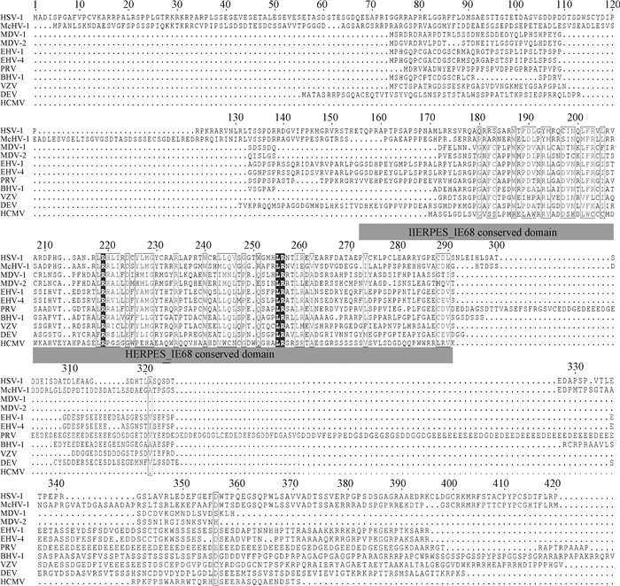
2 疱疹病毒ICP22蛋白的翻译后修饰序列比对发现已有报道的疱疹病毒ICP22蛋白含有一个涉及蛋白转录调控等重要功能的保守结构域IE_68(图 1)。已有报道的ICP22蛋白合成加工过程涉及多种修饰,各类修饰往往导致相对分子质量变大。例如,HSV-1 UL13与US3病毒蛋白以及未知的细胞蛋白可以磷酸化ICP22,其中,Thr-116和Thr-193位点的磷酸化与病毒毒力相关[26-27],另外还存在经细胞酪蛋白激酶介导的核苷酸化修饰,众多修饰导致ICP22表观相对分子质量为68 ku[28]。VZV ORF63基因编码的IE63蛋白可被细胞酪蛋白和ORF47激酶以及CDK 1、CDK 2磷酸化,最终呈现45 ku的表观相对分子质量[29]。其中,CDK 1磷酸化Ser-224涉及IE63细胞定位以及转录调控功能,CDK 2磷酸化Thr-171、Ser-181和Ser-186从而介导IE63与ASF-1的相互作用[30]以及VZV潜伏感染的建立[31]。近期研究表明,DEV ICP22相对分子质量比预期偏大22 ku,生物信息学分析结果显示,DEV ICP22含众多磷酸化和糖基化位点,推测其表观相对分子质量偏大是各类细胞激酶作用所致[24]。

|
保守域HERPES_IE68以灰色突出显示,保守氨基酸以黑色显示 The conserved HERPES_IE68 domain is highlighted in grey, and the conserved motifs are shown in black 图 1 疱疹病毒ICP22蛋白序列比对 Fig. 1 Sequence analysis of herpesvirus ICP22 amino acids |
不同疱疹病毒ICP22细胞内定位不尽相同,如MDV ICP22在DEF中定位于细胞质[3],而HSV-1 ICP22在感染细胞和转染细胞中均定位细胞核。HSV-1 ICP22含有两个独立的核定位信号(nuclear localization sequence, NLS),NLS1位于ICP22 16—31AA,NLS2位于ICP22 118—131AA[32]。然而N端缺失1—146AA的US1.5定位于核,推测ICP22还有未被鉴定出或入核能力较弱的核定位信号[33]。潜伏感染阶段的IE63定位于细胞质,体外裂解性感染时则定位细胞核,突变磷酸化位点后定位由核转变为质,表明激酶的磷酸化作用对IE63定位很重要[34]。研究表明,PRV ICP22定位于细胞核,其41—60AA为典型单分核定位信号,其49—50AA(KR)为关键氨基酸。同时,Cai等[8]发现PRV ICP22是通过Ran、importinα1-和α7介导途径靶向细胞核。最新研究发现,DEV ICP22在感染或转染情况下均定位于核,进一步发现305—314AA为一典型单分核定位信号,其中,309R为关键位点,然而突变该位点的重组病毒感染DEF后ICP22仍定位于核,推测感染情况下有其他病毒蛋白或ICP22本身还未鉴定出的NLS带动其入核[24]。
4 疱疹病毒ICP22功能研究进展 4.1 ICP22影响病毒潜伏感染的建立潜伏感染是病毒持续性感染的状态,病毒基因存在于组织或细胞中,既不产生感染性疾病也不出现临床症状,在某些条件下可被激活,急性发作而出现症状,此时可以检测出病毒[35]。HSV-1 ICP22在潜伏感染阶段不表达,但是缺失ICP22可显著降低病毒复制水平以及在感染小鼠三叉神经中建立潜伏感染的能力[3]。目前,已确定HSV-1 ICP22 Thr-193、Tyr-116的磷酸化与病毒毒力相关[27],Ser-34或Tyr-116单突变即可减弱HSV-1引起的眼疾严重程度。最新研究发现,ICP22与CD80启动子直接结合可抑制眼部免疫反应,同时该抑制能力取决于再激活病毒的复制水平,因此作者推测,Ser-34或Tyr-116位点突变不影响ICP22与CD80启动子的结合,ICP22对CD80的抑制作用可保护宿主免受病原引起的病理侵害[3]。
VZV可表达VZV-潜伏相关转录本(VZV latency transcript, VLT)和ORF63两种转录本[4]。ORF63是VZV复制必需基因,其编码蛋白IE63不仅作为激活E基因的转录调节因子,还能改变神经元对细胞凋亡的敏感性以及减弱Ⅰ型干扰素(interferon, IFNs)信号传导的免疫反应,在潜伏期作为主要表达产物可被检测到[36],ORF63缺失导致VZV在人黑素瘤细胞中的复制以及啮齿动物中潜伏期的建立受到损害[29]。此外,ORF61编码蛋白IE61是豚鼠肠道神经元中IE63入核的必需蛋白,推测VLT通过对ORF61转录和翻译的阻遏作用将IE63拦截在细胞质并阻止病毒的反式激活[37],这可能是潜伏感染期间IE63定位于细胞质的原因之一。
MDV ICP22参与病毒潜伏感染的研究已有较多报道。潜伏期间MDV-miR-M1-5P和MDV-miR-M5-3p能下调潜伏期MDV ICP22 mRNA水平,而MDV-miR-M4-5P和细胞同源物GGA mir-155-3p可以上调ICP22表达[6, 38]。Boumart等[6]提出一个假设模型,首先是GGA miR-155-3P促进ICP22表达,稍后GGA miR-155-3P以尚不清楚的分子机制被抑制表达。然后,被感染细胞中MDV-miR-M4-5P促进ICP22表达,由于MDV-miR-M4-5P在感染的各个阶段都表达,因此,潜伏阶段MDV-miR-M1-5P和MDV-miR-M5-3P会抑制ICP22表达从而保证潜伏感染细胞的存活[6]。
4.2 ICP22的转录调控作用细胞RNA聚合酶Ⅱ (RNA polymerase Ⅱ, RNA Pol Ⅱ)大亚基的保守C端结构域(C-terminal domain, CTD)由Tyr1-Ser2-Pro3-Thr4-Ser5-Pro6-Ser7 7肽序列重复组成,在所有真核生物中均保守,能为RNA Pol Ⅱ复合物招募mRNA加工和染色质修饰所需因子从而参与基因转录[39]。
正向转录延伸因子b(positive transcription elongation factor b, P-TEFb)是细胞周期蛋白依赖性蛋白激酶9(cyclin-dependent kinases, CDK 9)和某些周期蛋白形成的复合物,可被募集到转录起始的启动子区域磷酸化RNA Pol Ⅱ CTD Ser-2,进而促进转录延长。HSV-1感染后,ICP22与P-TEFb亚基CDK 9直接结合,阻断其募集从而抑制基因转录,该过程还可能有US3的参与[40]。RNA Pol Ⅱ可与ICP22、CDK 9共定位[41],猜测ICP22以某种未知方式调节CDK 9活性。此外,ICP22表达可使RNA Pol Ⅱ CTD Tyr-1、Ser-2磷酸化丢失[42],推测ICP22可参与HSV-1感染细胞中RNA Pol Ⅱ的修饰进而抑制病毒与宿主基因的转录。针对上文所述的转录抑制情况,HSV-1病毒蛋白VP16能够联合宿主细胞因子1(host cell factor-1, HCF-1)、八聚体转录因子(octamer transcription factor, Oct-1)等识别IE基因启动子上游区域的TAATGARAT特定序列元件,从而打破ICP22对IE基因转录的限制,有利于E和L基因的转录表达[40]。此外,ICP22可导致促染色质转录因子(facilitates chromatin transcription, FACT)在细胞核中重新定位,还可招募其与转录延伸因子Spt5(Supt5 h)、Spt6(Supt6 h)等至病毒基因组以促进病毒γ基因的转录表达[43]。综上表明,ICP22能参与HSV-1劫持宿主细胞RNA Pol Ⅱ以高效转录病毒基因。然而关于病毒感染后宿主基因转录受抑制模型仍需更多讨论,1)许多细胞系中HSV-1病毒基因转录以及L基因表达不绝对需要ICP22,例如在Vero细胞系中ICP22缺失株与野生株表型几乎一致。2)无论是否发生磷酸化修饰,RNA Pol Ⅱ均可被募集到病毒的转录/复制区室[44]。3)常规转录因子TFⅡE是未感染细胞中转录起始所必需的,感染HSV-1后在IE蛋白作用下丢失,有可能因此而导致细胞基因的转录受到抑制[45]。但是ICP22缺失株并不能消除这一现象[45],说明ICP22可能不参与这一路径。
VZV IE63可以与病毒反式激活因子IE62、RNA Pol Ⅱ和感染细胞核中的细胞延伸因子EF-1α启动子相互作用而参与VZV基因转录调控,其中,IE63可以结合与EF-1α启动子相互作用的细胞因子,从而反式激活其表达[46]。IE63强烈抑制IE62表达,也可以与IE62结合上调gI糖蛋白启动子的表达,还可以作为VZV和异源病毒许多启动子的转录阻遏物[29]。已经明确IE63可通过1—142AA序列与IE62相互作用并在感染细胞中部分共定位,还可与RNA Pol Ⅱ复合存在于感染细胞中,但是IE63-ORF47复合物能否改变RNA Pol Ⅱ的磷酸化进而影响VZV基因调控尚不清楚[47]。此外,干扰素(interferon,IFNs)在VZV致病机制中起重要作用,研究发现,IE63可通过抑制真核翻译起始因子2(eukaryotic initiation factor 2, eIF2)的磷酸化从而抑制IFN下游信号通路,同时,IE63还可介导eIF2脱磷酸化,具体作用机制也还未知[45]。这些发现表明,VZV可能已进化出复杂机制来逃避细胞保护路径,IE63在VZV致病过程中具有重要作用。
EHV-1病毒基因组仅表达一个IE蛋白(IE protein, IEP),可反式激活E和一些L基因以及异源病毒基因启动子[48]。早期蛋白EICP22是不结合DNA的非必需基因,对EHV-1基因启动子的反式激活作用很弱,不过EICP22 142-239AA序列可介导两者相互作用形成EICP22P-IEP复合物提高IEP与其靶序列的结合速率以促进E以及L基因的表达,但是任何形式的EICP22P都不能克服IEP对自身启动子的抑制作用[49]。此外,纯化的GST-EICP22融合蛋白能够恢复某些IEP突变体的DNA结合能力,推测EICP22与IEP的相互作用有助于招募转录因子和/或促进转录起始的细胞因子,增强IEP和病毒基因启动子的结合活性以提高转录效率。另外,研究表明EICP22 124—143AA可介导自身相互作用形成二聚体甚至高阶复合物,但仅有全长EICP22能与IEP协同作用促进反式激活[50]。Derbigny等[51]证明EICP22突变体的稳定性,因此,猜测EICP22P突变体蛋白构象发生改变使得与IEP的相互作用受损,相互作用的非必需序列有助于招募特定细胞因子到蛋白复合物中。然而EICP22调节病毒基因表达的确切机制仍然不清楚,后续可探究EICP22P与细胞转录因子有无相互作用和EICP22P-IEP复合物反式激活过程中EICP22参与调节的作用机制。
复制机制研究较为深入的是HSV-1和VZV,结果表明两者都已进化出篡夺细胞基因复制系统以大量产生病毒后代的机制,均编码与RNA Pol Ⅱ相互作用的IE蛋白ICP22和IE63。已经明确ICP22蛋白及其同源物在基因调控中具有重要作用,但如何参与劫持细胞基因表达系统以促进病毒基因复制以及转录本的翻译,如何在不同细胞系、不同病毒中发挥独特作用还知之甚少,关于α疱疹病毒基因调控仍需更多探索。
4.3 ICP22调节病毒核出芽疱疹病毒核衣壳太大不能自由穿过核膜。因此,细胞核内的子代核衣壳首先通过内核膜进入核周进行初级囊膜包膜,然后与外核膜融合进行去囊膜化从而进入细胞质[52-55]。就HSV-1而言,由UL31和UL34构成的核出芽复合体(nuclear egress complex, NEC)对病毒核衣壳的初次包装很关键[56]。ICP22可与UL31、UL34共定位于细胞核膜,缺失ICP22后UL31和UL34定位发生改变,反之UL31缺失亦可影响ICP22细胞内定位[57]。此外,ICP22缺失可导致细胞核周初级囊膜包被病毒粒子数量大量减少,核中积累大量衣壳。以上资料表明ICP22是病毒核出芽的重要非必需蛋白,可能通过与UL31和/或UL34相互作用来发挥作用[58]。
4.4 ICP22参与细胞凋亡一些病毒能致宿主细胞凋亡[59-61],细胞凋亡是一种必不可少的复杂天然免疫机制,可以有效清除感染或衰老细胞[62]。HSV-1感染后期ICP22可通过US1.5激活半胱天冬酶3(caspase-3)而实现促凋亡作用[52];随后发现ICP22蛋白具有抗细胞凋亡的作用,进一步研究发现ICP22缺失可下调US5基因(具有抗细胞凋亡功能)表达水平,推测ICP22可通过影响US5表达进而发挥抗凋亡作用[63],因而推断ICP22参与细胞凋亡过程且发挥双重作用。You等[52]、游韶平等[64]发现单独表达HSV-2 ICP22蛋白只能较低程度地抵抗放线菌素D(act-dactinomycin D, ActD)诱导的细胞凋亡,因而推测ICP22不是直接作用于细胞,而是通过调节病毒基因表达以及与病毒和/或细胞蛋白相互作用而发挥抗细胞凋亡作用,各类调节路径相互作用导致HSV-1 ICP22对细胞凋亡有双重作用。另一方面,p53可通过激活Bax或抑制Bcl-2来调节各种细胞途径并控制细胞凋亡[65],还可在感染后期可抑制ICP0表达,不过该抑制作用可被ICP22拮抗,从而促进细胞存活和病毒有效复制[28]。
IE63缺失病毒感染细胞的凋亡程度明显增加,此外单独表达IE63即可降低细胞凋亡水平,表明IE63具有抗细胞凋亡作用[66]。已经明确磷酸化eIF2可以介导细胞凋亡信号转导[67],而IE63能抑制eIF2的磷酸化以及介导eIF2脱磷酸化[52],因而推测IE63抑制细胞凋亡的作用路径可能与eIF2(脱)磷酸化有关。不过还未发现IE63有HSV-1 ICP22的促凋亡作用,表明即使同源蛋白之间也可能存在不同功能与作用机制[52]。
4.5 ICP22参与细胞自噬与抗病毒反应自噬是一种溶酶体降解系统,可促进细胞内蛋白质和细胞器的分解,传统上被认为是一种保护机制,然而在特定条件下也可以用作促死亡途径[68-69]。HSV-1感染细胞中,核仁蛋白(protein interacting with carboxyl terminus 1, PICT-1)通过抑制核糖体RNA(ribosomal RNA, rRNA)转录和失活AKT/mTOR/p70S6K通路而触发促死亡性自噬[70],病毒蛋白伴侣ICP0和ICP22与PICT-1相互作用参与到上述过程,然而ICP22的具体作用机制尚不清楚[71]。
HSV-1感染后,细胞蛋白会发生剧烈的空间重组并错误折叠到细胞核中,形成病毒诱导分子伴侣富集域(virus-induced chaperone-enriched, VICE),其中热休克同源蛋白70(heat shock cognate protein 70, Hsc70)可介导抗原递呈与细胞自噬过程,参与细胞抗病毒作用[72]。单独表达ICP22即可形成ICP22、Hsc70和错误折叠蛋白质构成的核内复合物,感染ICP22缺失病毒的情况下不会诱导VICE结构域的形成,表明ICP22是诱导VICE所必需的[73]。最新研究表明,ICP22作为一种J样蛋白,可能通过Hsc70与Hsp40/ Hsp70复合物相互作用,促使错误折叠蛋白向核聚集以降低细胞毒性来发挥抗病毒作用[74]。不过ICP22完整作用途径以及为何Hsc70只能与ICP22突变体而非全长免疫共沉淀还不清楚[74]。总之,作为第一个病毒J样分子伴侣蛋白以及抗病毒药物开发的靶标[72],ICP22在抗病毒科学研究与临床应用中具有重大意义。
5 小结疱疹病毒ICP22蛋白是一个翻译后修饰的多功能蛋白,可以参与病毒潜伏感染的建立、调控病毒基因转录以及成熟病毒粒子出芽,也涉及细胞凋亡、自噬和抗病毒等生命周期重要过程,但其机制大多不明确,对其开展深入研究将有利于疱疹病毒感染疾病的治疗与防控。此外,有关ICP22蛋白的报道大多源于人疱疹病毒,对动物疱疹病毒、特别是以ICP22作为靶标的新药研究,应该成为今后的研究重点之一。
| [1] | OWEN D J, CRUMP C M, GRAHAM S C. Tegument assembly and secondary envelopment of alphaherpesviruses[J]. Viruses, 2015, 7(9): 5084–5114. DOI: 10.3390/v7092861 |
| [2] | LEFKOWITZ E J, DEMPSEY D M, HENDRICK-SON R C, et al. Virus taxonomy:the database of the International Committee on Taxonomy of Viruses (ICTV)[J]. Nucleic Acids Res, 2018, 46(D1): D708–D717. DOI: 10.1093/nar/gkx932 |
| [3] | MATUNDAN H H, JAGGI U, WANG S H, et al. Loss of ICP22 in HSV-1 elicits immune infiltration and maintains stromal keratitis despite reduced primary and latent virus infectivity[J]. Invest Ophthalmol Vis Sci, 2019, 60(10): 3398–3406. DOI: 10.1167/iovs.19-27701 |
| [4] | DEPLEDGE D P, OUWENDIJK W J D, SADAOKA T, et al. A spliced latency-associated VZV transcript maps antisense to the viral transactivator gene 61[J]. Nat Commun, 2018, 9(1): 1167. DOI: 10.1038/s41467-018-03569-2 |
| [5] | HART J, MACHUGH N D, SHELDRAKE T, et al. Identification of immediate early gene products of bovine herpes virus 1 (BHV-1) as dominant antigens recognized by CD8 T cells in immune cattle[J]. J Gen Virol, 2017, 98(7): 1843–1854. DOI: 10.1099/jgv.0.000823 |
| [6] | BOUMART I, FIGUEROA T, DAMBRINE G, et al. GaHV-2 ICP22 protein is expressed from a bicistronic transcript regulated by three GaHV-2 microRNAs[J]. J Gen Virol, 2018, 99(9): 1286–1300. DOI: 10.1099/jgv.0.001124 |
| [7] | CYMERYS J, SŁOŃSKA A, BRZEZICKA J, et al. Replication kinetics of neuropathogenic and non-neuropathogenic equine herpesvirus type 1 (EHV-1) strains in primary murine neurons and ED cell line[J]. Pol J Vet Sci, 2016, 19(4): 777–784. DOI: 10.1515/pjvs-2016-0098 |
| [8] | CAI M S, JIANG S, ZENG Z C, et al. Probing the nuclear import signal and nuclear transport molecular determinants of PRV ICP22[J]. Cell Biosci, 2016, 6: 3. DOI: 10.1186/s13578-016-0069-7 |
| [9] | GUO Y F, CHENG A C, WANG M S, et al. Purification of anatid herpesvirus 1 particles by tangential-flow ultrafiltration and sucrose gradient ultracentrifugation[J]. J Virol Methods, 2009, 161(1): 1–6. DOI: 10.1016/j.jviromet.2008.12.017 |
| [10] | JIA R Y, CHENG A C, WANG M S, et al. Development and evaluation of an antigen-capture ELISA for detection of the UL24 antigen of the duck enteritis virus, based on a polyclonal antibody against the UL24 expression protein[J]. J Virol Methods, 2009, 161(1): 38–43. DOI: 10.1016/j.jviromet.2009.05.011 |
| [11] | ZHAO L C, CHENG A C, WANG M S, et al. Characterization of codon usage bias in the dUTPase gene of duck enteritis virus[J]. Prog Natl Sci, 2008, 18(9): 1069–1076. DOI: 10.1016/j.pnsc.2008.03.009 |
| [12] | CHANG H, CHENG A C, WANG M S, et al. Complete nucleotide sequence of the duck plague virus gE gene[J]. Arch Virol, 2009, 154(1): 163–165. DOI: 10.1007/s00705-008-0284-6 |
| [13] |
马云潮, 程安春, 汪铭书, 等. 重组鸭肠炎病毒载体疫苗研究进展[J]. 畜牧兽医学报, 2017, 48(11): 2015–2022.
MA Y C, CHENG A C, WANG M S, et al. Progress of recombinant duck enteritis virus-vectored vaccines[J]. Acta Veterinaria et Zootechnica Sinica, 2017, 48(11): 2015–2022. DOI: 10.11843/j.issn.0366-6964.2017.11.002 (in Chinese) |
| [14] | DEMBOWSKI J A, DREMEL S E, DELUCA N A. Replication-Coupled recruitment of viral and cellular factors to herpes simplex virus type 1 replication forks for the maintenance and expression of viral genomes[J]. PLoS Pathog, 2017, 13(1): e1006166. DOI: 10.1371/journal.ppat.1006166 |
| [15] | WIDENER R W, WHITLEY R J. Herpes simplex virus[J]. Handb Clin Neurol, 2014, 123: 251–263. |
| [16] | GINN S L, ALEXANDER I E, EDELSTEIN M L, et al. Gene therapy clinical trials worldwide to 2012-an update[J]. J Gene Med, 2013, 15(2): 65–77. DOI: 10.1002/jgm.2698 |
| [17] | RICE S A, DAVIDO D J. HSV-1 ICP22:hijacking host nuclear functions to enhance viral infection[J]. Future Microbiol, 2013, 8(3): 311–321. DOI: 10.2217/fmb.13.4 |
| [18] | MAJIMA R, SHINDOH K, YAMAGUCHI T, et al. Characterization of a thienylcarboxamide derivative that inhibits the transactivation functions of cytomegalovirus IE2 and varicella zoster virus IE62[J]. Antiviral Res, 2017, 140: 142–150. DOI: 10.1016/j.antiviral.2017.01.024 |
| [19] | ZERBONI L, SEN N, OLIVER S L, et al. Molecular mechanisms of varicella zoster virus pathogenesis[J]. Nat Rev Microbiol, 2014, 12(3): 197–210. DOI: 10.1038/nrmicro3215 |
| [20] | LI M L, ZHAO Z Y, CHEN J H, et al. Characterization of synonymous codon usage bias in the pseudorabies virus US1 gene[J]. Virol Sin, 2012, 27(5): 303–315. DOI: 10.1007/s12250-012-3270-9 |
| [21] | ROBINSON K E, MEERS J, GRAVEL J L, et al. The essential and non-essential genes of bovine herpesvirus 1[J]. J Gen Virol, 2008, 89(11): 2851–2863. DOI: 10.1099/vir.0.2008/002501-0 |
| [22] | AHN B, ZHANG Y F, OSTERRIEDER N, et al. Properties of an equine herpesvirus 1 mutant devoid of the internal inverted repeat sequence of the genomic short region[J]. Virology, 2011, 410(2): 327–335. DOI: 10.1016/j.virol.2010.11.020 |
| [23] | WU Y, CHENG A C, WANG M S, et al. Complete genomic sequence of Chinese virulent duck enteritis virus[J]. J Virol, 2012, 86(10): 5965. DOI: 10.1128/JVI.00529-12 |
| [24] | LI Y G, WU Y, WANG M S, et al. Duplicate US1 genes of duck enteritis virus encode a non-essential immediate early protein localized to the nucleus[J]. Front Cell Infect Microbiol, 2020, 9: 463. DOI: 10.3389/fcimb.2019.00463 |
| [25] | WU Y, CHENG A C, WANG M S, et al. Comparative genomic analysis of duck enteritis virus strains[J]. J Virol, 2012, 86(24): 13841–13842. DOI: 10.1128/JVI.01517-12 |
| [26] | LIN F S, DING Q, GUO H, et al. The herpes simplex virus type 1 infected cell protein 22[J]. Virol Sin, 2010, 25(1): 1–7. DOI: 10.1007/s12250-010-3080-x |
| [27] | OSTLER J B, HARRISON K S, SCHROEDER K, et al. The glucocorticoid receptor (GR) stimulates herpes simplex virus 1 productive infection, in part because the infected cell protein 0 (ICP0) promoter is cooperatively transactivated by the GR and Krüppel-like transcription factor 15[J]. J Virol, 2019, 93(6): e02063–18. |
| [28] | MARUZURU Y, FUJⅡ H, OYAMA M, et al. Roles of p53 in herpes simplex virus 1 replication[J]. J Virol, 2013, 87(16): 9323–9332. DOI: 10.1128/JVI.01581-13 |
| [29] | AMBAGALA A P N, COHEN J I. Varicella-zoster virus IE63, a major viral latency protein, is required to inhibit the alpha interferon-induced antiviral response[J]. J Virol, 2007, 81(15): 7844–7851. DOI: 10.1128/JVI.00325-07 |
| [30] | AMBAGALA A P, BOSMA T, ALI M A, et al. Varicella-zoster virus immediate-early 63 protein interacts with human antisilencing function 1 protein and alters its ability to bind histones h3. 1 and h3. 3[J]. J Virol, 2009, 83(1): 200–209. DOI: 10.1128/JVI.00645-08 |
| [31] | MUELLER N H, WALTERS M S, MARCUS R A, et al. Identification of phosphorylated residues on varicella-zoster virus immediate-early protein ORF63[J]. J Gen Virol, 2010, 91(Pt 5): 1133–1137. |
| [32] | STELZ G, RVCKER E, ROSORIUS O, et al. Identification of two nuclear import signals in the α-Gene product ICP22 of herpes simplex virus 1[J]. Virology, 2002, 295(2): 360–370. DOI: 10.1006/viro.2002.1384 |
| [33] | BASTIAN T W, RICE S A. Identification of sequences in herpes simplex virus type 1 ICP22 that influence RNA polymerase Ⅱ modification and viral late gene expression[J]. J Virol, 2009, 83(1): 128–139. DOI: 10.1128/JVI.01954-08 |
| [34] | LEISENFELDER S A, KINCHINGTON P R, MOFFAT J F. Cyclin-dependent kinase 1/Cyclin B1 phosphorylates varicella-zoster virus IE62 and is incorporated into virions[J]. J Virol, 2008, 82(24): 12116–12125. DOI: 10.1128/JVI.00153-08 |
| [35] | REESE T A. Coinfections:another variable in the herpesvirus latency-reactivation dynamic[J]. J Virol, 2016, 90(12): 5534–5537. DOI: 10.1128/JVI.01865-15 |
| [36] | DEPLEDGE D P, SADAOKA T, OUWENDIJK W J D. Molecular aspects of varicella-zoster virus latency[J]. Viruses, 2018, 10(7): 349. DOI: 10.3390/v10070349 |
| [37] | BAIRD N L, ZHU S Y, PEARCE C M, et al. Current in vitro models to study varicella zoster virus latency and reactivation[J]. Viruses, 2019, 11(2): 103. DOI: 10.3390/v11020103 |
| [38] | YAO Y X, VASOYA D, KGOSANA L, et al. Activation of gga-miR-155 by reticuloendotheliosis virus T strain and its contribution to transformation[J]. J Gen Virol, 2017, 98(4): 810–820. DOI: 10.1099/jgv.0.000718 |
| [39] | SHERIDAN R M, FONG N, D'ALESSANDRO A, et al. Widespread backtracking by RNA pol Ⅱ is a major effector of gene activation, 5' pause release, termination, and transcription elongation rate[J]. Mol Cell, 2019, 73(1): 107–118. DOI: 10.1016/j.molcel.2018.10.031 |
| [40] | LEI G, WU W J, LIU L D, et al. Herpes simplex virus 1 ICP22 inhibits the transcription of viral gene promoters by binding to and blocking the recruitment of P-TEFb[J]. PLoS One, 2012, 7(9): e45749. DOI: 10.1371/journal.pone.0045749 |
| [41] | OU M, SANDRI-GOLDIN R M. Inhibition of cdk9 during herpes simplex virus 1 infection impedes viral transcription[J]. PLoS One, 2013, 8(10): e79007. DOI: 10.1371/journal.pone.0079007 |
| [42] | ZABOROWSKA J, BAUMLI S, LAITEM C, et al. Herpes simplex virus 1 (HSV-1) ICP22 protein directly interacts with cyclin-dependent kinase (CDK)9 to inhibit RNA polymerase Ⅱ transcription elongation[J]. PLoS One, 2014, 9(9): e107654. DOI: 10.1371/journal.pone.0107654 |
| [43] | FOX H L, DEMBOWSKI J A, DELUCA N A. A herpesviral immediate early protein promotes transcription elongation of viral transcripts[J]. mBio, 2017, 8(3): e00745–17. |
| [44] | RICE S A, LONG M C, LAM V, et al. Herpes simplex virus immediate-early protein ICP22 is required for viral modification of host RNA polymerase Ⅱ and establishment of the normal viral transcription program[J]. J Virol, 1995, 69(9): 5550–5559. DOI: 10.1128/JVI.69.9.5550-5559.1995 |
| [45] | VAN OPDENBOSCH N, VAN DEN BROEKE C, DE REGGE N, et al. The IE180 protein of pseudorabies virus suppresses phosphorylation of translation initiation factor eIF2α[J]. J Virol, 2012, 86(13): 7235–7240. DOI: 10.1128/JVI.06929-11 |
| [46] | ZERBONI L, SOBEL R A, RAMACHANDRAN V, et al. Expression of varicella-zoster virus immediate-early regulatory protein IE63 in neurons of latently infected human sensory ganglia[J]. J Virol, 2010, 84(7): 3421–3430. DOI: 10.1128/JVI.02416-09 |
| [47] | STOEGER T, ADLER H. "Novel" triggers of herpesvirus reactivation and their potential health relevance[J]. Front Microbiol, 2019, 9: 3207. DOI: 10.3389/fmicb.2018.03207 |
| [48] | CHARVAT R A, BREITENBACH J E, AHN B, et al. The UL4 protein of equine herpesvirus 1 is not essential for replication or pathogenesis and inhibits gene expression controlled by viral and heterologous promoters[J]. Virology, 2011, 412(2): 366–377. DOI: 10.1016/j.virol.2011.01.025 |
| [49] |
高俊.马疱疹病毒1型潜伏相关转录体的转录调控机制研究[D].呼和浩特: 内蒙古农业大学, 2016.
GAO J. Transcriptional regulation mechanism research of equine herpesvirus 1 latency associated transcripts[D]. Hohhot: Inner Mongolia Agricultural University, 2016. (in Chinese) |
| [50] | ZHANG Y F, CHARVAT R A, KIM S K, et al. The EHV-1 UL4 protein that tempers viral gene expression interacts with cellular transcription factors[J]. Virology, 2014, 449: 25–34. DOI: 10.1016/j.virol.2013.11.005 |
| [51] | DERBIGNY W A, KIM S S, JANG H K, et al. EHV-1 EICP22 protein sequences that mediates its physical interaction with the immediate-early protoin are not sufficient to enhance the trans-activation activity of the IE protein[J]. Virus Res, 2002, 84(1-2): 1–15. DOI: 10.1016/S0168-1702(01)00377-X |
| [52] | YOU Y, CHENG A C, WANG M S, et al. The suppression of apoptosis by α-herpesvirus[J]. Cell Death Dis, 2017, 8(4): e2749. DOI: 10.1038/cddis.2017.139 |
| [53] | JING Y C, WU Y, SUN K F, et al. Role of duck plague virus glycoprotein C in viral adsorption:absence of specific interactions with cell surface heparan sulfate[J]. J Integr Agric, 2017, 16(5): 1145–1152. DOI: 10.1016/S2095-3119(16)61550-2 |
| [54] | ZHANG D X, LAI M Y, CHENG A C, et al. Molecular characterization of the duck enteritis virus US10 protein[J]. Virol J, 2017, 14(1): 183. DOI: 10.1186/s12985-017-0841-2 |
| [55] | YOU Y, LIU T, WANG M S, et al. Author correction:duck plague virus glycoprotein J is functional but slightly impaired in viral replication and cell-to-cell spread[J]. Sci Rep, 2018, 8(1): 6488. DOI: 10.1038/s41598-018-24845-7 |
| [56] | FUNK C, OTT M, RASCHBICHLER V, et al. The herpes simplex virus protein pUL31 escorts nucleocapsids to sites of nuclear egress, a process coordinated by its N-terminal domain[J]. PLoS Pathog, 2015, 11(6): e1004957. DOI: 10.1371/journal.ppat.1004957 |
| [57] | ARⅡ J, TAKESHIMA K, MARUZURU Y, et al. Roles of the interhexamer contact site for hexagonal lattice formation of the herpes simplex virus 1 nuclear egress complex in viral primary envelopment and replication[J]. J Virol, 2019, 93(14): e00498–19. |
| [58] | MARUZURU Y, SHINDO K, LIU Z M, et al. Role of herpes simplex virus 1 immediate early protein ICP22 in viral nuclear egress[J]. J Virol, 2014, 88(13): 7445–7454. DOI: 10.1128/JVI.01057-14 |
| [59] | ZHAO C K, HE T Q, XU Y, et al. Molecular characterization and antiapoptotic function analysis of the duck plague virus Us5 gene[J]. Sci Rep, 2019, 9(1): 4851. DOI: 10.1038/s41598-019-41311-0 |
| [60] | LAI Y L, ZENG N, WANG M S, et al. The VP3 protein of duck hepatitis A virus mediates host cell adsorption and apoptosis[J]. Sci Rep, 2019, 9(1): 16783. DOI: 10.1038/s41598-019-53285-0 |
| [61] | SUN D, WEN X J, WANG M S, et al. Apoptosis and autophagy in picornavirus infection[J]. Front Microbiol, 2019, 10: 2032. DOI: 10.3389/fmicb.2019.02032 |
| [62] | JORGENSEN I, RAYAMAJHI M, MIAO E A. Programmed cell death as a defence against infection[J]. Nat Rev Immunol, 2017, 17(3): 151–164. DOI: 10.1038/nri.2016.147 |
| [63] | YU X L, HE S D. The interplay between human herpes simplex virus infection and the apoptosis and necroptosis cell death pathways[J]. Virol J, 2016, 13(1): 77. DOI: 10.1186/s12985-016-0528-0 |
| [64] |
游韶平, 刘岩, 樊建勇, 等. 单纯疱疹病毒Ⅱ型感染细胞蛋白22在Vero细胞中的表达特性及其对细胞凋亡的影响[J]. 实用医学杂志, 2014, 30(3): 364–366.
YOU S P, LIU Y, FAN J Y, et al. Expression characteristics of herpes simplex virus type Ⅱ infected cell protein 22 in Vero cells and its effect on cell apoptosis[J]. The Journal of practical medicine, 2014, 30(3): 364–366. DOI: 10.3969/j.issn.1006-5725.2014.03.009 (in Chinese) |
| [65] | WU J X, LIU H H, HUANG H, et al. p53-dependent pathway and the opening of mPTP mediate the apoptosis of co-cultured sertoli-germ cells induced by microcystin-LR[J]. Environ Toxicol, 2019, 34(10): 1074–1084. DOI: 10.1002/tox.22808 |
| [66] | JAMES S F, MAHALINGAM R, GILDEN D. Does apoptosis play a role in varicella zoster virus latency and reactivation?[J]. Viruses, 2012, 4(9): 1509–1514. DOI: 10.3390/v4091509 |
| [67] | COLLIER A E, WEK R C, SPANDAU D F. Translational repression protects human keratinocytes from UVB-induced apoptosis through a discordant eIF2 kinase stress response[J]. J Invest Dermatol, 2015, 135(10): 2502–2511. DOI: 10.1038/jid.2015.177 |
| [68] | DENTON D, XU T Q, KUMAR S. Autophagy as a pro-death pathway[J]. Immunol Cell Biol, 2015, 93(1): 35–42. DOI: 10.1038/icb.2014.85 |
| [69] | WU X L, JIA R Y, WANG M S, et al. Downregulation of microRNA-30a-5p contributes to the replication of duck enteritis virus by regulating beclin-1-mediated autophagy[J]. Virol J, 2019, 16(1): 144. DOI: 10.1186/s12985-019-1250-5 |
| [70] | WANG S H, XU X L, HU Y L, et al. Sotetsuflavone induces autophagy in non-small cell lung cancer through blocking PI3K/Akt/mTOR signaling pathway in vivo and in vitro[J]. Front Pharmacol, 2019, 10: 1460. DOI: 10.3389/fphar.2019.01460 |
| [71] | CHEN H B, DUO Y H, HU B, et al. PICT-1 triggers a pro-death autophagy through inhibiting rRNA transcription and AKT/mTOR/p70S6K signaling pathway[J]. Oncotarget, 2016, 7(48): 78747–78763. DOI: 10.18632/oncotarget.12288 |
| [72] | SALAS-CÁRDENAS S P, OLAYA-GALÁN N N, FERNÁNDEZ K, et al. Decreased rotavirus infection of MA104 cells via probiotic extract binding to Hsc70 and ẞ3 integrin receptors[J]. Univ Sci, 2018, 23(2): 219–239. DOI: 10.11144/Javeriana.SC23-2.drio |
| [73] | MOSTAFA H H, DAVIDO D J. Herpes simplex virus 1 ICP22 but not US1. 5 is required for efficient acute replication in mice and VICE domain formation[J]. J Virol, 2013, 87(24): 13510–13519. DOI: 10.1128/JVI.02424-13 |
| [74] | ADLAKHA M, LIVINGSTON C M, BEZSONOVA I, et al. The Herpes simplex virus 1 immediate early protein ICP22 is a functional mimic of a cellular J protein[J]. J Virol, 2020, 94(4): e01564–19. |


