生活在高寒低氧地区的牦牛在生理、生化和形态学上已获得了适应高原低氧的稳定遗传学特性[1-2]。本团队以往的研究发现,不同年龄阶段的牦牛肺动脉中膜会出现不同程度的重塑现象[2]。血小板源性生长因子受体-β(platelet-derived growth factor receptor-beta,PDGFR-β)是酪氨酸蛋白激酶家族重要成员,在细胞的增殖分化[3]和侵袭迁移[4]、肺纤维化[5-6]、肺血管重塑[7]过程中具有促进血管形成等作用。本试验拟采用免疫组织化学、Western-blot技术和qRT-PCR检测方法首次对PDGFR-β在不同年龄牦牛肺中的准确分布位置和表达进行研究比较,为牦牛肺低氧适应性结构的形成机理提供部分理论基础,也为呼吸系统相关疾病的治疗和高原医学研究提供部分理论资料。
1 材料与方法 1.1 试验动物与材料牦牛肺样品分别来自于甘南藏族自治州(新生组)、青海省西宁市(1岁和6岁组)、临夏自治州(3岁组)。健康牦牛经颈动脉放血处死后,迅速采集肺膈叶组织,分别于4%多聚甲醛溶液固定和液氮冻存。
1.2 免疫组织化学PDGFR-β抗体(Rabbit Anti-PDGFR-β,bs-0323R,北京博奥森生物科技有限公司,1:400稀释)和SP检测试剂盒(同上)。组织样品切4 μm,按照试剂盒说明进行处理。空白对照用0.01 mol·L-1的PBS代替一抗,DAB显色,苏木精复染。结果深棕黄色为强阳性表达,浅棕黄色为阳性表达,接近背景色或者无色为阴性。使用Olympus DP71显微照相系统进行拍摄。
1.3 Western-blot检测称取0.1 g不同年龄组牦牛肺组织,分别加入1 mL RIPA裂解液和10 μL PMSF,置于摇床2.5 h,4 ℃, 12 000 r·min-1离心10 min,吸取上清液备用。向蛋白质样品中按照比例加入蛋白上样缓冲液混匀后置于金属浴100 ℃放置10 min。通过制备分离胶和浓缩胶、电泳、转膜、封闭、一抗孵育、二抗孵育和化学发光操作对PDGFR-β蛋白表达量进行检测。为确定目的基因相对表达量的稳定性,该试验重复3次。
1.4 qRT-PCR检测按照TRIzol试剂(Invitrogen, 美国)总RNA提取试剂盒说明提取各年龄组肺脏总RNA。按照GoScriptTM Reverse Transcription System反转录试剂盒说明合成cDNA,-80 ℃保存。根据牛PDGFR-β基因序列,用Primer Premier 6.0软件设计一对目的片段为131 bp荧光定量PCR引物。引物序列为:PDGFR-β-F: 5′-ATCAGACGTCAGGGCACAAG-3′;PDGFR-β-R:5′-GGACTAGACCCTGGGCAAAC-3′。内参片段为141 bp荧光定量PCR引物,β-actin-F: 5′-GCAATGAGCGGTTCC-3′; β-actin-R: 5′- CCGTGTTGGCGTAGAG-3′。使用Lightcycler96(Roche,美国)qRT-PCR检测PDGFR-β在肺脏中mRNA的表达。反应体系和反应条件参照TB GreenⓇ Fast qPCR Mix荧光定量PCR试剂盒的说明进行。
1.5 数据分析SPSS19.0分析PDGFR-β在不同年龄牦牛肺表达量的差异,以P < 0.01表示差异性极显著、P < 0.05表示差异性显著, P>0.05表示差异性不显著。
2 结果 2.1 免疫组织化学检测不同年龄牦牛肺中PDGFR-β分布PDGFR-β在不同年龄组牦牛肺中均有不同程度的分布与表达(图 1)。主要分布在气道上皮细胞、肺动脉血管内皮细胞及管壁平滑肌细胞中;少量分布在气道平滑肌细胞与肺泡隔细胞中。以新生组气道上皮细胞、肺动脉内皮细胞和平滑肌细胞的表达最强。

|
a, b.新生组;c, d.1岁组;e, f.3岁组;g, h.6岁组(400×);i.空白对照(200×)。B.细支气管;TB.终末细支气管;PA.肺动脉;RB.呼吸性细支气管;AS.肺泡囊 a, b. Newborn group; c, d. 1-year-old group; e, f. 3-year-old group; g, h. 6-year-old group (400 ×); i. Blank control (200 ×). B. Bronchioles; TB. Terminal bronchioles; PA. Pulmonary artery; RB. Respiratory bronchioles; AS. Alveolar sac 图 1 不同年龄段牦牛肺脏内PDGFR-β的分布 Fig. 1 Distribution of PDGFR-β in the yak lungs at different ages |
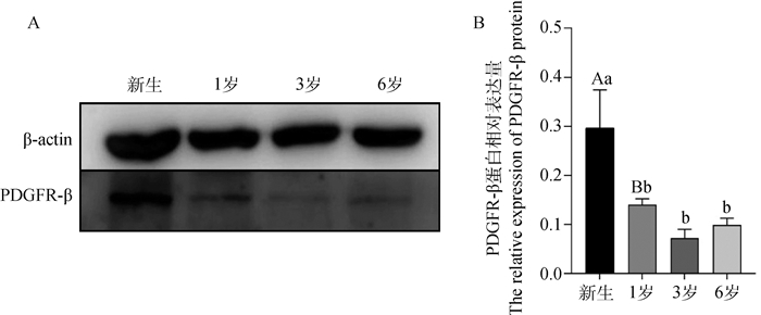
PDGFR-β在不同年龄组牦牛肺中均能检测到表达。新生组表达量极显著高于其他3组(P < 0.01);1岁龄组、3岁龄组、6岁龄组两两比较,差异性不显著(P>0.05, 图 2)。

|
A. Western-blot结果;B. PDGFR-β灰度值分析。大写字母不同表示差异极显著(P < 0.01),小写字母不同表示差异显著(P < 0.05), 相同字母表示差异不显著(P>0.05)。下同 A. The result of Western-blot; B. PDGFR-β gray value analysis. Different uppercase letters mean extremely significant differences (P < 0.01), different lowercase letters mean significant differences (P < 0.05), same letters mean no significant difference (P>0.05). The same as below 图 2 不同年龄牦牛肺脏中PDGFR-β蛋白的表达 Fig. 2 PDGFR-β protein expression in yak lungs at different ages |
PDGFR-β mRNA水平在新生组牦牛肺中最高,极显著高于其他年龄组(P < 0.01);其次为1岁组,1岁组与3岁组、6岁组相比较,差异性同样极显著(P < 0.01);而6岁组显著高于3岁组(P < 0.05,图 3)。

|
图 3 不同年龄牦牛肺脏中PDGFR-β mRNA的分析 Fig. 3 Analysis of PDGFR-β mRNA in lungs of yak at different ages |
低氧环境下可引起肺动脉高压,继而造成肺结构的适应性改建,以肺动脉结构的变化最为明显。
我们观察到不同年龄段牦牛肺中PDGFR-β的分布和表达与Kishi等[7-10]在小鼠、大鼠和猪的肺中研究结果一致,即低氧条件下PDGFR-β受体表达水平上调。说明牦牛生活在低氧环境下,PDGFR-β对牦牛肺的低氧适应性调节是至关重要的。
随着牦牛的成长,PDGFR-β的表达呈现先下降后上升的趋势。PDGFR-β的蛋白及mRNA的表达水平在新生组表达量最高,其次为1岁组、6岁组,而3岁组的表达水平最弱。说明牦牛能通过自身的调节能力和适应环境的本能,逐渐适应了高寒低氧环境。而PDGFR-β的表达在6岁时会升高,可能由于年龄的增长,肺机能下降,为了维持机体稳态,PDGFR-β的水平可能处于动态平衡而有所升高。本试验表明PDGFR-β对牦牛肺结构起到持续调控的作用,而这种调控在低年龄阶段更为显著。
4 结论PDGFR-β主要分布在牦牛肺的细支气管及其分支气道的上皮细胞,少量分布在各级肺动脉内皮细胞中;以新生到1岁的表达最为明显。PDGFR-β在不同年龄牦牛气道和肺动脉发育及低氧适应性的形成中均发挥作用。
| [1] |
陈秋生, 冯霞, 姜生成. 牦牛肺脏高原适应性的结构研究[J]. 中国农业科学, 2006, 39(10): 2107–2113.
CHEN Q S, FENG X, JIANG S C. Structural study on plateau adaptability of yak lung[J]. Scientia Agricultura Sinica, 2006, 39(10): 2107–2113. DOI: 10.3321/j.issn:0578-1752.2006.10.024 (in Chinese) |
| [2] |
何俊峰, 余四九, 崔燕. 不同年龄高原牦牛肺脏的组织结构特征[J]. 畜牧兽医学报, 2009, 40(5): 748–755.
HE J F, YU S J, CUI Y. Structural features of lungs of plateau yak at different ages[J]. Acta Veterinaria et Zootechnica Sinica, 2009, 40(5): 748–755. DOI: 10.3321/j.issn:0366-6964.2009.05.023 (in Chinese) |
| [3] |
王文玲, 张振庭, 王书华, 等. 血小板衍生生长因子B及其受体的表达对肾癌ACHN细胞增殖的影响[J]. 中华肿瘤杂志, 2015, 37(3): 170–174.
WANG W L, ZHANG Z T, WANG S H, et al. Effect of platelet derived growth factor-B and its receptor expression on the proliferation of renal cell carcinoma ACHN cells[J]. Chinese Journal of Cancer, 2015, 37(3): 170–174. DOI: 10.3760/cma.j.issn.0253-3766.2015.03.003 (in Chinese) |
| [4] | SATO H, ISHⅡ Y, YAMAMOTO S, et al. PDGFR-β plays a key role in the ectopic migration of neuroblasts in cerebral stroke[J]. Stem Cells, 2015, 34(3): 685–698. |
| [5] | WILSON C L, STEPHENSON S E, HIGUERO J P, et al. Characterization of human PDGFR-β-positive pericytes from IPF and non-IPF lungs[J]. Am J Physiol Lung Cell Mol Physiol, 2018, 315(6): L991–L1002. DOI: 10.1152/ajplung.00289.2018 |
| [6] | FAN H, MA L, FAN B, et al. Role of PDGFR-β/PI3K/AKT signaling pathway in PDGF-BB induced myocardial fibrosis in rats[J]. Am J Transl Res, 2014, 6(6): 714–723. |
| [7] | KISHI M, AONO Y, SATO S, et al. Blockade of platelet-derived growth factor receptor-β, not receptor-α ameliorates bleomycin-induced pulmonary fibrosis in mice[J]. PLoS One, 2018, 13(12): e0209786. DOI: 10.1371/journal.pone.0209786 |
| [8] | ZHOU L, SUN X, HUANG Z J, et al. Imatinib Ameliorated Retinal Neovascularization by Suppressing PDGFR-α and PDGFR-β[J]. Cell Physiol Biochem, 2018, 48: 263–273. DOI: 10.1159/000491726 |
| [9] | SCHERMULY R T, DONY E, GHOFRANI H A, et al. Reversal of experimental pulmonary hypertension by PDGF inhibition[J]. J Clin Invest, 2005, 115(10): 2811–2821. DOI: 10.1172/JCI24838 |
| [10] | LANNÉR M C, RAPER M, PRATT W M, et al. Heterotrimeric G proteins and the platelet-derived growth factor receptor-beta contribute to hypoxic proliferation of smooth muscle cells[J]. Am J Respir Cell Mol Biol, 2005, 33(4): 412–419. DOI: 10.1165/rcmb.2005-0004OC |


