


2. 中国科学院微生物研究所,北京 100101
2. Institute of Microbiology, Chinese Academy of Sciences, Beijing 100101
卵黄蛋白原(Vitellogenin,Vg),分子量约200 kD,在卵生动物体中是一种重要的营养蛋白。在卵形成过程中,Vg携带修饰在蛋白上的营养物质一并经运输入卵,在卵内被加工成为成熟的卵黄蛋白(Vitellin,Vn)。Vn在卵内积累,为后期胚胎的发育提供氨基酸、脂肪、碳水化合物、磷、硫及微量元素等[1-4]。作为一种生殖营养蛋白,Vg一般在卵黄发育期受到激素的诱导[5-7],在昆虫雌性成虫的脂肪体、鱼类的肝脏或者其他的动物的特定组织中特异性地大量表达,并通过定位于卵巢的Vg受体(Vg receptor,VgR)所介导的内吞作用入卵[8-11]。Vg准确的时空特异性表达决定了卵的正常发育[12]。Vg在卵巢外组织中翻译为前体蛋白之后,会经历一系列加工过程,包括蛋白修饰、剪切、分泌、运输、受体识别等。不同的Vg剪切形式及其时空表达,能介导不同的生物学功能。例如,在Vg运输过程中,在血淋巴内,特定剪切形式的Vg被证明与病毒表面蛋白互作[13],将病毒携带入卵实现其垂直传播的过程;对于病原微生物,Vg通过与病原蛋白的分子互作直接杀菌,或间接介导昆虫对微生物的免疫识别[14-16]。本文将综述Vg的基本结构、加工运输及近年来在Vg非营养功能方面的新进展,旨为后续Vg的功能研究提供指导。
1 Vg的基本结构不同来源的Vg在结构上具有高度的保守性:其N端为引导蛋白分泌的信号肽序列,从N端到C端依次编码3个结构域:卵黄蛋白原N端结构域(Vitellogenin N domain,VitN),未知功能结构域(Unknown function domain,DUF)和血管性血友病因子结构域(von Willebrand domain,vWD)[8]。VitN结构域的主要特点包括:(1)多数Vg同源蛋白在该结构域内编码两个由连续丝氨酸组成的丝氨酸聚集区,是Vg的主要磷酸化位点,两个丝氨酸聚集区之间具有保守的Arg-X-Arg/Lys-Arg(RXR/KR)酶切位点,几乎所有Vg均能在该位点被特异的蛋白酶剪切形成N端小亚基和C端大亚基,剪切后的Vg亚基以聚合体形式一起分泌至血淋巴中;(2)该结构域内具有分泌信号肽和Vg与VgR的互作位点,因此单独的Vg小亚基能够完成向卵母细胞内的运输功能,丝氨酸的磷酸化对Vg的剪切及Vg-VgR的识别具有重要作用[17-18];(3)该结构域是重要的蛋白修饰区域,高达16%(W/W)位置被酯化,能结合25-40个脂肪;2/3的区域被磷酸化[19-20]。在蜜蜂和灰飞虱体内均发现大量的Vg小亚基单独由脂肪体分泌并向卵巢运输的现象,Vg在该区域的高度修饰有可能在其介导的营养输送中发挥主要功能。DUF和vWD两个结构域均具有与病毒或细菌互作的性质,对微生物进行识别并介导微生物的清除或垂直传播[13, 21]。在三维结构上,位于N末端的VitN的主要结构是N-折叠;其后是一段α螺旋结构,其和位于C端结构域的序列,形成一个“脂穴”结构,是脂肪分子结合的主要部位[12]。
2 Vg的表达、加工及运输 2.1 Vg的表达Vg传统上被认为是在雌性个体处于卵巢发育期时特异性地诱导表达[6],卵生脊椎动物的肝脏和昆虫的脂肪体是Vg的主要合成加工部位。以昆虫为例,在幼虫期Vg不表达或表达水平很低,或Vg在非脂肪体细胞中进行表达;变成成虫后进入了生殖生长阶段,脂肪体中的Vg开始跳跃式地高表达,并且在产卵过程中一直维持很高的水平。对于没有交配的雌虫,Vg的表达则明显降低[22-23]。除了脂肪体,在卵巢发育过程中,卵巢上的滤泡细胞也会合成部分Vg,为卵发育提供营养物质[24]。
近年来对多个物种的Vg研究发现,除在卵巢发育期为了供给营养使得Vg大量表达之外,在幼虫、雄虫、或者各龄期昆虫的其它组织中也发现有一定量的Vg表达。在功能性不育的蜜蜂的工蜂体内,其下咽腺及与之毗邻的头部脂肪体细胞均能合成Vg,暗示了Vg用于育雏食物的功能;Bombus hypocrita的蜂后、工蜂及雄蜂从蛹到成虫的各个发育阶段Vg都能以不同的水平表达[25];Camponotus festinatus的Vg在蜂后及工蜂体内以不同丰度进行表达,在成虫羽化前Vg的合成水平较低[26]。Leucophaea maderae的雌性和雄性脂肪体细胞均有Vg表达[27];我们近期对灰飞虱的研究发现在灰飞虱体内,雄虫和幼虫都能稳定表达Vg,与雌虫不同,非雌性昆虫的Vg仅表达于其血细胞内,而在Vg主要合成组织脂肪体内则完全没有表达[28]。
2.2 Vg的表达调节Vg通常在特定的发育时期大量表达,其产生、加工甚至运输均依赖激素的调节作用[2-3, 10]。调节Vg产生的激素主要为保幼激素(Juvenile hormone,JH),某些物种中蜕皮素和神经肽也能协助JH调节Vg[29-30]。在调节卵巢发育及Vg产生相关的基因的上游通常结合有重要的应答原件,包括激素受体。在卵巢发育期,JH增多并作为配体分子与激素受体结合,进而诱导相应基因的表达。飞蝗脂肪体中的伴侣蛋白Grp78(78 kD glucose-regulated protein)能协助Vg在内质网上翻译产生时正确折叠,JH能够诱导Grp78基因的表达,从而对Vg的表达产生调控[31]。JH同时能调节蝗虫的Cdc6(cell-division-cycle 6)基因,促使脂肪体和卵泡细胞的多倍体化,诱导Vg的表达和卵巢的正常发育[32]。对于大多数昆虫,JH能通过调节多种基因的表达并最终促进Vg的产生,而蜜蜂中,JH对Vg的产生是负调控作用—JH抑制Vg的表达[33]。利用激素对Vg的调节作用,人为干扰生物体内的激素水平,甚至可以导致生物的“变性”,在雄性个体中诱发Vg的表达。当在雄性蟑螂体内注射入JH时,也能诱发Vg的产生,尽管产生的Vg后期加工与雌虫不同[34]。这类情况不止发生在昆虫中,在鱼类也会发生:受水体到外界污染时,雄鱼体内JH会上调,能诱发Vg的表达[35-36]。
昆虫的卵黄发育需要营养物质供给,营养状态决定了卵黄发育水平,尤其在蚊子这类吸血昆虫中,只有在摄取了血液后,才能开启Vg表达和卵巢发育[37]。研究表明无论JH对Vg是正调控还是负调控,营养物通过调控激素水平间接地正向调控Vg的表达。褐飞虱中,氨基酸营养能通过促进JH的合成来促进Vg的合成[38]。而在蜜蜂中,缺乏营养时,IIS(Insulin-IGF-1 signaling)表达上调,IIS和受体结合,激发下游的激酶活性,磷酸化FOXO,从而解除其对JH的抑制功能,导致JH上调,从而抑制Vg的表达[39]。
2.3 Vg的剪切Vg的剪切通过枯草芽孢杆菌酶及其同工酶完成,该类酶具有furin结构域,识别RXXR位点并在第四位R之后发生剪切[40]。大多数Vg同源蛋白具有一个或以上的RXXR位点,其中最保守的RXR/KR位于N端两个丝氨酸聚集区之间,在该位点的酶切产生N端小亚基和C端大亚基[8]。对具有多个RXXR位点的Vg同源蛋白,其酶切发生于部分RXXR位点,需要通过实验对酶切位点进行验证。例如,褐飞虱Vg有3个RXXR位点,仅在最保守的位点进行剪切[41];蟑螂Vg有5个酶切位点,剪切发生在其中3个位点之后[42]。除位点特异性剪切外,Vg也会被组织特异性或发育阶段特异性剪切。例如淡水虾的Vg在脂肪体中被枯草芽孢杆菌酶切产生大小两个亚基,并分泌至血淋巴中,在血淋巴中,Vg大亚基进一步被剪切为两个中等大小的亚基[43];蟑螂雌性和雄性血淋巴中含有种类不同的Vg亚基[29]。经剪切的Vg亚基在进入卵巢之后会进一步加工并最终形成成熟的卵黄蛋白。本实验室最近的研究发现灰飞虱Vg有5个RXXR位点,酶切可以发生在其中两个位点之后,N端保守RXRR位点的酶切发生在入卵之前,C端大亚基的二次酶切则发生在入卵之后[28];在雄性灰飞虱体内,Vg仅由血细胞表达且不发生剪切,以全长形式分布于血细胞和血淋巴中,在水稻条纹病毒RSV的水平传播中发挥功能[28]。由于Vg是多亚基组成的蛋白,剪切产生的含有不同亚基组成的Vg片段是否发挥不同的生物学功能是未来Vg研究的重要内容之一。我们对灰飞虱Vg的不同分子形式进行功能鉴定,发现其脂肪体加工产生的Vg N端小亚基可以分泌并入卵,但是由于缺乏与病毒RSV互作的vWD及DUF结构域,不能介导病毒的传播;而其血细胞产生的Vg含有C端RSV互作结构域,因而在体内发挥传播RSV的功能[28]。
2.4 Vg的运输卵巢到达卵黄发育期,VgR在激素诱导下表达,开始介导Vg向卵内的运输[44-45]。Vg通过卵泡细胞的间隙到达卵母细胞表面,其VitN结构域与VgR结合,通过内吞作用将Vg运输到卵内[17]。Vg被VgR介导内吞进入卵细胞,快速被运输到初级内涵体,在内涵体中,v-ATPase介导的酸性环境,使Vg和VgR分离[10]。内涵体的酸性环境活化其内的组织蛋白酶活性,对Vg进行切割,加工成为成熟的Vn,Vn在卵内聚集在光镜可见的卵黄体中,后期,为胚胎发育提供营养。此外,除了直接被卵细胞上的VgR识别,有些物种内,卵巢的其他种类细胞也可以吸收Vg,如在灰飞虱中,Vg除直接被卵细胞吸收之外,还可以进入滋养区的营养细胞内,通过营养细胞与卵细胞相连的营养丝间接运输至卵母细胞内[13]。
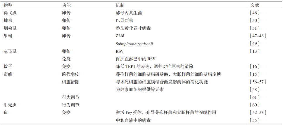
3 Vg的非营养功能 3.1 Vg介导微生物的垂直传播卵生动物及昆虫体内均携带多种共生或病原微生物,包括细菌与真菌性共生菌,病毒病原菌等。作为卵生动物最重要的营养运输蛋白,Vg被微生物劫持实现自身入卵传播的研究得到了越来越多的例证(表 1)。如在褐飞虱的血淋巴及卵巢中,酵母类共生菌周围有大量的Vg聚集,暗示其入卵运输与Vg的密切关系[46];果蝇体内的ZAM病毒是一种卵传病毒,在感染ZAM病毒的卵巢中,ZAM出现在含Vg的内吞小泡中,干扰果蝇VgR同时影响了Vg及ZAM病毒的入卵过程[47-48];果蝇细胞内共生细菌Spiroplasma poulsonii在血淋巴中与Vg互作并因此被运输至卵黄体中[49];干扰蜱虫的VgR会影响其卵巢发育,进而影响Vg及巴贝西虫的入卵能力[50]。
在植物病毒经介体昆虫传播的过程中,病毒经卵传播至子代,在幼虫体内过冬并在来年侵染植物,增加了植物病毒病害的防治难度,也使得对病毒在介体昆虫体内的垂直传播机理成为重要的研究内容。近年来在植物病毒经介体昆虫卵传的研究取得了一系列重要的成果。其中之一为RSV在灰飞虱体内的垂直传播过程。研究发现RSV的入卵传播路径与灰飞虱Vg进入滋养区营养细胞并进而进入卵母细胞的过程高度一致,机理研究表明RSV的结构蛋白能与灰飞虱Vg的DUF和vWD结构域发生体外分子互作,在体内,RSV与Vg的C端大亚基在血细胞中结合并分泌至血淋巴[28],运输至卵巢滋养区,RSV-Vg复合体通过Vg与营养细胞表面VgR的分子互作,突破卵巢屏障,并最终进入到卵母细胞内,垂直传播至子代灰飞虱[13]。水稻黑条矮缩病毒也能经灰飞虱进行水平传播,但不能垂直传播至子代,我们的工作发现该病毒的结构蛋白不能与灰飞虱Vg发生分子互作。
这种植物病毒借助Vg运输途径进行垂直传播的机制在植物病毒的传播中有一定的共性。最新近的研究证明了烟粉虱Vg在双生病毒垂直传播过程中的功能,为昆虫Vg介导的病毒卵传提供了新的例证[51]。番茄黄化卷叶病毒TYLCV能以持久循环方式经由其介体烟粉虱进行传播,TYLCV进入烟粉虱生殖器官的能力与烟粉虱卵巢的发育阶段密切相关,随着烟粉虱成虫虫龄的增加,TYLCV垂直传播至子代昆虫的几率大大增加。机理研究表明了该病毒外壳蛋白与昆虫Vg的分子互作,干扰Vg表达水平能抑制病毒的入卵几率。与灰飞虱传水稻黑条矮缩病毒相似,烟粉虱的Vg与中国番木瓜曲叶病毒的外壳蛋白不发生互作,该病毒也因此不能被烟粉虱垂直传播至子代。
3.2 Vg的免疫功能及介导隔代免疫血液或淋巴液是动物的循环系统,也是免疫作用的主要场所之一,Vg在通过血液或血淋巴进行运输的过程中发挥免疫功能,直接或间接清除病原微生物的性质首先在鱼类研究中得到了较多的例证。鱼的Vg可以作为模式识别分子,识别细菌细胞壁脂多糖和脂磷壁酸,直接促使细菌裂解[52];在体外实验中,将鱼的Vg添加至细菌生长液中,抑制细菌的生长[53];鱼的Vg也可以作为调理素分子激活Fcγ受体介导的吞噬作用对病原菌进行间接的清除[54];在与病毒互作的研究中,Vg被发现能结合至病毒表面,通过对病毒粒子进行交联而将其中和[55]。昆虫Vg的免疫相关功能首先在蚊子中得到解析,蚊子的含硫酯键蛋白(Thioester-containing protein 1,TEP1)介导疟原虫的免疫清除,Vg通过降低TEP1的表达间接调控了蚊子对疟原虫的清除效力[16];蜜蜂的Vg受伤口诱导,并与坏死细胞的细胞膜结合激发溶酶体的消化功能,加速细胞的清除过程[56-57];蜜蜂血细胞的生长需要重要的微量元素锌,Vg作为营养物质能和健康的血细胞互作,为其提供锌离子,以维持血细胞的生长及功能[58]。
Vg经血淋巴传播至子代的过程中,与其结合的微生物分子被携带入卵,则会介导免疫信号的隔代传递[15]。跨代免疫素(Trans-generational immune priming,TGIP)将母体内的免疫记忆传给后代,以对后代起到初始保护作用。在脊椎动物中,母体以抗体作为TGIP传递免疫记忆,而昆虫则将其体内诱发先天免疫的病原微生物抗原作为TGIP,通过卵将免疫记忆传给后代。以蜜蜂为例,其Vg与芽孢杆菌的细胞壁脂磷壁酸及大肠杆菌的细胞壁脂多糖互作,携带这些微生物碎片入卵,促进子代昆虫的免疫系统初建[15]。
3.3 Vg的行为调节功能在对社会型昆虫的研究中发现,Vg对昆虫的觅食行为有重要的调节作用[59]。对甲壳虫研究发现,其大脑能表达VgR,Vg因此能运输到大脑中,通过调节神经系统来实现对昆虫行为的调节[60],这种机制对于了解Vg在多种完全社会型及半社会型昆虫中的行为调节能力具有指导意义。Vg对自由基的结合,能保护健康细胞免于伤害,从而对蜜蜂的寿命起到重要的调节作用,Vg表达水平的不同,导致夏季生工蜂的寿命仅有6周,而冬季生工蜂能存活数月[61]。在蜜蜂的社会分工中,蜂王的功能是繁育后代,社会地位高,获得比工蜂和雄蜂更多的营养供给。充分的营养物质能降低IIS(Insulin-IGF-1 signaling),抑制保佑激素的产生,解除JH对Vg的抑制功能,提高Vg表达水平及蜂王的产卵能力[39]。
4 总结及讨论Vg作为卵黄蛋白的前体,其产生、加工及运输过程在不同的动物间比较保守:在动物肝脏或昆虫脂肪体细胞中,Vg的mRNA翻译合成Vg蛋白前体,通过糖基化、磷酸化、酯化等修饰及枯草芽孢杆菌酶剪切,形成亚基,分泌至血淋巴并运输至卵巢,其N端的VgR识别位点与VgR结合,通过内吞作用运输到卵细胞中,为胚胎发育提供营养。Vg在从血淋巴向卵巢运输过程中,会与微生物发生多种形式的互作,通过直接或间接的免疫功能清除病原微生物,或通过与微生物的互作携带微生物进行垂直传播,或通过与微生物碎片的互作,将免疫信号传递给子代(图 1)。对Vg非营养功能的研究集中于Vg与微生物互作及由此引发的促进微生物传播或通过免疫清除微生物的过程,深入了解Vg的非营养功能,需要了解Vg在卵巢外的多种剪切策略;由此产生的不同Vg分子形式,将介导多样化的生物学功能。

|
| 图 1 卵黄蛋白原的产生、运输及功能 1:在脂肪体或者肝脏细胞中,卵黄蛋白原Vg在激素诱导下表达、翻译并进行翻译后修饰;2:Vg的蛋白蛋白结构,包括N端信号肽,从N至C端的VitN、DUF及vWD三个主要结构域,VitN结构域内包含丝氨酸聚集区及保守的RXXR酶切位点;3:Vg在其产生细胞中被枯草芽孢杆菌酶切成Vgcu(Vg cutting unit,也称为Vg亚基);4:Vg亚基释放至血淋巴中;5:血淋巴中的Vgcu介导的免疫,营养,病原运输及行为调节等功能;6:Vgcu被卵细胞上的受体VgR识别,通过内吞作用运输到卵内 |
| [1] |
Schwabl H. Yolk is a source of maternal testosterone for developing birds[J]. Proc Natl Acad Sci USA, 1993, 90: 11446-11450. DOI:10.1073/pnas.90.24.11446 |
| [2] |
Ho SM, Kleis S, Mcpherson R, et al. Regulation of vitellogenesis in reptiles[J]. Hrepetologica, 1982, 3840-3450. |
| [3] |
Robin A, Wallace E, Bergink EW. Amphibian vitellogenin:properties, hormonal regulation of hepatic synthesis and ovarian uptake, and conversion to yolk proteins[J]. Soc Integ Comp Biol, 1974, 14: 1159-1175. |
| [4] |
Tsutomu N, Kenneth HF, Bert LV. Zinc, iron, and copper contents of Xenopus laevis oocytes and embryos[J]. Molecular Reproduction and Development, 1993, 36: 419-423. DOI:10.1002/(ISSN)1098-2795 |
| [5] |
Bownes M. The roles of juvenile hormone, ecdysone and the ovary in the control of Drosophila vitellogenesis[J]. J Insect Physiol, 1989, 35: 409-413. DOI:10.1016/0022-1910(89)90115-7 |
| [6] |
Campbell DRI C.M.. Hormonal control of vitellogenesis in hypophysectomized winter flounder(Pseudopleuronectes americanus Walbaum)[J]. Gen Comp Endocrinol, 1976, 28: 413-150. DOI:10.1016/0016-6480(76)90149-0 |
| [7] |
Tufail M, Nagaba Y, Elgendy AM, et al. Regulation of vitellogenin genes in insects[J]. Entomological Science, 2014, 17(3): 269-282. DOI:10.1111/ens.2014.17.issue-3 |
| [8] |
Tufail M, Takeda M. Molecular characteristics of insect vitellogenins[J]. J Insect Physiol, 2008, 54(12): 1447-1458. DOI:10.1016/j.jinsphys.2008.08.007 |
| [9] |
Sappington TW, Raikhel AS. Molecular characteristics of insect vitellogenins and vitellogenin receptors[J]. Insect Biochemistry and Molecular Biology, 1998, 28: 277-300. DOI:10.1016/S0965-1748(97)00110-0 |
| [10] |
Tufail M, Takeda M. Insect vitellogenin/lipophorin receptors:molecular structures, role in oogenesis, and regulatory mechanisms[J]. J Insect Physiol, 2009, 55(2): 87-103. DOI:10.1016/j.jinsphys.2009.01.009 |
| [11] |
Bai H, Qiao H, et al. Molecular characterization and developmental expression of vitellogenin in the oriental river prawn Macrobrachium nipponense and the effects of RNA interference and eyestalk abla-tion on ovarian maturation[J]. Gene, 2015, 562(1): 22-31. DOI:10.1016/j.gene.2014.12.008 |
| [12] |
Veerana M, Kubera A, Ngernsiri L. Analysis of the vitellogenin gene of rice moth, Corcyra cephalonica stainton[J]. Archives of Insect Biochemistry and Physiology, 2014, 87(3): 126-147. DOI:10.1002/arch.v87.3 |
| [13] |
Huo Y, Liu W, Zhang F, et al. Transovarial transmission of a plant virus is mediated by vitellogenin of its insect vector[J]. PLoS Pathogens, 2014, 10(3): e1003949. DOI:10.1371/journal.ppat.1003949 |
| [14] |
Milutinovic B, Kurtz J. Immune memory in invertebrates[J]. Seminars in Immunology, 2016, 28(4): 328-342. DOI:10.1016/j.smim.2016.05.004 |
| [15] |
Salmela H, Amdam GV, Freitak D. Transfer of immunity from mother to offspring is mediated via egg-yolk protein vitellogenin[J]. PLoS Pathogens, 2015, 11(7): e1005015. DOI:10.1371/journal.ppat.1005015 |
| [16] |
Rono MK, Whitten MM, Oulad-Abdelghani M, et al. The major yolk protein vitellogenin interferes with the anti-plasmodium response in the malaria mosquito Anopheles gambiae[J]. PLoS Biology, 2010, 8(7): e1000434. DOI:10.1371/journal.pbio.1000434 |
| [17] |
Roth Z, Weil S, Aflalo ED, et al. Identification of receptor-interacting regions of vitellogenin within evolutionarily conserved beta-sheet structures by using a peptide array[J]. Chembiochem, 2013, 14(9): 1116-1122. DOI:10.1002/cbic.201300152 |
| [18] |
Romans P, Tu Z, et al. Analysis of a vitellogenin gene of the mosq-uito, aedes aegypti and comparisons to vitellogenins from other org-anisms[J]. Insect Biochem Mol Biol, 1995, 25(8): 939-958. DOI:10.1016/0965-1748(95)00037-V |
| [19] |
Ziegler R, Van AR. Lipid uptake by insect oocytes[J]. Insect Biochemistry and Molecular Biology, 2006, 36(4): 264-272. DOI:10.1016/j.ibmb.2006.01.014 |
| [20] |
Anderson TA, Leivitt DG, Banaszak LJ. The structural basisi of lipid interactions in lipovitellin, a soluble liporotein[J]. Current Biology, 1998, 6: 895-909. |
| [21] |
Sun C, Hu L, Liu S, et al. Functional analysis of domain of unknown function(DUF)1943, DUF1944 and von Willebrand factor type D domain(vWD)in vitellogenin2 in zebrafish[J]. Deve Comp Immunol, 2013, 41(4): 469-476. DOI:10.1016/j.dci.2013.07.005 |
| [22] |
Thompson JR, Leonard JB. Lipid-protein interactions in lipovitellin[J]. Biochemistry, 2002, 41: 9398-9409. DOI:10.1021/bi025674w |
| [23] |
Tufail M, Takeda M. Molecular cloning and developmental expression pattern of the vitellogenin receptor from the cockroach, Leucophaea maderae[J]. Insect Biochemistry and Molecular Biology, 2007, 37: 235-245. DOI:10.1016/j.ibmb.2006.11.007 |
| [24] |
Kitano H, Nagano N, Sakaguchi K, et al. Two vitellogenins in the loliginid squid Uroteuthis edulis:Identification and specific expression in ovarian follicles[J]. Molecular Reproduction and Development, 2017, 84(5): 363-375. DOI:10.1002/mrd.v84.5 |
| [25] |
Li JL, Huang JX, et al. The vitellogenin of the bumblebee, Bombus hypocrita:studies on structural analysis of the cDNA and express-ion of the mRNA[J]. J Comp Physiol B, 2010, 180(2): 161-170. DOI:10.1007/s00360-009-0434-5 |
| [26] |
Martinez T, Wheeler D. Identification of vitellogenin in the ant, camponotus-festinatus-changes in hemolymph-proteins and fat-body development in workers[J]. Archives of Insect Biochemistry and Physiology, 1991, 17(2-3): 143-155. DOI:10.1002/(ISSN)1520-6327 |
| [27] |
Chen JS, Raikhel AS. Subunit cleavage of mosquito pro-vitellogenin by a subtilisin-like convertase[J]. Proc Natl Acad Sci USA, 1996, 93: 6186-6190. DOI:10.1073/pnas.93.12.6186 |
| [28] |
Huo Y, Yu Y, et al. Insect tissue-specific vitellogenin facilitates transmission of plant virus[J]. PLoS Pathogens, 2018, 14(2): e1006909. DOI:10.1371/journal.ppat.1006909 |
| [29] |
Martin D, Piulachs MD, Belles X. Production and extraovarian processing of vitellogenin in ovariectomized Blattella germanica(L.)(Dictyoptera, Blattellidae)[J]. J Insect Physiol, 1996, 42(2): 101-105. DOI:10.1016/0022-1910(95)00100-X |
| [30] |
Nagaba Y, Tufail M, Inui H, Takeda M. Hormonal regulation and effects of four environmental pollutants on vitellogenin gene transcription in the giant water bug, Lethocerus deyrollei(Hemiptera:Belostomatidae)[J]. J Insect Conserv, 2011, 15: 421-431. DOI:10.1007/s10841-010-9315-1 |
| [31] |
Luo M, Li D, Wang Z, et al. Juvenile hormone differentially regulates two Grp78 genes encoding protein chaperones required for insect fat body cell homeostasis and vitellogenesis[J]. The Journal of Biological Chemistry, 2017, 292(21): 8823-8834. DOI:10.1074/jbc.M117.780957 |
| [32] |
Wu Z, Guo W, Xie Y, Zhou S. Juvenile hormone activates the transcription of cell-division-cycle 6(cdc6)for polyploidy-dependent insect vitellogenesis and oogenesis[J]. The Journal of Biological Chemistry, 2016, 291(10): 5418-5427. DOI:10.1074/jbc.M115.698936 |
| [33] |
Seehuus SC, Norberg K, Gimsa U, et al. Reproductive protein protects functionally sterile honey bee workers from oxidative stress[J]. Proc Natl Acad Sci USA, 2006, 103(4): 962-967. DOI:10.1073/pnas.0502681103 |
| [34] |
Don-Wheeler G, Egelmann F. The biosynthesis and processing of vitellogenin in the fat bodies of females and males of the cockroach Leucophaea maderae[J]. Insect Biochemistry and Molecular Biology, 1997, 27: 908-917. |
| [35] |
Del GG, Prisco M, Agnese M, et al. Effects of nonylphenol on vitellogenin synthesis in adult males of the spotted ray Torpedo marmorata[J]. J Fish Biol, 2012, 80(5): 2112-2121. DOI:10.1111/jfb.2012.80.issue-5 |
| [36] |
Verderame M, Scudiero R. Estrogen-dependent, extrahepatic synthesis of vitellogenin in male vertebrates:A mini-review[J]. Comptes Rendus Biologies, 2017, 340(3): 139-144. DOI:10.1016/j.crvi.2017.01.005 |
| [37] |
Gonzales KK, et al. Blood serum and BSA, but neither red blood cells nor hemoglobin can support vitellogenesis and egg production in the dengue vector Aedes aegypt[J]. Peer J, 2015, 3: e938. DOI:10.7717/peerj.938 |
| [38] |
Lu K, et al. Nutritional signaling regulates vitellogenin synthesis and egg development through juvenile hormone in Nilaparvata lugens(Stål)[J]. Int J Mol Sci, 2016, 17(3): 269. |
| [39] |
Seehuus SC, Norberg K, Gimsa U, et al. Reproductive protein protects functionally sterile honey bee workers from oxidative stress[J]. Proc Natl Acad Sci USA, 2006, 103(4): 962-967. DOI:10.1073/pnas.0502681103 |
| [40] |
Rouille Y, Duguay SJ, Lund K, et al. Proteolytic processing mechanisms in the biosynthesis of neuroendocrine peptides:the subtilisin-like proprotein convertases[J]. Frontiers In Neuroendocrinology, 1995, 16: 322-361. DOI:10.1006/frne.1995.1012 |
| [41] |
Tufail M, Naeemullah M, Elmogy M, et al. Molecular cloning, transcriptional regulation, and differential expression profiling of vitellogenin in two wing-morphs of the brown planthopper, Nilaparvata lugens Stål(Hemiptera:Delphacidae)[J]. Insect Mol Biol, 2010, 19(6): 787-798. DOI:10.1111/j.1365-2583.2010.01035.x |
| [42] |
Tufail M, Takeda M. Vitellogenin of the cockroach, Leucophaea maderae:nucleotide sequence, structure and analysis of processing in the fat body and oocytes[J]. Insect Biochemistry and Molecular Biology, 2002, 32: 1469-1476. DOI:10.1016/S0965-1748(02)00067-X |
| [43] |
Okuno A, Yang WJ, Jayasankar V, et al. Deduced primary structure of vitellogenin in the giant freshwater prawn, Macrobrachium rosenbergii, and yolk processing during ovarian maturation[J]. The Journal of Experimental Zoology, 2002, 292(5): 417-429. DOI:10.1002/(ISSN)1097-010X |
| [44] |
Tufail M, Takeda M. Molecular cloning and developmental expression pattern of the vitellogenin receptor from the cockroach, Leucophaea maderae[J]. Insect Biochemistry and Molecular Biology, 2007, 37: 235-245. DOI:10.1016/j.ibmb.2006.11.007 |
| [45] |
Tufail M, Takeda M. Molecular cloning, characterization and regulation of the cockroach vitellogenin receptor during oogenesis[J]. Insect Mol Biol, 2005, 14(4): 389-401. DOI:10.1111/imb.2005.14.issue-4 |
| [46] |
Cheng DJ, Hou RF. Determination and distribution of a female-specific protein in the brown planthopper, Nilaparvata lugens Stål(Homoptera:Delphacidae)[J]. Tissue & Cell, 2005, 37(1): 37-45. |
| [47] |
Leblanc P, Desset S, Giorgi F, et al. Life Cycle of an endogenous retrovirus, ZAM, in Drosophila melanogaster[J]. Journal of Virology, 2000, 74(22): 10658-10669. DOI:10.1128/JVI.74.22.10658-10669.2000 |
| [48] |
Brasset E, Taddei AR, Arnaud F, et al. Viral particles of the endogenous retrovirus ZAM from Drosophila melanogaster use a pre-existing endosome/exosome pathway for transfer to the oocyte[J]. Retrovirology, 2006, 3: 25. DOI:10.1186/1742-4690-3-25 |
| [49] |
Herren JK, Paredes JC, Schupfer F, et al. Vertical transmission of a Drosophila endosymbiont via cooption of the yolk transport and internalization machinery[J]. MBio, 2013, 4(2): e00532. |
| [50] |
Boldbaatar D, et al. Tick vitellogenin receptor reveals critical role in oocyte development and transovarial transmission of Babesia parasite[J]. Biochem Cell Biol, 2008, 86(4): 331-344. DOI:10.1139/O08-071 |
| [51] |
Wei J, He YZ, Guo Q, et al. Vector development and vitellogenin determine the transovarial transmission of begomoviruses[J]. Proc Natl Acad Sci USA, 2017, 14(26): 6746-6751. |
| [52] |
Li Z, Zhang S, et al. Vitellogenin is a cidal factor capable of killing bacteria via interaction with lipopolysaccharide and lipoteichoic acid[J]. Mol Immunol, 2009, 46(16): 3232-3239. DOI:10.1016/j.molimm.2009.08.006 |
| [53] |
Tong Z, Li L, Pawar R, et al. Vitellogenin is an acute phase protein with bacterial-binding and inhibiting activities[J]. Immunobiology, 2010, 215(11): 898-902. DOI:10.1016/j.imbio.2009.10.001 |
| [54] |
Liu M, Pan J, Ji H, et al. Vitellogenin mediates phagocytosis through interaction with FcγR[J]. Molecular Immunology, 2011, 49(1-2): 211-218. DOI:10.1016/j.molimm.2011.08.011 |
| [55] |
Garcia J, Munro ES, Monte MM, et al. Atlantic salmon(Salmo salar L.)serum vitellogenin neutralises infectivity of infectious pancreatic necrosis virus(IPNV)[J]. Fish & Shellfish Immunology, 2010, 29(2): 293-297. |
| [56] |
Havukainen H, Munch D, Baumann A, et al. Vitellogenin recognizes cell damage through membrane binding and shields living cells from reactive oxygen species[J]. The Journal of Biological Chemistry, 2013, 288(39): 28369-28381. DOI:10.1074/jbc.M113.465021 |
| [57] |
Salmela H, Stark T, Stucki D, et al. Ancient duplications have led to functional divergence of vitellogenin-like genes potentially involved in inflammation and oxidative stress in honey bees[J]. Genome Biology Evol, 2017, 8(3): 495-506. |
| [58] |
Amdam GV, Simoes ZL, Hagen A, et al. Hormonal control of the yolk precursor vitellogenin regulates immune function and longevity in honeybees[J]. Exp Gerontol, 2004, 39(5): 767-773. DOI:10.1016/j.exger.2004.02.010 |
| [59] |
Nelson CM, Ihle KE, Fondrk MK, et al. The gene vitellogenin has multiple coordinating effects on social organization[J]. PLoS Biology, 2007, 5(3): e62. DOI:10.1371/journal.pbio.0050062 |
| [60] |
Roy-Zokan EM, Cunningham CB, Hebb LE, et al. Vitellogenin and vitellogenin receptor gene expression is associated with male and female parenting in a subsocial insect[J]. Proceedings Biological Sciences, 2015, 282(1809): 20150787. DOI:10.1098/rspb.2015.0787 |
| [61] |
Munch D, Amdam GV. The curious case of aging plasticity in honey bees[J]. FEBS Letters, 2010, 584(12): 2496-2503. DOI:10.1016/j.febslet.2010.04.007 |