扩展功能
文章信息
- 孙烁, 庄新明, 汪振宇, 曲志刚, 宋清旭, 张琪, 刘一
- SUN Shuo, ZHUANG Xinming, WANG Zhenyu, QU Zhigang, SONG Qingxu, ZHANG Qi, LIU Yi
- 间盘切除前椎间隙撑开在颈椎前路手术中的应用
- Application of distraction before discectomy in anterior cervical surgery
- 吉林大学学报(医学版), 2018, 44(01): 131-136
- Journal of Jilin University (Medicine Edition), 2018, 44(01): 131-136
- 10.13481/j.1671-587x.20180125
-
文章历史
- 收稿日期: 2017-09-05
颈椎前路间盘切除植骨融合内固定术(anterior cervical discectomy and fusion, ACDF)是治疗颈椎病的经典术式[1-2]。为了获得更好的减压视野,术中利用椎间撑开技术扩大病变间隙是必做的操作。通常的做法是在切除纤维环和部分间盘后进行椎间撑开操作, 即间盘切除后撑开(distraction after discectomy, DAD)。由于失去了间盘的抗张力作用,椎间隙比较容易撑开,但同时,撑开程度也容易失控,过度撑开往往导致椎间融合器型号选择失当,超过一定间隙高度的融合固定可产生术后颈肩痛[3-5]。术中如何通过简便、可靠的方法适度撑开椎间隙,进而选择适宜高度的椎间融合器,目前鲜有报道。Brenke等[6]利用计算机软件辅助椎间融合器高度的选择。在椎间盘切除术前实施撑开病变椎间隙的操作,即间盘切除前撑开(distraction before discectomy, DBD)。目前,DBD的临床应用尚未见报道。本研究拟通过对因脊髓型颈椎病行ACDF的患者症状和临床影像学进行观察、分析,探讨DBD在颈椎前路手术中应用的可行性。
1 资料与方法 1.1 一般资料选择2016年8月—2017年8月在本院因脊髓型颈椎病行ACDF患者31例,其中男性18例,女性13例,年龄(51.42±8.81)岁(35~74岁)。其中,颈4-5节段12例,颈5-6节段17例,颈6-7节段2例。
纳入标准:符合脊髓型颈椎病诊断标准,术前无明显颈肩痛症状,术前颈椎MRI提示病变间隙上下邻近节段间盘无明显退行性改变,椎间高度无明显丢失。排除标准:术前有明确颈肩痛,既往颈椎创伤、感染、肿瘤及手术病史。
手术方法:手术均由同一高年资术者完成。术中采用DBD撑开病变椎间隙,即在保证前纵韧带和纤维环完整的情况下,对椎间隙进行一次性撑开。当感到撑开力骤然增加,且很难跨越Caspar撑开器下一棘齿,以此作为撑开操作结束的标准。在保持此间隙高度的状态下切除椎间盘的前2/3部分,然后用试模选择匹配的椎间融合器型号。在确定融合器型号后,继续完成余下的减压和融合固定操作。固定方式采用钛板加Cage(Cervios,瑞士辛迪思公司)或零切迹颈椎前路椎间融合器(Zero-P, 瑞士辛迪思公司)。
1.2 临床评价术后颈肩痛评分采用视觉模拟评分法(VAS)0~10分的评分方式。颈肩痛评价时间为术后3 d。根据术后患者颈肩痛VAS评分进行分组,VAS评分≥4分的患者为有颈肩痛组,VAS评分 < 4分的患者为无颈肩痛组。
1.3 影像学资料测量方法:患者术前、术中及术后均取仰卧位拍摄颈椎侧位X线片。进入手术室后患者取仰卧位,全麻后摆放手术体位,颈椎轻度后伸,手术开始前拍摄X光片,测得术前椎间隙高度。手术开始后不再改变手术体位及颈椎屈伸角度,并以此为基础进行摄片。术后患者取仰卧位拍摄X光片,去除佩戴的颈托,颈椎略后伸拍摄X光片。由于患者术前及术中的X光片是由手术室中的可移动C臂机拍摄的,而术后的X光片是由影像科的X线机拍摄的,由于机器自带测量软件的精度及单位不一致,为了减少由此带来的偏倚均选用游标卡尺进行测量。使用数字型游标卡尺(工PD-151, 台湾宝工实业股份有限公司,0~150mm,误差±0.02mm)在胶片或显示屏上进行测量,再根据胶片或显示屏的缩放比例计算出所测值的真实大小。2名脊柱外科住院医师经过培训后,使用同一个校准后的电子游标卡尺,分别对同一个患者术前、术中及术后椎间隙的高度进行测量,取测量值的平均值作为最终值。结果精确到小数点后2位,所有相关高度测量单位均为毫米(mm)。
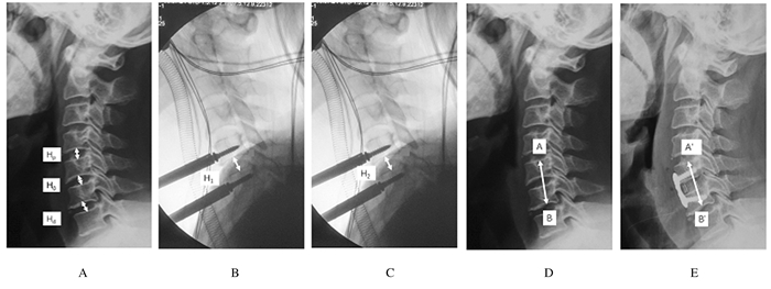
测量指标:术前,分别测量病变椎间隙上端椎体下终板弧顶最高点至下端椎体上终板中点连线的距离,即病变节段椎间隙的高度H0,以及相邻近、远端椎间隙的高度Hp和Hd(图 1A)。计算出Hp和Hd的平均值H,以此作为病变椎间隙预期恢复高度的参考值。术中, 测量DBD撑开后病变椎间隙高度H1(图 1B)和间盘切除后的病变椎间隙高度H2(图 1C)。术后,分别测量手术前、后病变节段高度(病变椎间隙相邻近端椎体上终板中点至临近远端椎体下终板中点的距离)AB和A′B′(图 1D和E)。手术前后病变节段高度变化值ΔH=A′B′-AB。术后病变椎间隙高度H3=H0+ΔH。分别计算术中病变椎间隙高度的变化值:ΔH1=H1-H0, ΔH2=H2-H1, ΔH3=H3-H2。

|
| A:Illustration of intervertebral space height (Hp:proximal intervertebral space height; H0:pre-operative index space height; Hd:distal intervertebral space height); B:Index intervertebral space height with DBD (H1); C:Index intervertebral space height with distraction after discectomy (H2); D:Illustration of pre-operative segment height AB; E:Illustration of post-operative segment height A′B′. 图 1 患者X光片上椎间隙高度及病变节段高度测量方法图 Figure 1 Illustrations of measurement methods of intervertebral space height and segment height on X-ray photos of patient |
|
|
采用SPSS 23.0统计软件进行统计学分析。H0、H1、H2、H、ΔH1、ΔH2、ΔH3和ΔH等测量数据经正态性检验,均符合正态分布,各指标均以x±s表示。组内比较采用配对样本t检验, 组间比较采用两独立样本t检验。以P < 0.05表示差异有统计学意义。在GraphPad Prism 7 for Mac软件中,采用Bland-Altman法进行一致性分析,以x±2s为允许范围,做比值-均值散点图。
2 结果31例患者ACDF术中均采用DBD,观察指标测量数据见表 1。
| (n=31, l/mm) | ||
| Parameter | Value (x±s) | Range |
| H0 | 5.94±0.82 | 3.96-7.43 |
| H | 6.93±0.69 | 5.10-8.24 |
| H1 | 7.13±1.00 | 5.11-8.98 |
| H2 | 7.51±1.07 | 5.24-9.46 |
| H3 | 7.90±1.16 | 5.34-10.18 |
| ΔH | 1.96±0.73 | 0.51-3.40 |
| ΔH1 | 1.19±0.51 | 0.28-2.47 |
| ΔH2 | 0.37±0.14 | 0.08-0.56 |
| ΔH3 | 0.39±0.20 | 0.10-0.89 |
术后有6例患者出现颈肩痛(VAS评分≥4分),颈肩痛发生率为19.35%。有颈肩痛组患者(6例)术后VAS评分为(4.83±0.75)分,无颈肩痛组患者(25例)术后VAS评分为(2.64±0.64)分。
2.2 采用DBD后2组患者椎间隙高度无颈肩痛组患者H1与H比较差异无统计学意义(P=0.80),H2与H1比较差异有统计学意义(P < 0.01);有颈肩痛组患者H2与H1比较差异有统计学意义(P < 0.01)。见表 2。ΔH2的95%可信区间(CI)为0.32~0.43 mm。
| (x±s, l/mm) | |||||
| Group | n | H | H1 | H2 | H3 |
| Neck pain | 6 | 6.72±0.65 | 7.87±1.35 | 8.33±1.39* | 8.93±1.52 |
| Non-neck pain | 25 | 6.98±0.70 | 6.95±0.84 | 7.31±0.90* | 7.65±0.93 |
| * P < 0.01 compared with H1. | |||||
有颈肩痛组患者ΔH为(3.04±0.42)mm,无颈肩痛组患者ΔH为(1.70±0.51)mm,组间比较差异有统计学意义(P<0.01)。见表 3。
| (x±s, l/mm) | |||||
| Group | n | ΔH | ΔH1 | ΔH2 | ΔH3 |
| Neck pain | 6 | 3.04±0.42 | 1.98±0.31 | 0.46±0.06 | 0.60±0.26 |
| Non-neck pain | 25 | 1.70±0.51* | 1.00±0.34* | 0.35±0.15* | 0.34±0.14* |
| * P < 0.01 compared with neck pain group. | |||||
Bland-Altman一致性分析显示:以x±2s为允许范围,做比值-均值散点图(图 2),3张图中分别只有1个点在允许范围外,即95%的数据在允许范围内,表明无颈肩痛组患者中H1、H2和H3与H一致性较好。

|
| A: H1 and H ratio vs average; B: H2 and H ratio vs average; C: H3 and H ratio vs average. 图 2 Bland-Altman法分析无颈肩痛组患者椎间隙高度(H1、H2和H3)与参考高度(H)比值-均值散点图 Figure 2 Scatter diagrams of intervertebral space height(H1, H2, H3) and reference height(H) ratios-averages of patients in non-neck pain group analyzed by Bland-Altman method |
|
|
目前DAD已经常规应用于ACDF手术当中,方便对狭窄的病变间隙进行减压[7-10]。椎间隙的撑开高度取决于DAD撑开力的大小。Ha等[11]的临床研究表明:为防止过度撑开导致术后颈痛的产生,术中椎间撑开力不宜超过6 kgf·cm。Wen等[12-13]的体外研究显示:采用DAD极易撑开椎间隙,当撑开高度由1 mm增大到6 mm时,所需的撑开力变化很小,且均小于15 N。由于手术中所使用的Caspar撑开器齿间距相对较大(1.6~2.5 mm),而切除间盘后撑开椎间隙所需要的力量又很小,且随着撑开高度的增加,所需撑开力的变化并不明显,因而极易造成过度撑开椎间隙,最终造成植入的融合器高度过高,影响手术效果。因此,由于切除部分纤维环后椎间隙缺乏张力限制,DAD撑开力的大小难以掌控。
而采用DBD撑开椎间隙所需要的力会明显增大:撑开1 mm需要(26.1±11.04)N,当撑开高度分别为1、2、3和4 mm时,所需撑开力的增幅基本一致,撑开高度同撑开力接近线性变化。撑开5 mm需要的力则要大于250 N[12]。因此,术中在使用DBD撑开时,术者能够清晰感知撑开力随撑开高度的变化,根据需要适度撑开椎间隙,避免过度撑开。但DBD的临床应用尚未见报道。
Sasso等[14]和Wilson等[15]认为:颈椎病变椎间隙的邻近间隙高度,可以作为病变椎间隙理想撑开高度的预期值。如何做到这一点,临床上通常是凭经验,或者通过不同型号的试模反复透视比对进行选择[16-18], 尚未见有简便、有效和易行方法报道。本研究中ACDF术后无颈痛组患者,DBD撑开后病变椎间隙高度H1同病变邻近椎间隙高度H比较差异无统计学意义,且H1同H的一致性良好;间盘切除后的病变椎间隙高度H2和术后病变椎间隙高度H3与H也有良好的一致性。说明DBD可以一步到位达到理想撑开高度,不用术中反复透视确认,简便易行。所以,采用DBD操作,完整的病变间隙纤维环自身的张力,既可以实现病变椎间隙的理想撑开,又具有限制过度撑开的作用。
本研究结果显示:ACDF术中采用DBD撑开椎间隙,切除间盘前、后的椎间隙高度H1和H2比较差异有统计学意义,ΔH2的95%CI为0.32~0.43 mm。分析原因,可能是切除间盘后,颈椎前柱张力带破坏,失去限制,由于Caspar撑开器预应力的作用,椎间隙会有进一步的小幅撑开。本研究结果提示:试模型号的选择要小于H2为佳,以试模能够轻松植入为标准,试模与椎间隙完全贴合即可,然后用该试模选择匹配的椎间融合器型号。避免用外力强行插入试模去选择融合器型号,否则融合器选择过大可导致术后病变间隙过度撑开。如果H2的宽度不便于减压操作,可以进一步撑开后再减压,但最后固定时融合器的型号不能改变。
本组患者ACDF术中均采用DBD操作,术后有6例患者出现颈肩痛症状,颈肩痛发生率为19.35%,虽然明显低于以往DAD法的颈肩痛发生率[19-20],但由于不同研究当中对于颈肩痛和轴性症状的具体标准不同,导致统计出的颈肩痛发生率有差异,因此,本研究结果尚不能证明在减少术后颈肩痛方面DBD优于DAD。有颈痛组和无颈痛组患者椎间隙高度变化值ΔH比较差异有统计学意义,说明DBD导致有颈痛组患者出现了一定程度的过度撑开。分析本研究DBD法出现过度撑开的原因,可能与Caspar撑开器的设计缺陷有关。
综上所述,在ACDF术中DBD可以把病变椎间隙撑开高度有效控制在2mm以内,便于将病变椎间隙撑开到与邻近节段相似的高度,且简便易行。
| [1] | Fountas KN, Kapsalaki EZ, Nikolakakos LG, et al. Anterior cervical discectomy and fusion associated complications[J]. Spine, 2007, 32(21): 2310–2317. DOI:10.1097/BRS.0b013e318154c57e |
| [2] | Spetzger U, Frasca M, König SA. Surgical planning, manufacturing and implantation of an individualized cervical fusion titanium cage using patient-specific data[J]. Eur Spine J, 2016, 25(7): 2239–2246. DOI:10.1007/s00586-016-4473-9 |
| [3] | Buttermann GR. Anterior cervical discectomy and fusion outcomes over 10 years[J]. Spine, 2017, 43(3): 207–214. |
| [4] | Bai J, Zhang X, Zhang D, et al. Impact of over distraction on occurrence of axial symptom after anterior cervical discectomy and fusion[J]. Int J Clin Exp Med, 2015, 8(10): 19746–19756. |
| [5] | 罗春山, 欧阳北平, 梁栋柱, 等. 颈椎前路融合术中植骨块的高度对邻近节段关节突压力及椎间位移的影响[J]. 中国临床解剖杂志, 2016, 34(3): 338–342. |
| [6] | Brenke C, Carolus A, Fischer S, et al. Radiological and clinical results of patients after ACDF with and without preoperative software-assisted cage selection[J]. Clin Neurol Neurosurg, 2016, 142: 38–42. DOI:10.1016/j.clineuro.2016.01.015 |
| [7] | Aryan HE, Newman CB, Lu DC, et al. Relaxation of forces needed to distract cervical vertebrae after discectomy:A biomechanical study[J]. J Spinal Disord Tech, 2009, 22(2): 100–104. DOI:10.1097/BSD.0b013e318168d9c0 |
| [8] | Bailey RW, Badgley CE. Stabilization of the cervical spine by anterior fusion[J]. J Bone Joint Surg Am, 1960, 42(A): 565–594. |
| [9] | Caspar W, Barbier DD, Klara PM. Anterior cervical fusion and Caspar plate stabilization for cervical trauma[J]. Neurosurgery, 1989, 25(4): 491–502. DOI:10.1227/00006123-198910000-00001 |
| [10] | Cloward RB. The anterior approach for removal of ruptured cervical disks[J]. J Neurosurg, 1958, 15(6): 602–617. DOI:10.3171/jns.1958.15.6.0602 |
| [11] | Ha SM, Kim JH, Oh SH, et al. Vertebral distraction during anterior cervical discectomy and fusion causes postoperative neck pain[J]. J Korean Neurosurg Soc, 2013, 53: 288–292. DOI:10.3340/jkns.2013.53.5.288 |
| [12] | Wen J, Xu J, Li L, et al. Factors affecting the non-linear force versus distraction height curves in an in vitro C5-6 anterior cervical distraction model[J]. Clin Spine Surg, 2017, 30(5): E510–E514. |
| [13] | Wen J, Xu J, Li L, et al. Development of a remodeled caspar retractor and its application in the measurement of distractive resistance in an in vitro anterior cervical distraction model[J]. J Spinal Disord Tech, 2017, 30(5): E592–E597. |
| [14] | Sasso RC, Mitchell MD. Cervical disc arthroplasty[A]. Shen FH, Samartzis D. Textbook of the cervical spine[M]. St. Louis: WB Saunders Ltd, 2014: 299-303. |
| [15] | Wilson AS, Samartzis D. Anterior cervical disectomy and fusion[A]. Shen FH, Samartzis D. Textbook of the cervical spine[M]. St. Louis: WB Saunders Ltd., 2014: 285-293. |
| [16] | Tetreault L, Ibrahim A, Côté P, et al. A systematic review of clinical and surgical predictors of complications following surgery for degenerative cervical myelopathy[J]. J Neurosurg Spine, 2016, 24(1): 77–99. DOI:10.3171/2015.3.SPINE14971 |
| [17] | Chen Y, Chen H, Cao P, et al. Anterior cervical interbody fusion with the Zero-P spacer:mid-term results of two-level fusion[J]. Eur Spine J, 2015, 24(8): 1666–1672. DOI:10.1007/s00586-015-3919-9 |
| [18] | Chong E, Pelletier MH, Mobbs RJ, et al. The design evolution of interbody cages in anterior cervical discectomy and fusion:a systematic review[J]. BMC Musculoskelet Disord, 2015, 16(1): 99. DOI:10.1186/s12891-015-0546-x |
| [19] | 于淼, 王少波, 刘忠军. 颈前路椎间过度撑开与术后颈肩痛关系的探讨[J]. 中国脊柱脊髓杂志, 2008, 18(4): 257–260. |
| [20] | Kawakami M, Tamaki T, Yoshida M, et al. Axial symptoms and cervical alignments after cervical anterior spinal fusion for patients with cervical myelopathy[J]. J Spinal Disord, 1999, 12(1): 50–56. |
 2018, Vol. 44


