扩展功能
文章信息
- 裴颖, 邹莹, 巴秀敏, 李旭
- PEI Ying, ZOU Ying, BA Xiumin, LI Xu
- 鼻内镜下改良泪前隐窝入路术式治疗泪囊原发性恶性黑色素瘤1例报告及文献复习
- Modified anterior lacrimal recess approach under nasal endoscope in treatment of primary malignant melanoma of lacrimal sac: A case report and literature review
- 吉林大学学报(医学版), 2020, 46(04): 863-866
- Journal of Jilin University (Medicine Edition), 2020, 46(04): 863-866
- 10.13481/j.1671-587x.20200432
-
文章历史
- 收稿日期: 2019-08-30
原发性泪囊恶性黑色素瘤比较罕见,肿瘤细胞来自泪囊上皮的黑色素细胞,约占泪道肿瘤的5.0%,眼部黑色素瘤的0.7%,主要经血液转移,可以危及患者的生命。原发性泪囊恶性黑色素瘤发病隐匿,病程长,溢泪和流脓等症状及体征极易与眼科常见的慢性泪囊炎混淆[1-3]。错误地选择手术方式如术中应用泪道插管,易导致恶性黑色素瘤细胞向鼻腔扩散和种植,不仅不能彻底切除肿瘤,还会造成医源性局部及远处转移[2, 4-6]。本文作者采用鼻内镜下改良泪前隐窝入路术式治疗1例泪囊区原发性恶性黑色素瘤患者,随访3年,患者生命体征良好,无局部及远处肿瘤转移,现报道如下。
1 临床资料 1.1 一般资料患者,女性,53岁。因右眼流泪2年、内眦处肿物6个月,于2016年1月5日来本院就诊,门诊以“泪囊肿物”收入院。2015年1月患者因右眼流泪就诊于当地医院,给予泪道冲洗,泪道通畅,未见异常,自行抗生素眼液点眼治疗,流泪症状未见好转。入院前6个月,右眼内眦部触及肿物,伴内眦部红肿、压痛、皮肤溃破和流脓。经全身及局部抗炎治疗1周,红肿、疼痛症状消失,但内眦部肿物持续增大,流泪加重。
1.2 临床表现和相关检查全身检查未见异常。眼科检查:视力,右0.6矫正不应,左0.5矫正不应;右眼内眦部触及肿物,肿物大小约1.7 cm×1.2 cm×1.0 cm,质韧,边界不清,压痛,与周围皮肤紧密黏连,挤压泪囊区,少许血性分泌物自泪点溢出。冲洗泪道,原泪点返流。上睑轻度回缩,迟落征阳性。双眼结膜无明显充血,角膜透明,前房深度正常,房水清,瞳孔对光反射灵敏,晶状体轻度混浊。眼底检查,视盘界清,色淡红,C/D=0.3, 血管走行大致正常。
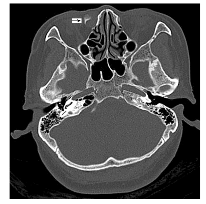
1.3 影像学检查眼眶CT检查结果显示:右侧泪囊内高密度影,右侧泪囊区、鼻泪管内高密度影,右侧泪囊明显增大;双侧眼球大小和形态未见异常,其内未见异常密度影,双眼晶状体存在,各眼肌及视神经无明显增粗,眶壁骨质结构完整(图 1)。诊断提示:右侧泪囊及鼻泪管占位性病变。

|
| Note:The arrow indicated malignant melanoma. 图 1 泪囊原发性恶性黑色素瘤患者眼眶CT影像 Fig. 1 Orbital CT image of patient with primary malignant melanoma of lacrimal sac |
|
|
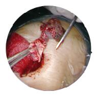
初步诊断:泪囊肿物(右)(性质待查)。2016年1月11日全麻下行右眼鼻内镜下泪囊肿物摘除术。术中见泪囊肿物大小约为1.5cm ×1.8cm×1.2cm,呈咖啡色,与周围组织紧密黏连。手术通过改良泪前隐窝入路,完整摘除肿物及泪囊,剪断深部鼻泪管(图 2, 见插页十)。手术过程顺利,患者术后7d出院。

|
| (seen on page 864 in paragraph) 图 2 鼻内镜下改良泪前隐窝人路术式切除泪囊区原发性恶性黑色素瘤 Fig. 2 Completely removed primary malignant melanoma with modified anterior lacrimal recess approach approach nasal nasal |
|
|
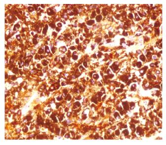
右侧泪囊区恶性黑色素瘤(梭形细胞形),累及周围肌肉组织。免疫组织化学MelanA染色结果:HMB45(+),S-100(-),MelanA(+)、K167(阳性率20%)、CK(AE1/AE3)(-),Vimentin(+)。见图 3(插页十)。

|
| (seen on page 864 in paragraph) 图 3 泪囊区原发性恶性黑色素瘤肿瘤组织MelanA染色结果(× 400) Fig. 3 MelanA staining results of tumor tissue of primary malignant melanoma in lacrimal sac area(× 400) |
|
|
术后随访3年,患者生命体征稳定,肿瘤无复发,无全身转移。
2 讨论泪道系统原发性恶性黑色素瘤非常罕见[6],而且泪囊区肿瘤常并发流泪、泪道阻塞及分泌物等体征和症状,易与泪囊狭窄和慢性泪囊炎等疾病相混淆,因此临床经常会发生误诊的情况。很多患者在泪囊炎复发进行泪囊活检后才诊断为肿瘤。如果怀疑有泪道系统恶性肿瘤,需要进行包括血液学检查(全血计数、常规生化、乳酸脱氢酶、癌胚抗原)和胸部及腹部CT等全身检查[7-9]。
原发于泪道系统的肿瘤一般根据生长过程可分为慢性泪囊炎期、泪囊肿块期、局部扩展期和肿瘤转移期[10-12]。临床大多数泪囊区肿瘤表现为内眦肿块和流泪等继发性鼻泪管阻塞的典型症状,容易被误诊为泪囊炎。泪囊病理检查有重要诊断意义,可以指导治疗方法的选择。本例患者尚属于泪囊肿块期。
黑色素瘤大多数是上皮样细胞类型[13-16],是泪腺上皮内层和下方的黑色素细胞产生,也可以继发于结膜黑色素瘤,结膜黑色素瘤沿着泪道引流管“播种”到泪囊[17]。因此,建议对疑似患者进行同侧结膜检查。本例患者系泪囊原发恶性黑色素瘤,检查患侧结膜未发现相关病变。
原发性泪囊黑色素瘤的治疗取决于肿瘤大小、局部浸润范围和患者的健康状况。治疗的目的是彻底切除肿瘤,防止复发及远处转移[18]。传统泪囊区肿物切除采用局部浸润麻醉,在距内眦1cm处行弧形切口,切口深度达内眦韧带前,切开泪骨前嵴处骨膜后,将泪囊与骨膜一同自泪囊窝游离.暴露泪骨达后泪嵴,切除泪囊区肿物。该方法损伤大,愈合慢,术后颜面部瘢痕明显。随着手术方式和器械的发展与演变,传统的手术方式逐渐被鼻内镜手术替代。2007年周兵等[19]首次报道经鼻腔外侧壁切开入路上颌窦手术, 目前鼻内镜下经典泪前隐窝入路术式逐渐作为泪囊肿瘤首选术式[20-21],对泪囊区病灶处理越来越彻底,同时手术的副损伤也越来越小,患者术后生活质量不断提高。经典泪前隐窝入路术式是在骨性梨状孔缘内侧的下鼻甲前端做纵形切口,以下甲骨在上颌骨下甲嵴附着处为中心,向上和向下延长切口至鼻泪管上端水平和鼻底。去除下甲骨前端部分骨质和上颌骨下甲嵴及部分上颌窦内侧壁和骨性鼻泪管骨壁,形成鼻泪管-下鼻甲黏膜瓣并向内侧推移,形成进入上颌窦的通路并进行后续的手术操作。改良泪前隐窝入路的手术方法:①切口的设计。沿下鼻甲前缘下方鼻腔外侧壁,自上而下至鼻底做纵形切口; 若下鼻道空间较窄,可将切口向鼻底延长呈“L”型。该切口与经典泪前隐窝入路切口不同,未暴露下鼻甲骨质,而是以下鼻甲附着处为上界,向下纵形切开,延长至鼻底形成以下鼻甲附着缘为顶、鼻底为底边的三角形操作入口。②暴露下鼻道骨壁。沿切口用剥离子分离黏骨膜暴露下鼻道骨壁,根据上颌窦内病变位置,确定下鼻道黏膜分离的长度。③切除病灶。采用剥离子、骨凿或咬骨钳、骨钻等手术器械去除下鼻道骨质,形成下鼻道骨窗。开窗口位置依据上颌窦内病变位置而确定。若病变根蒂位于上颌窦前壁,则可将开窗口向前扩大,利用角度内镜对病变进行观察,利用角度器械清除上颌窦前壁病变。④复位下鼻道黏膜。清理术腔并复位下鼻道黏膜,黏膜切口对位缝合或填塞止血材料将黏膜切口对位固定。此术式具有术野暴露好、微创、易于操作和低风险等优势[22]。
本例患者采用鼻内镜下改良泪前隐窝入路,完整地切除泪囊区肿瘤,颜面部无切口、损伤小、愈合快。术后需对肿物进行组织病理学检查,组织病理学检查是确诊肿瘤类型和进一步选择治疗方案的关键。成功治疗泪囊肿瘤需要以下几点:①减少误诊率,提高对黑色素瘤的诊断认知,如任其发展可导致致命性的肿瘤转移;②早期发现,早期治疗,彻底切除肿瘤,防止复发及远处转移;③长期随访,即使手术完整切除肿瘤,多年后,仍然有复发及转移的可能[3, 23]。本例患者术后随访3年,患者生命体征稳定,未发现肿瘤复发及远处转移。
综上所述,改良泪前隐窝入路是对既往传统泪囊手术的延伸和创新,在临床中依据病变特点和累及范围灵活选择正确的术式,对充分清除病灶、减少手术并发症和保护鼻腔生理功能均具有非常重要的意义,是一种具有较高临床推广价值的治疗泪囊区肿瘤的手术方式。
| [1] |
HEINDL L M, SCHICK B, KÄMPGEN E, et al. Malignant melanoma of the lacrimal sac[J]. Ophthalmologe, 2008, 105(12): 1146-1149. DOI:10.1007/s00347-008-1740-0 |
| [2] |
REN M, ZENG J H, LUO Q L, et al. Primary malignant melanoma of lacrimal sac[J]. Int J Ophthalmol, 2014, 7(6): 1069-1070. |
| [3] |
MAEGAWA J, YASUMURA K, IWAI T, et al. Malignant melanoma of the lacrimal sac:a case report[J]. Int J Dermatol, 2014, 53(2): 243-245. |
| [4] |
LI Y J, ZHU S J, YAN H, et al. Primary malignant melanoma of the lacrimal sac[J]. BMJ Case Rep, 2012, 2012: bcr2012006349. |
| [5] |
SHAO J W, YIN J H, XIANG S T, et al. CT and MRI findings in relapsing primary malignant melanoma of the lacrimal sac: a case report and brief literature review[J/OL].BMC Ophthalmol, 2020.DOI: 10.1186/s12886-020-01356-6.
|
| [6] |
SITOLE S, ZENDER C A, AHMAD A Z, et al. Lacrimal sac melanoma[J]. Ophthalmic Plast Reconstr Surg, 2007, 23(5): 417-419. |
| [7] |
NAM J H, KIM S M, CHOI J H, et al. Primary malignant melanoma of the lacrimal sac:a case report[J]. Korean J Intern Med, 2006, 21(4): 248-251. DOI:10.3904/kjim.2006.21.4.248 |
| [8] |
YOSHIZAKI S, INOKUCHI A, HAMADA T, et al. Primary extradural malignant melanoma of the spine:a case report[J]. J Orthop Sci, 2019, 24(4): 757-760. |
| [9] |
MONICA K, CVJETKO L, BOZENA S, et al. Primary melanoma of th urinary bladder:case report[J]. Acta Clin Croatica, 2019, 58(1): 180-182. |
| [10] |
KOH J, LEE J, JUNG S Y, et al. Primary malignant melanoma of the breast:a report of two cases[J]. J Pathol Transl Med, 2019, 53(2): 119-124. DOI:10.4132/jptm.2018.10.18 |
| [11] |
RICHTIG E, LANGMANN G, MÜLLNER K, et al. Ocular melanoma:epidemiology, clinical presentation and relationship with dysplastic nevi[J]. Ophthalmologica, 2004, 218(2): 111-114. |
| [12] |
KWAN M Y, CHAN W K, SOWOON K, et al. Primary malignant melanoma of the small intestine[J]. Ann Surg Treatment Res, 2018, 94(5): 274-278. DOI:10.4174/astr.2018.94.5.274 |
| [13] |
ALP E, BIJL D, BLEICHRODT R P, et al. Surgical smoke and infection control[J]. J Hosp Infect, 2006, 62(1): 1-5. DOI:10.1016/j.jhin.2005.01.014 |
| [14] |
ISMAIL A S. Nasal base narrowing:the alar flap advancement technique[J]. Otolaryngol Head Neck Surg, 2011, 144(1): 48-52. |
| [15] |
DAIX M, DILLIES P, GUEUNING A, et al. Primary malignant melanoma of vagina[J]. Rev Med Liege, 2018, 73(7/8): 413-418. |
| [16] |
KOGA N, KUBO N, SAEKI H, et al. Primary amelanotic malignant melanoma of the esophagus:a case report[J]. Surg Case Rep, 2019, 5(1): 4. DOI:10.1186/s40792-019-0564-2 |
| [17] |
MILIARAS S, ZIOGAS I A, MYLONAS K S, et al. Primary malignant melanoma of the ascending colon[J]. BMJ Case Rep, 2018, 2018: bcr-2017-223282. |
| [18] |
ABU-ZAID A, AL-ZAHER N. Primary oral malignant melanoma of the tongue[J]. N Z Med J, 2018, 131(1470): 87-88. |
| [19] |
周兵, 韩德民, 崔顺九, 等. 鼻内镜下鼻腔外侧壁切开上颌窦手术[J]. 中华耳鼻咽喉头颈外科杂志, 2007, 42(10): 743-748. DOI:10.3760/j.issn:1673-0860.2007.10.006 |
| [20] |
黄谦, 周兵, 崔顺九, 等. 经鼻内镜治疗蝶窦炎性疾病的手术方式和策略[J]. 临床耳鼻咽喉头颈外科杂志, 2016, 30(16): 1265-1270. |
| [21] |
周兵. 慢性鼻窦炎围手术期处理意义的再认识[J]. 临床耳鼻咽喉头颈外科杂志, 2016, 30(16): 1261-1265. |
| [22] |
CARBAJO-RODRÍGUEZ H, AGUAYO-ALBASINI J L, SORIA-ALEDO V, et al. Surgical smoke:risks and preventive measures[J]. Cir Esp, 2009, 85(5): 274-279. DOI:10.1016/j.ciresp.2008.10.004 |
| [23] |
GLEIZAL A, KODJIKIAN L, LEBRETON F, et al. Early CT-scan for chronic lacrimal duct symptoms-case report of a malignant melanoma of the lacrimal sac and review of the literature[J]. J Craniomaxillofac Surg, 2005, 33(3): 201-204. DOI:10.1016/j.jcms.2005.01.012 |
 2020, Vol. 46


