扩展功能
文章信息
- 文庆一, 焦苗苗, 谢潇, 彭敬予, 董凤歌, 魏雪, 杨明
- WEN Qingyi, JIAO Miaomiao, XIE Xiao, PEN Jingyu, DONG Fengge, WEI Xue, YANG Ming
- 双硫仑联合顺铂对三阴性乳腺癌MDA-MB-231细胞的增殖抑制作用及其机制
- Inhibitory effect of disulfiram combined with cisplatin on triple negative breast cancer MDA-MB-231 cells and its mechanism
- 吉林大学学报(医学版), 2020, 46(03): 523-529
- Journal of Jilin University (Medicine Edition), 2020, 46(03): 523-529
- 10.13481/j.1671-587x.20200316
-
文章历史
- 收稿日期: 2019-08-28
2005年由BRENTON等[1]首次提出三阴性乳腺癌(triple-negative breast cancer,TNBC)的概念,TNBC是指一组不表达雌激素受体(estrogen receptor, ER)、孕激素受体(progesterone receptor, PR)和表皮生长因子受体(human epidermal growth factor, HER2)的复杂乳腺癌亚型,TNBC患者发病年龄较轻,且恶性程度高,侵袭性强,病情进展比较迅速,容易出现局部复发和远处转移,患者总体生存期短。TNBC患者常伴发乳腺癌易感基因1(breast cancer susceptibility gene-1, BRCA-1)突变,当BRCA-1基因发生突变时失去了断裂DNA双链的修复功能[2-4]。在疾病的早期和晚期,化疗是TNBC患者的主要全身治疗方法。目前临床TNBC的药物治疗主要以紫衫类和蒽环类联合化疗为主,铂类是除紫衫及蒽环类外的重要选择[5]。顺铂(cis- diamineichloroplain,CDDP)等铂类药物可与DNA双链发生交联,致使DNA双链断裂,无法完成DNA复制和转录,最终导致细胞死亡[6]。临床研究[7]的初步结果也证实:CDDP单药新辅助治疗TNBC有效,可明显提高病理完全缓解(pathological complete response,PCR)率。但是,CDDP靶向肿瘤细胞的能力差、体内代谢时间短,虽能杀死肿瘤细胞,但同样对正常细胞的功能也造成了严重损害。TNBC患者缺乏靶向疗法,这促使科研工作者努力探索可治疗的分子靶标以治疗TNBC患者[8]。双硫仑(disulfiram, DSF)能通过抑制乙醛脱氢酶阻断酒精氧化代谢,在临床上应用于戒酒治疗达60年。近些年,DSF被证实具有抗肿瘤潜能[9-13],说明DSF可作为治疗TNBC的潜力药物,可单独或与其他一线或二线化疗药物联合应用。然而,目前尚无研究表明同时使用CDDP和DSF具有抗肿瘤作用。
本研究旨在通过体外实验评估CDDP与DSF联合应用对MDA-MB-231细胞增殖的作用,并分析其可能的分子机制,为未来临床转化应用提供依据。
1 材料与方法 1.1 细胞、主要试剂和仪器TNBC MDA-MB-231细胞购于上海博古生物科技有限公司。CDDP购于中国科学院长春应用化学研究所,DSF、四甲基偶氮唑盐(methyl thiazolyl tetrazolium,MTT)噻唑蓝和二甲基亚砜(dimethyl sulphoxide, DMSO)购于美国Sigma公司,DMEM干粉、青霉素、链霉素、0.25%胰蛋白酶消化液和0.25%EDTA-胰蛋白酶购于美国Gibco公司,胎牛血清(fetal bovine serum, FBS)购于美国Clark公司,AnnexinⅤ-FITC/PI试剂盒和DCFH-DA购于美国KeyGEN公司。Matrigel基质胶和流式细胞仪购于美国BD公司,CO2培养箱和高速离心机购于美国Thermo公司,倒置显微镜购于日本Nikon公司,酶标仪购于美国BioYek公司。
1.2 MDA-MB-231细胞培养将MDA-MB-231细胞培养于含有10%FBS、100 U·mL-1青霉素和100 U·mL-1链霉素的DMEM培养液中。置于37℃、5%CO2培养箱中常规培养,传代3代以上并取呈对数生长期的细胞用于后续实验。
1.3 MTT法检测各组MDA-MB-231细胞存活率取呈对数期生长的MDA-MB-231细胞并调整细胞密度,细胞密度调至8000个/孔后快速铺板,每孔200 μL接种于96孔板。设定CDDP终浓度为200 μmol·L-1,同时将60μmol·L-1 CDDP分别与1、2和4μmol·L-1 DSF联合应用,细胞分为60 μmol·L-1 CDDP组、联合1组(60 μmol·L-1 CDDP+1 μmol·L-1 DSF)、联合2组(60 μmol·L-1 CDDP+2 μmol·L-1 DSF)和联合3组(60 μmol·L-1 CDDP+4 μmol·L-1 DSF)。置于5% CO2、37℃条件下孵育24 h,3倍稀释至6个浓度,每组设置4个复孔,分别培养72 h,倒置显微镜下观察。72 h后,每孔加入20 μL MTT(5 g·L-1,即质量分数为0.5% MTT)溶液,继续置于培养箱中培养4 h。终止培养,小心吸干净含MTT的孔内培养液。每孔加入150 μL DMSO,置入酶标仪中振荡5 min,使结晶物充分溶解。采用酶联免疫检测仪测量各孔在波长为490 nm处的吸光度(A)值,计算MDA-MB-231细胞存活率。细胞存活率=(样品A值/对照品A值)× 100%。实验重复3次。以药物浓度的对数作为横坐标,细胞存活率作为纵轴应用Orgin 9.2软件绘制细胞生长抑制效应曲线。
1.4 流式细胞术检测各组MDA-MB-231细胞凋亡率采用0.25% EDTA-胰蛋白酶消化呈对数生长的贴壁细胞,制成均匀细胞悬液,显微镜下计数,以2.5×10⁵个/孔接种于6孔板。24 h培养后,分别加入浓度为160 μmol·L-1 CDDP后继续于5% CO2、37℃条件下培养24、48和72 h。吸弃孔内培养液,采用2 mL磷酸盐缓冲液(phosphate buffer solution,PBS)清洗1次。采用0.5 mL 0.5%胰蛋白酶消化细胞,将MDA-MB-231细胞分为空白对照组、2 μmol·L-1 DSF组、60 μmol·L-1 CDDP组和联合组(60 μmol·L-1 CDDP+2 μmol·L-1 DSF)。以0.5 mL DMEM培养液中和,移入1.5 mL EP管中,2 000 r·min-1离心5 min,吸弃上清液,再用PBS洗涤细胞2次(2 000 r·min-1离心5 min)收集细胞。加入500 μL Binding Buffer悬浮细胞,加入5 μL Annexin Ⅴ-FITC混匀后,加入5 μL PI,混匀, 室温避光反应15 min。1 h内进行流式细胞术检测。结果判定标准:AnnexinⅤ-FITC(-)PI(-)为活细胞,AnnexinⅤ-FITC(+)PI(-)为早期凋亡细胞,AnnexinⅤ-FITC(+)PI(+)为中晚期凋亡细胞,AnnexinⅤ-FITC(-)PI(+)为死亡细胞,计算各组MDA-MB-231细胞凋亡率。细胞凋亡率=(早期凋亡细胞数+中晚期凋亡细胞和死亡细胞数)/细胞总数×100%。
1.5 流式细胞术检测各组MDA-MB-231细胞中活性氧(reactive oxygen species,ROS)水平采用0.25% EDTA-胰蛋白酶消化呈对数生长的贴壁细胞,制成均匀细胞悬液,显微镜下细胞板计数,以5×10⁴个/孔接种于12孔板,培养24 h。分为空白对照组、2 μmol·L-1 DSF组、60 μmol·L-1 CDDP组、联合组(60μmol·L-1 CDDP+2 μmol·L-1 DSF),置于5% CO2、37℃条件下培养3 h。3 h后,吸弃培养液,采用PBS清洗3次,以1 mL PBS定容。配制10 mmol·L-1 DCFH-DA储备液(4.87 g·L-1,以DMSO溶解),调至终浓度为5 μmol·L-1,每孔加入2 μLDCFH-DA,充分摇匀。37℃培养20 min,弃去孔内液体,以PBS洗涤3次。每孔加入1 mL PBS后置于荧光显微镜下迅速观察并拍照,吸弃PBS,采用0.25%EDTA-胰蛋白酶消化液处理细胞后收集,采用流式细胞术检测各组MDA-MB-231细胞中ROS水平。采用Origin 9.2软件绘图并分析ROS水平变化。
1.6 电感耦合等离子体质谱仪(inductively coupled plasma-mass spectrometry,ICP-MS)检测各组MDA-MB-231细胞中铂(Pt)水平采用0.25% EDTA-胰蛋白酶消化呈对数生长的贴壁细胞,制成均匀细胞悬液,显微镜下细胞板计数,以每个培养皿4.0×10⁵个细胞接种于50 mm培养皿,培养24 h。实验分为空白对照组、20 μmol·L-1 CDDP组、联合组(20 μmol·L-1 CDDP+0.625 μmol·L-1 DSF),每组设置3个平行样,加入各组药物后置于5% CO2、37℃条件下作用4和24 h,吸弃皿内液体,采用PBS洗涤3次。采用0.6 mL0.25% EDTA-胰蛋白酶消化液消化细胞,1 000 r·min-1离心5 min,以2 mL PBS重悬细胞后制成细胞悬液,移入5 mL EP管中,倒置显微镜下进行细胞计数。5 mL EP管中加入硝酸(体积分数为68%),在70℃条件下煮12 h。采用ICP-MS检测各组MDA-MB-231细胞中Pt水平。
1.7 统计学分析采用SPSS 19.0统计软件进行统计学分析。各组MDA-MB-231细胞存活率、MDA-MB-231细胞凋亡率、MDA-MB-231细胞中ROS水平和MDA-MB-231细胞中Pt水平符合正态分布,以x ±s表示,MDA-MB-231细胞存活率和凋亡率组间比较采用χ2检验,MDA-MB-231细胞中ROS水平和Pt水平组间比较采用SNK-q检验。以P < 0.05为差异有统计学意义。
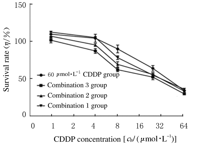
2 结果 2.1 各组MDA-MB-231细胞存活率以不同浓度DSF联合CDDP作用于MDA-MB-231细胞72 h后可见:CDDP浓度一定的情况下,DSF浓度越大,细胞存活率越低。与60 μmol·L-1 CDDP组比较,联合1组、联合2组和联合3组MDA-MB-231细胞存活率降低(P < 0.05)。在72 h时,小分子药物CDDP以及按60:1、60:2、60:4配比与DSF联合应用的最小抑制浓度(IC50)分别为32.012、29.107、26.258和16.901 μmol·L-1。加入DSF后,IC50随着DSF浓度的增加而减小。在达到细胞相同抑制效果时,CDDP浓度随着DSF浓度的增加而降低。72 h后各组MDA-MB-231细胞的生长抑制效应曲线见图 1。

|
| 图 1 作用72 h后各组MDA-MB-231细胞生长抑制效应曲线 Fig. 1 Growth inhibition effect curves of MDA-MB-231 cells in various groups after treated for 72 h |
|
|
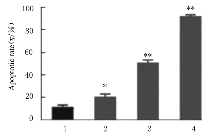
相同浓度CDDP(当量)作用于MDA-MB-231细胞后,时间越长,凋亡细胞数越多。CDDP作用于细胞24 h后,发生明显的细胞凋亡,作用72 h后MDA-MB-231细胞凋亡率为92.4%。CDDP对MDA-MB-231细胞的抑制作用呈剂量和时间依赖性。见图 2。

|
| 1:Blank control group; 2-4: 60 μmol·L-1 CDDP group(24, 48, and 72 h).*P < 0.05, **P < 0.01 vs blank control group. 图 2 60 μmol·L-1 CDDP作用MDA-MB-231细胞24、48和72 h后细胞凋亡率 Fig. 2 Apoptotic rates of MDA-MB-231 cells after treated with 60 μmol·L-1 CDDP for 24, 48 and 72 h |
|
|
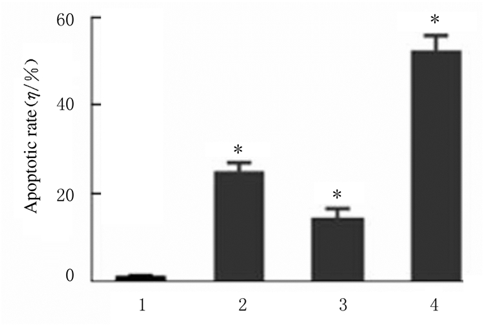
作用24 h后,各组MDA-MB-231细胞均有凋亡发生。与60 μmol·L-1 CDDP组和2 μmol·L-1 DSF组比较,联合组(60 μmol·L-1 CDDP+2 μmol·L-1 DSF)MDA-MB-231细胞凋亡率升高(P < 0.05)。见图 3。

|
| 1:Control group; 2: 60 μmol·L-1 CDDP group; 3: 2 μmol·L-1 DSF group; 4:Combination group (60 μmol·L-1 CDDP+2 μmol·L-1 DSF).*P < 0.05 vs control group. 图 3 作用24h后各组MDA-MB-231细胞凋亡率 Fig. 3 Apoptotic rates of MDA-MB-231 cells in various groups after treated for 24 h |
|
|
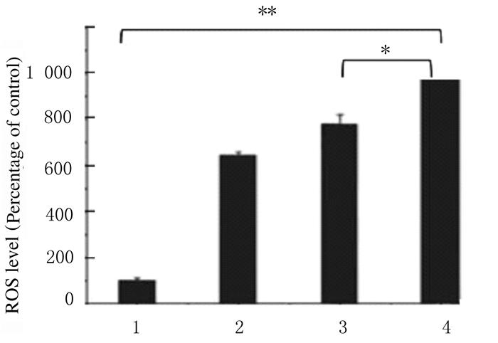
DCFH-DA荧光染色后,与空白对照组比较,其他各组MDA-MB-231细胞中均可见DCF绿色荧光,即DSF、CDDP单独及联合应用均可明显引起MDA-MB-231细胞中ROS水平升高。作用3h后联合组(60 μmol·L-1 CDDP+2 μmol·L-1 DSF)MDA-MB-231细胞中ROS水平高于2 μmol·L-1 DSF组和60 μmol·L-1 CDDP组,约为空白对照组MDA-MB-231细胞中ROS水平的10倍(P < 0.05或P < 0.01)。见图 4。

|
| 1:Blank control group; 2:2 μmol·L-1 DSF group; 3:60 μmol·L-1 CDDP group; 4:Combination group(60 μmol·L-1 CDDP+2 μmol·L-1 DSF).*P < 0.05, * *P<0.01 vs blank control group. 图 4 流式细胞术检测各组MDA-MB-231细胞中ROS水平 Fig. 4 Levels of ROS in MDA-MB-231 cells in various groups detected by flow |
|
|
作用4 h后,20 μmol·L-1 CDDP组MDA-MB-231细胞中Pt水平为(9.760±0.875) ng/106个细胞,联合组(20 μmol·L-1 CDDP+0.625 μmol·L-1 DSF)MDA-MB-231细胞中Pt水平为(9.374±1.125) ng/106细胞,二者比较差异无统计学意义(P>0.05)。作用24 h后,20 μmol·L-1 CDDP组MDA-MB-231细胞中Pt水平为(22.062±1.229)ng/106个细胞,联合组(20 μmol·L-1 CDDP+0.625 μmol·L-1 DSF)MDA-MB-231细胞中Pt水平为(28.607±1.380)ng/106个细胞,二者比较差异有统计学意义(P < 0.01)。
3 讨论作为一种特殊的乳腺癌亚型,TNBC具有特殊的生物学行为和临床病理特征。TNBC患者的全身治疗仍以化疗为主。目前,美国食品药品监督管理局(Food and Drug Administration, FDA)尚未批准任何专门针对TNBC的药物。多年以来,研究主要集中在化疗药物的选择上,如蒽环类药、紫衫类药和铂类药物等。近些年的临床前研究和临床研究[14-16]已经证实:乳腺癌易感基因(BRCA)相关性乳腺癌对紫衫醇和多西他赛存在耐药现象,而对CDDP比较敏感,尤其是对于BRCA-1突变的TNBC患者,PCR率可高达72%~90% [17-18],但仍需要大样本随机对照研究进一步评估CDDP单药及其与铂类联合化疗新辅助治疗TNBC的疗效。关于CDDP治疗TNBC尚存在大量问题,首先是其严重的不良反应,恶心、呕吐和累积性及剂量相关性肾损伤是CDDP的主要限制性毒性。研究[18-19]证实:CDDP只对存在BRCA-1突变的TNBC有效,而存在BRCA-1突变的患者只约占TNBC患者20% [20],因此CDDP尚未被批准用于TNBC的一线治疗。
寻找能增强TNBC细胞对传统化疗药物的化疗敏感性、降低化疗药物的用药剂量、减轻正常机体遭受损害的安全无毒药物是研究人员探索的新方向。DSF作为经FDA批准的安全戒酒药物,应用于临床已有60余年。研究者[21]发现:该药物还具有抗白内障和抗肿瘤的作用,临床试验已证实DSF安全无毒。DSF不仅能有效杀伤包括乳腺癌在内的多种肿瘤细胞,而且对TNBC细胞有极强的杀伤作用且对传统化疗药物具有增敏作用。ROBINSON等[22]发现:DSF能有效抑制包括MDA-MB-231在内的多种TNBC细胞系的生长,半数抑制浓度(IC50)为300 nmol·L-1。ROBINSON等[22]研究显示:DSF能与阿霉素、紫杉醇和吉西他滨等产生协同效应。
LIU等[23]对具有乳腺癌干细胞特性的耐药TNBC细胞系MDA-MB-231PAC10进行了细胞毒性实验及相关耐药性研究发现:MDA-MB-231PAC10对DSF非常敏感,暴露于DSF仅4 h便完全失去了成球囊能力,作用24 h后可检测到大量凋亡细胞,而对紫杉醇、多西他赛、阿霉素和CDDP均表现出极强的抗药性。
本研究结果显示:不同浓度DSF辅助CDDP作用于MDA-MB-231细胞72 h后,在达到细胞相同抑制效果时,CDDP浓度会随DSF浓度的增加而降低。与其他研究不同点:本研究所采用的DSF浓度相对较低,以DSF:CDDP摩尔比为4:60的比例即可产生明显的抑制效应。微量DSF能有效地辅助CDDP抑制MDA-MB-231细胞的增殖,在达到相同抑制效果的情况下,DSF的添加大大降低了CDDP的应用浓度,从而降低了CDDP对机体产生的毒性。
目前虽证实DSF能增强传统化疗药物的敏感性,但是其具体机制尚不清楚。很多研究[24-26]已经证实:高水平ROS能够破坏细胞的DNA和磷脂等结构,并诱导细胞凋亡及坏死。与正常细胞比较,癌细胞本身便存在较高水平ROS [27-28]。诱导癌细胞产生更多的ROS,使肿瘤细胞比正常细胞提早达到并超过ROS耐受阈值,最终导致癌细胞的选择性凋亡,也是肿瘤治疗方向之一。以往有研究[29]显示:DSF-Cu可能是通过刺激ROS的产生诱导凋亡效应。本研究结果显示:荧光显微镜下,DSF和CDDP以摩尔比2:60联合应用作用于MDA-MB-231细胞3 h后产生的ROS荧光强度大于CDDP单独应用,流式细胞术检测结果也验证了该结论,由此证明:与单独应用CDDP比较,相对微量DSF与CDDP联合应用能明显增加细胞中ROS水平,并诱导细胞凋亡。
综上所述,小分子CDDP利用其扩散作用可以快速进入细胞,当添加DSF作为辅助药物后,细胞内Pt水平明显增加,说明DSF能促进CDDP更多地进入细胞,增加细胞中CDDP浓度,从而增强CDDP对细胞的毒性作用。
| [1] |
BRENTON J D, CAREY L A, Ahmed A A, et al. Molecular classification and molecular forecasting of breast cancer:Ready for clinical application?[J]. J Clin Oncol, 2005, 23(29): 7350-7360. DOI:10.1200/JCO.2005.03.3845 |
| [2] |
SCHNEIDER B P, WINER E P, FOULKES W D, et al. Triple-negative breast cancer:risk factors to potential targets[J]. Clin Cancer Res, 2008, 14(24): 8010-8018. DOI:10.1158/1078-0432.CCR-08-1208 |
| [3] |
CLEATOR S, HELLER W, COOMBES R C. Triple-negative breast cancer:therapeutic options[J]. Lancet Oncol, 2007, 8(3): 235-244. DOI:10.1016/S1470-2045(07)70074-8 |
| [4] |
PUPPE J, OPDAM M, SCHOUTEN P C, et al. EZH2 is overexpressed in BRCA1-like breast tumors and predictive for sensitivity to high-dose platinum-based chemotherapy[J]. Clin Cancer Res, 2019, 25(14): 4351-4362. DOI:10.1158/1078-0432.CCR-18-4024 |
| [5] |
RODLER E, KORDE L, GRALOW J. Current treatment options in triple negative breast cancer[J]. Breast Dis, 2010, 32(1/2): 99-122. |
| [6] |
REEDIJK J, LOHMAN P H. Cisplatin:synthesis, antitumour activity and mechanism of action[J]. Pharm Weekbl Sci, 1985, 7(5): 173-180. DOI:10.1007/BF02307573 |
| [7] |
VETTER M, FOKAS S, BISKUP E, et al. Efficacy of adjuvant chemotherapy with carboplatin for early triple negative breast cancer:a single center experience[J]. Oncotarget, 2017, 8(43): 75617-75626. DOI:10.18632/oncotarget.18118 |
| [8] |
BIANCHINI G, BALKO J M, MAYER I A, et al. Triple-negative breast cancer:challenges and opportunities of a heterogeneous disease[J]. Nat Rev Clin Oncol, 2016, 13(11): 674-690. DOI:10.1038/nrclinonc.2016.66 |
| [9] |
YANG Y P, ZHANG K F, WANG Y N, et al. Disulfiram chelated with copper promotes apoptosis in human breast cancer cells by impairing the mitochondria functions[J]. Scanning, 2016, 38(6): 825-836. DOI:10.1002/sca.21332 |
| [10] |
JIVAN R, DAMELIN L H, BIRKHEAD M, et al. Disulfiram/copper-disulfiram damages multiple protein degradation and turnover pathways and cytotoxicity is enhanced by metformin in oesophageal squamous cell carcinoma cell lines[J]. J Cell Biochem, 2015, 116(10): 2334-2343. DOI:10.1002/jcb.25184 |
| [11] |
CHERIYAN V T, WANG Y, MUTHU M, et al. Disulfiram suppresses growth of the malignant pleural mesothelioma cells in part by inducing apoptosis[J]. PLoS One, 2014, 9(4): e93711. DOI:10.1371/journal.pone.0093711 |
| [12] |
JIAO Y, HANNAFON B N, DING W Q. Disulfiram's anticancer activity:evidence and mechanisms[J]. Anti-Cancer Agent Med Chem, 2016, 16(11): 1378-1384. DOI:10.2174/1871520615666160504095040 |
| [13] |
VIOLA-RHENALS M, PATEL K R, JAIMES-SANTAMARIA L, et al. Recent advances in antabuse (disulfiram):the importance of its metal-binding ability to its anticancer activity[J]. Curr Med Chem, 2018, 25(4): 506-524. DOI:10.2174/0929867324666171023161121 |
| [14] |
ROTTENBERG S, NYGREN A O, PAJIC M, et al. Selective induction of chemotherapy resistance of mammary tumors in a conditional mouse model for hereditary breast cancer[J]. Proc Natl Acad Sci U S A, 2007, 104(29): 12117-12122. DOI:10.1073/pnas.0702955104 |
| [15] |
KRIEGE M, JAGER A, HOONING M J, et al. The efficacy of taxane chemotherapy for metastatic breast cancer in BRCA1 and BRCA2 mutation carriers[J]. Cancer, 2012, 118(4): 899-907. |
| [16] |
AKASHI-TANAKA S, WATANABE C, TAKAMARU T, et al. BRCAness predicts resistance to taxane-containing regimens in triple negative breast cancer during neoadjuvant chemotherapy[J]. Clin Breast Cancer, 2015, 15(1): 80-85. |
| [17] |
BYRSKI T, HUZARSKI T, DENT R, et al. Pathologic complete response to neoadjuvant cisplatin in BRCA1-positive breast cancer patients[J]. Breast Cancer Res Treat, 2014, 147(2): 401-405. |
| [18] |
BYRSKI T, GRONWALD J, HUZARSKI T, et al. Pathologic complete response rates in young women with BRCA1-positive breast cancers after neoadjuvant chemotherapy[J]. J Clin Oncol, 2010, 28(3): 375-379. DOI:10.1200/JCO.2008.20.7019 |
| [19] |
VOLLEBERGH M A, LIPS E H, NEDERLOF P M, et al. An aCGH classifier derived from BRCA1-mutated breast cancer and benefit of high-dose platinum-based chemotherapy in HER2-negative breast cancer patients[J]. Ann Oncol, 2011, 22(7): 1561-1570. DOI:10.1093/annonc/mdq624 |
| [20] |
HARTMAN A R, KALDATE R R, SAILER L M, et al. Prevalence of BRCA mutations in an unselected population of triple-negative breast cancer[J]. Cancer, 2012, 118(11): 2787-2795. DOI:10.1002/cncr.26576 |
| [21] |
JOHANSSON B. A review of the pharmacokinetics and pharmacodynamics of disulfiram and its metabolites[J]. Acta Psychiatr Scand Suppl, 1992, 369: 15-26. |
| [22] |
ROBINSON T J, PAI M, LIU J C, et al. High-throughput screen identifies disulfiram as a potential therapeutic for triple-negative breast cancer cells:interaction with IQ motif-containing factors[J]. Cell Cycle, 2013, 12(18): 3013-3024. DOI:10.4161/cc.26063 |
| [23] |
LIU P, KUMAR I S, BROWN S, et al. Disulfiram targets cancer stem-like cells and reverses resistance and cross-resistance in acquired paclitaxel-resistant triple-negative breast cancer cells[J]. Brt J Cancer, 2013, 109(7): 1876-1885. DOI:10.1038/bjc.2013.534 |
| [24] |
SAITO S, LIN Y C, TSAI M H, et al. Emerging roles of hypoxia-inducible factors and reactive oxygen species in cancer and pluripotent stem cells[J]. Kaohsiung J Med Sci, 2015, 31(6): 279-286. DOI:10.1016/j.kjms.2015.03.002 |
| [25] |
DE MIGUEL M P, ALCAINA Y, DE LA MAZA D S, et al. Cell metabolism under microenvironmental low oxygen tension levels in stemness, proliferation and pluripotency[J]. Curr Mol Med, 2015, 15(4): 343-359. DOI:10.2174/1566524015666150505160406 |
| [26] |
OZBEN T. Oxidative stress and apoptosis:impact on cancer therapy[J]. J Pharm Sci, 2007, 96(9): 2181-2196. DOI:10.1002/jps.20874 |
| [27] |
CABELLO CM, BAIR W B, WONDRAK G T. Experimental therapeutics:targeting the redox Achilles heel of cancer[J]. Curr Opin Invest Dr, 2007, 8(12): 1022-1037. |
| [28] |
TRACHOOTHAM D, ALEXANDRE J, HUANG P. Targeting cancer cells by ROS-mediated mechanisms:a radical therapeutic approach?[J]. Nat Rev Drug Discov, 2009, 8(7): 579-591. DOI:10.1038/nrd2803 |
| [29] |
YIP N C, FOMBON I S, LIU P, et al. Disulfiram modulated ROS-MAPK and NFkappaB pathways and targeted breast cancer cells with cancer stem cell-like properties[J]. Brit J Cancer, 2011, 104(10): 1564-1574. DOI:10.1038/bjc.2011.126 |
 2020, Vol. 46


