扩展功能
文章信息
- 胡雪, 李天博, 李雪洋, 王倩, 张颖丽
- HU Xue, LI Tianbo, LI Xueyang, WANG Qian, ZHANG Yingli
- 右上颌第二磨牙牙内陷1例报告及文献复习
- Dens invaginatus of right maxillary second molar: A case report and literature review
- 吉林大学学报(医学版), 2019, 45(05): 1173-1176
- Journal of Jilin University (Medicine Edition), 2019, 45(05): 1173-1176
- 10.13481/j.1671-587x.20190534
-
文章历史
- 收稿日期: 2019-03-22
2. 吉林大学口腔医院修复科, 吉林 长春 130021
 面平龈,牙冠巨大,
 面正中见凹陷及龋坏,未达髓腔,影像学检查可见17
  面类牙釉质密度影像向根方凹陷,深度约7.7mm,确诊为牙内陷。初诊时去除腐质,采用安抚治疗,复诊时发现患牙仍有自发痛,且未能解决食物嵌塞痛,考虑到术后恢复效果不佳,故患者同意拔除患牙。离体牙做病理磨片,显微镜下观察可见内陷处牙釉质及牙本质结构异常。结论:
发生于磨牙区的牙内陷患者较为罕见,诊疗过程中应结合影像学检查明确疾病类型及治疗计划,建议治疗效果不佳的患牙应及时拔除。
面类牙釉质密度影像向根方凹陷,深度约7.7mm,确诊为牙内陷。初诊时去除腐质,采用安抚治疗,复诊时发现患牙仍有自发痛,且未能解决食物嵌塞痛,考虑到术后恢复效果不佳,故患者同意拔除患牙。离体牙做病理磨片,显微镜下观察可见内陷处牙釉质及牙本质结构异常。结论:
发生于磨牙区的牙内陷患者较为罕见,诊疗过程中应结合影像学检查明确疾病类型及治疗计划,建议治疗效果不佳的患牙应及时拔除。2. Department of Prosthodontics, Stomatology Hospital, Jilin University, Changchun 130021, China
牙内陷(dens invaginatus,DI)是一种牙齿发育异常,通常表现为釉质覆盖的牙冠或牙根表面出现深凹陷,也可伴随其他发育异常,如过大牙、过小牙、牛牙症、融合牙及釉质发育不全等[1],常发生于上颌侧切牙、尖牙、前磨牙、多生的切牙、下颌前牙和前磨牙[2],其中上颌侧切牙发生率最高[3]。磨牙区的牙内陷病例十分罕见,ALANI等[4]报道:14090颗牙内陷牙齿中,只有6.5%为后牙。国外少数文献[5-6]报道了乳磨牙区牙内陷的病例,国内相关报道较少。本文作者报道1例恒磨牙区牙内陷病例,分析其影像学表现及病理学特征与临床症状之间的联系,为临床治疗方案的制定提供参考。
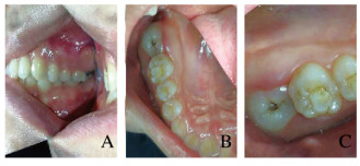
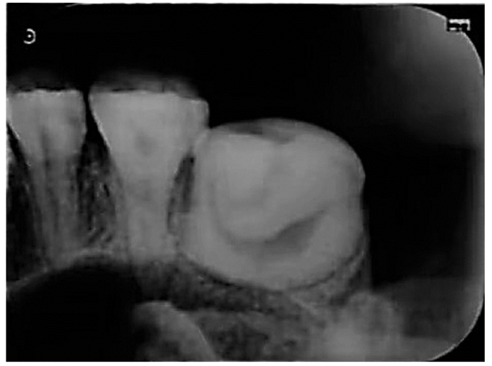
1 临床资料 1.1 一般资料患者,女性,28岁,因右上后牙发现有洞6个月于2018年11月26日就诊于吉林大学口腔医院。患者6个月前右上后牙牙龈红肿,炎症消除后发现右上后牙萌出,自觉有洞,影响咀嚼。否认既往病史、药物过敏史及常规血液检查在内的家族遗传病史,口外检查和全身性检查均未发现其他临床综合征。专科检查:颌面部基本对称,张口度及开口型正常,咬合关系正常,关节无弹响; 口内检查:牙合左右对称,17部分萌出,牙合面平龈,牙冠色正常,体积巨大,牙合面正中有明显凹陷,可探及龋坏,龋坏深达牙本质中层,探诊敏感,叩痛(-),冷水反应(-),牙齿无松动,牙龈轻度充血(图 1,见插页六)。根尖CR片(图 2):17牙合面向髓腔内陷入较深,位于牙齿中央,内陷处正中可见龋坏,未达髓腔;未见常规根管形态;根尖周未见明显异常。锥形束CT(CBCT)显示:17牙合面高密度影像向根方凹陷,深度约7.7mm,龋坏处下方可见髓角,髓腔形态呈“C”形,内陷形成的通道与牙髓腔不相通,牙周膜间隙增宽(图 3)。

|
| View of buccal side; B, C:View of occlusal surface 图 1 右上颌第二磨牙牙内陷患者口腔内照片 Fig. 1 Intraoral biew of patient with dens invaginatus of right maxillary second molar(seen on page 1174 in paragraph) |
|
|

|
| 图 2 右上颌第二磨牙牙内陷患者患牙根尖CR影像 Fig. 2 Periapical CR radiograph of patient with dens invaginatus of right maxillary second molar |
|
|

|
| A: Axial view; B: Sagital view; C: Coronal view. 图 3 上颌第二磨牙牙内陷患者患牙CBCT影像 Fig. 3 CBCT radiograph of patient with dens invaginatus of right maxillary second molar |
|
|
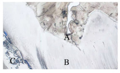
17龋坏深达牙本质中层,初诊时建议患者采用17安抚术。17牙合面去净腐质,制备洞形,拭干,隔湿,氢氧化钙双糊剂护髓,氧化锌安抚治疗;术后1周复诊,患牙自发痛及牙龈红肿状况未缓解,建议拔除。后经患者知情同意,拔除17。患牙做病理磨片,观察内陷釉质形态。显微镜下观察:牙釉质内可见裂隙延伸至牙本质;牙本质内可见牙本质小管排列不良;牙髓未见明显病理改变(图 4,见插页六)。

|
| A: Enamel; B: Dentine; C:Endodontic pulp. 图 4 牙内陷患者患牙病理表现 Fig. 4 Pathology of tooth of patient with dens invaginatus(seen on page 1174 in paragraph) |
|
|
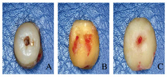
考虑到患牙近中食物嵌塞痛未能缓解且牙合面中央凹陷深,去净腐质难度较大,洞形制备不够理想,也可能出现填充不全,术后若出现继发龋时治疗难度较大,若出现牙髓症状时,由于髓腔形态特殊,根管治疗较难达到理想效果,远期预后不佳,最终拔除患牙(图 5,见插页六)。

|
| A: Occlusive view; B: Side view; C: Root view 图 5 牙内陷患者离体患牙照片 Fig. 5 Picture of isolated tooth of patient with dens invaginatus(seen on page 1175 in paragraph) |
|
|
牙内陷发生率较低,为0.04%~10.00%[7],磨牙区牙内陷的相关报道[8]更为少见。牙内陷的病因一直存有争议,有学者认为牙内陷是在牙发育过程中成釉器过度卷曲而形成的牙齿发育异常[9];还有学者认为牙内陷是釉质形成过程中内部釉质上皮的部分细胞生长异常,其余正常细胞继续增殖而异常细胞增殖突入牙乳头,导致牙釉质发育形态异常[10];还可能存在牙融合、感染、创伤或遗传[11-16]等多种因素影响。
目前国际广泛采用OEHLERS等[17]牙内陷分类法。该分类法根据内陷深度及内陷与根尖牙周相通情况将牙内陷分为3型:Ⅰ型内陷局限于牙冠内且不与髓腔相通;Ⅱ型内陷延伸至釉牙骨质界下方,下端终止于牙本质内形成一盲端,不与髓腔相通;Ⅲ(A)型内陷向下延伸在根尖或根侧形成通道,通道走行于牙根内,不与牙髓相通;Ⅲ(B)型内陷向下延伸在根尖或根侧形成“第二根尖孔”,并与牙髓牙周组织相通。有研究[12, 18-19]显示:Ⅰ型牙内陷在牙内陷中所占比例相对较高,为69.8%~93.8%,其次为Ⅱ型,占3.1%~26.6%;Ⅲ型占3.0%~12.5%。本研究中患者属于Ⅱ型牙内陷,该类型牙内陷的釉质外观上表现为釉质盲孔,此处结构薄弱,可能与牙髓存在交通支且细菌容易聚集,临床上应该对此结构进行预防性树脂充填以减少牙髓感染的可能[20]。该患者由于内陷部位积存食物已形成龋坏,而牙体形态因素又使得牙髓治疗预后较差,故而拔除。
目前临床常用的影像学检查手段为X线片,牙内陷通常表现为与牙釉质密度相同的高密度影像在舌侧由冠部向根部移行。但在诊断牙内陷时,通常需要明确牙齿的三维解剖结构,相比之下CBCT不仅能显示三维解剖结构,还能清楚观察根尖周骨质的缺损大小,查找轻度病变,同时避免根尖片投照条件限制导致的图像失真问题[21],更具有临床应用价值。有研究者通过CBCT观察分析了牙内陷患牙的解剖结构发现:①内陷位于主根管中央且主根管任何方向均为X线透射区;②内陷位于主根管一侧,主根管主要为C形透射区;③内陷将主根管分为两个相反的新月形透射区;④内陷位于牙根一侧,不与主根管交通,主根管影像为圆形、C形或不规则透射区等;⑤只有主根管透射影未检查到内陷形成的通道影像[22]。牙内陷复杂的解剖结构也使得患牙一旦发生牙体牙髓疾病时其治疗效果和远期预后不理想。对于牙内陷患者,临床常用治疗方案多为根管治疗,大多因患牙不能及时发现而导致牙髓症状,且因长期预后不佳,后期常建议拔除[23-25]。
综上所述,该患者右上颌磨牙区患牙由于未能尽早发现而导致龋坏程度较重,食物嵌塞情况未缓解,可能导致第一磨牙远中颈部龋坏,因此与患者沟通,经患者知情同意后拔除。随着影像学技术的发展,很多早期较难发现的疾病能够及早发现并得到及时的防护和治疗,故临床工作中应多关注患者就诊要求以外的牙列,对于还未有明显临床症状的患牙要尽量保留,而治疗效果不佳的患牙要及时拔除以免对临近组织造成不可逆的损伤,尽量做到早发现、早诊断和早治疗。
| [1] | PATIL P B, CHAUDHARI S G, GOEL A, et al. Rare association of dens invaginatus with impacted mesiodens-A case report[J]. J Oral Biol Craniofac Res, 2012, 2(2): 138–140. DOI:10.1016/j.jobcr.2012.05.003 |
| [2] | 郑治国, 杨健, 段晓磊. 上颌侧切牙Ⅲ型牙内陷根管治疗1例[J]. 实用口腔医学杂志, 2015, 31(2): 298–299. DOI:10.3969/j.issn.1001-3733.2015.02.048 |
| [3] | TEIXIDÓ, MIGUEL, ABELLA F, et al. The use of cone-beam computed tomography in the preservation of pulp vitality in a maxillary canine with type 3 dens invaginatus and an associated periradicular lesion[J]. J Endod, 2014, 40(9): 1501–1504. DOI:10.1016/j.joen.2014.01.033 |
| [4] | ALANI A, BISHOP K. Dens invaginatus. Part 1:classification, prevalence and aetiology[J]. Int Endod J, 2009, 41(12): 1123–1136. |
| [5] | BANSAL A V, BANSAL A, KULKARNI V K, et al. Dens invaginatus in primary maxillary molar:A rare case report and review of literature[J]. Int J Clin Pediatr Dent, 2012, 5(2): 139–141. DOI:10.5005/jp-journals-10005-1152 |
| [6] | BARBOSA BRADÃO E C, AQUIAR RIBEIRO A, AITO SEABRA L M. Rare condition of dens invaginatus in a maxillary primary molar and a birooted maxillary primary canine diagnosed during routine examination[J]. Int J Clin Pediatr Dent, 2017, 10(2): 193–195. DOI:10.5005/jp-journals-10005-1433 |
| [7] | ZHANG P, WEI X. Combined therapy for a rare case of type Ⅲ dens invaginatus in a mandibular central incisor with a periapical lesion:A case report[J]. J Endod, 2017, 43(8): 1378–1382. DOI:10.1016/j.joen.2017.03.002 |
| [8] | JAIKAILASH S, KAVITHA M, RANJANI M S, et al. Five root canals in peg lateral incisor with dens invaginatus:A case report with new nomenclature for the five canals[J]. J Conserv Dent, 2014, 17(4): 379–381. DOI:10.4103/0972-0707.136516 |
| [9] | AlMEIDA B, SILVA A, PEREIRA M, et al. Case report of a dilated odontome in the posterior mandible[J]. Int J Surg Case Rep, 2016, 20: 14–16. DOI:10.1016/j.ijscr.2015.12.048 |
| [10] | LUUKKO K, KETTUNEN P. Coordination of tooth morphogenesis and neuronal development through tissue interactions:lessons from mouse models[J]. Exp Cell Res, 2014, 325(2): 72–77. DOI:10.1016/j.yexcr.2014.02.029 |
| [11] | JAYA R, KUMAR R S M, SRINIVASAN R. Management of oehler's type Ⅲ dens invaginatus using cone beam computed tomography[J]. Case Rep Dent, 2016, 2016: 1–6. DOI:10.1155/2016/3573612 |
| [12] | RÓZYŁO T K, RÓZYŁO-KALINOWSKAI, PISKÓRZ M. Cone-beam computed tomography for assessment of dens invaginatus in the Polish population[J]. Oral Radiol, 2018, 34(2): 136–142. DOI:10.1007/s11282-017-0295-7 |
| [13] | WEN D, WEI D, HAIYANG Y. The role of fibroblast growth factors in tooth development and incisor renewal[J]. Stem Cells Int, 2018, 2018: 1–14. DOI:10.1155/2018/7549160 |
| [14] | LI C Y, HU J, LU H, et al. αE-catenin inhibits YAP/TAZ activity to regulate signalling centre formation during tooth development[J]. Nat Commun, 2016, 7: 12133. DOI:10.1038/ncomms12133 |
| [15] | SPALLAROSSA M, CANEVELLO C, BIAVATI F S, et al. Surgical orthodontic treatment of an impacted canine in the presence of dens invaginatus and follicular cyst[J]. Case Rep Dent, 2015, 2014: 643082. |
| [16] | ABAZARPOUR R, PARIROKH M, FARHADI A, et al. Successful ultra-conservative management of a mandibular premolar with dens invaginatus[J]. Iranian Endodontic J, 2017, 12(3): 390–395. DOI:10.22037/iej.v12i3.16559 |
| [17] | OEHLERS F A. Dens invaginatus (dilated composite odontome). Ⅰ. Variations of the invagination process and associated anterior crown forms[J]. Oral Surg Oral Med Oral Pathol, 1957, 10(11): 1204–1218. DOI:10.1016/0030-4220(57)90077-4 |
| [18] | CAPAR I D, ERTAS H, ARSLAN H, et al. A retrospective comparative study of cone-beam computed tomography versus rendered panoramic images in identifying the presence, types, and characteristics of dens invaginatus in a Turkish population[J]. J Endodontics, 2015, 41(4): 473–478. DOI:10.1016/j.joen.2014.12.001 |
| [19] | MARTINS J N R, COSTA R P D, ANDERSON C. Endodontic management of dens invaginatus type Ⅲb case series[J]. Eur J Dent, 2016, 10(4): 561–565. DOI:10.4103/1305-7456.195176 |
| [20] | 冯汝舟, 吕长海, 刘娟. 双侧上颌侧切牙Ⅲ型牙中牙1例[J]. 实用口腔医学杂志, 2015(6): 873–874. DOI:10.3969/j.issn.1001-3733.2015.06.033 |
| [21] | 渠薇, 李刚, 马绪臣. 锥形束CT在牙体牙髓病诊治中的研究进展[J]. 中华口腔医学研究杂志:电子版, 2014(2): 55–60. |
| [22] | RAJASEKHARAN S. In vitro analysis of extracted dens invaginatus using various radiographic imaging techniques[J]. Eur J Paediatr Dent, 2014, 15(3): 265–270. DOI:10.1186/1472-6831-14-111 |
| [23] | 杨云丹, 张彊弢, 黄瑾, 等. CBCT在口腔正畸学的应用[J]. 中国医学物理学杂志, 2017, 34(1): 48–52. DOI:10.3969/j.issn.1005-202X.2017.01.010 |
| [24] | 林娟颖, 刘晓颖, 吕建成, 等. 上颌第二磨牙生物力学有限元分析[J]. 中国医学物理学杂志, 2017, 34(8): 832–836. DOI:10.3969/j.issn.1005-202X.2017.08.016 |
| [25] | 马学娟, 陈旭. 牙内陷的诊断与治疗[J]. 中国实用口腔科杂志, 2016, 9(9): 519–522. |
 2019, Vol. 45


