扩展功能
文章信息
- 陈力, 鞠昊, 公柏娟, 李志民
- CHEN Li, JU Hao, GONG Baijuan, LI Zhimin
- 锥形束CT在青少年多生牙诊断中的应用
- Application of cone beam CT in diagnosis of supernumerary teeth of adolescents
- 吉林大学学报(医学版), 2019, 45(02): 435-438
- Journal of Jilin University (Medicine Edition), 2019, 45(02): 435-438
- 10.13481/j.1671-587x.20190240
-
文章历史
- 收稿日期: 2018-09-03
2. 吉林大学口腔医院正畸科, 吉林 长春 130021
2. Department of Orthodontic, Stomatology Hospital, Jilin University, Changchun 130021, China
多生牙是口腔科常见的疾病,多发生于上颌骨前部。多生牙一般常因乳牙不退、恒牙迟萌、牙齿间隙过大和牙列不齐拥挤就诊时被发现。少数是无任何症状,因治疗其他患牙偶然发现多生牙的存在。多生牙对乳牙的脱落、恒牙的萌出有影响,造成邻牙的移位、牙根吸收甚至形成牙源性囊肿。既往常采用传统X线拍照技术,通过咬合片、全景片或头颅正侧位片来诊断,但这些均为二维影像,存在失真和漏诊等情况,同时不能提供多生牙和邻牙及周围关系的三维影像[1-2]。螺旋CT弥补了这个缺点,但螺旋CT的放射剂量较大。目前,锥形束CT(cone beam CT, CBCT)的普遍应用,更有利于多生牙的定位和诊断[2-3]。以往研究[1, 4-6]多数是埋伏牙定位,研究对象年龄分布广。本研究选取正畸科青少年多生牙患者的CBCT数据资料进行回顾性分析,阐明本地区青少年多生牙的CBCT影像学和临床特点,为多生牙诊断和下一步临床治疗提供参考。
1 临床资料 1.1 一般资料收集2017年1月—2018年6月于吉林大学口腔医院正畸科就诊经CBCT检查诊断为多生牙的患者,根据2005年WHO对年龄的划分,筛选出10~19岁青少年多生牙患者共146例,203颗多生牙按照多生牙形态分为圆锥形组、结节形组和补充型组。纳入标准:①三维影像上显示有多生牙;②接受CBCT检查时,患者年龄为10~19岁。排除标准:①肿瘤;②唇腭裂;③有牙颌外伤史。
1.2 检测方法患者均采用CBCT(普兰梅卡ProMax 3D, 芬兰)设备扫描, 患者统一取坐位,头部直立,上下牙列处于咬合状态,扫描条件:电压90kV,电流7.1 mA,时间12 s,照射野:8 cm×8 cm,获得患者上、下颌骨的三维影像,图像无运动伪影。
1.3 图像观察和指标分析CBCT数据通过软件Invivo5.4.5(美国)重建,图像可在任意层面观察,记录患者多生牙的数目、位置、方向及其与周围邻牙和骨组织的位置关系,观察多生牙形态和邻牙牙根吸收情况,并利用软件中的测量工具测量牙齿的长度等数据信息。
1.4 统计学分析采用SPSS 17.0统计软件进行统计学分析。采用χ2检验评估多生牙形态分别与多生牙引起邻牙吸收的关联性。以P<0.05为差异有统计学意义。
2 结果 2.1 多生牙临床分布在146例患者中,男性97例,女性49例,男女比例为1.98:1;203颗多生牙中,97例男性共有137颗多生牙,平均每人有1.41颗多生牙;49例女性共有66颗多生牙,平均每人有1.34颗多生牙。
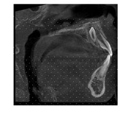
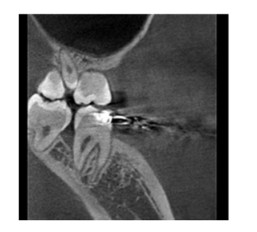
2.2 多生牙影像学特点 2.2.1 多生牙数目和部位在146例患者中,95例(66.07%)为单个多生牙,46例(31.51%)有2颗多生牙,4例(2.74%)有3颗多生牙,1例(0.68%)有4颗多生牙,平均每例患者有1.39颗多生牙。146例患者共有203颗多生牙,上颌前部有164颗(80.79%)多生牙(图 1),前磨牙区有36颗(17.73%)多生牙,磨牙区有3颗(1.48%)多生牙(图 2)。上颌多生牙有184颗(90.64%),下颌多生牙有19颗(9.36%)。23颗(11.33%)多生牙已萌出,未萌埋伏多生牙为180颗(88.64%)。

|
| 图 1 CBCT显示青少年多生牙患者上颌前部多生牙 Fig. 1 Supernumerary teeth in anterior maxilla of a adolescent patient with supernumerary teeth showed by CBCT |
|
|

|
| 图 2 CBCT显示青少年多生牙患者上颌磨牙区多生牙 Fig. 2 Supernumerary teeth in molar area of a adolescent patient with supernumerary teethshowed by CBCT |
|
|
多生牙生长方向:倒置多生牙112颗(55.17%),近远中方向的多生牙37颗(18.22%),唇腭向的多生牙28颗(13.80%),正常方向生长的多生牙为26颗(12.81%)。多生牙在正常牙列的唇腭/舌向位置关系:腭/舌侧有157颗(77.34%)多生牙,唇侧7颗(3.45%)多生牙,正常牙列内的多生牙为39颗(19.21%)。多生牙牙齿形状:圆锥形多生牙132颗(65.02%),结节形态多生牙42颗(20.69%),补充型多生牙29颗(14.29%)。
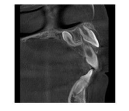
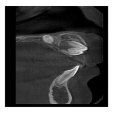
2.2.3 多生牙长度及其与周围组织关系多生牙长度和体积较正常恒牙短小,有34颗(16.75%)多生牙牙根未发育,169颗(85.25%)牙根发育完全。牙根弯曲的多生牙48颗(23.65%)(图 3),牙根不弯曲的多生牙155颗(76.35%)。引起邻牙吸收的多生牙31颗(15.27%)。多生牙形状与多生牙引起邻牙吸收的具体统计数据见表 1。突破鼻底的多生牙6颗(2.96%)(图 4),进入切牙管的多生牙4颗(1.97%), 伴随囊肿的多生牙7颗(3.35%)。

|
| 图 3 CBCT显示青少年多生牙患者多生牙牙根弯曲 Fig. 3 Root bending of supernumerary teeth of a adolescent patient with supernumerary teeth showed by CBCT |
|
|
| Shape | Number | Root resorption | Non-root resorption | Percentage |
| Conical | 132 | 18 | 132 | 8.87% |
| Tuberculate | 42 | 6 | 42 | 14.29% |
| Supplemental | 29 | 7 | 29 | 24.14% |
| Total | 203 | 31 | 203 | 15.27% |
| χ2 = 2.066,P=0.356. | ||||

|
| 图 4 CBCT显示青少年多生牙患者突破鼻底的多生牙 Fig. 4 Supernumerary teeth breaking through bottom of nose of a adolescent patient with supernumerary teeth showed by CBCT |
|
|
影像学检查为临床医师提供了清晰的图像。随着现代医学的不断发展,传统的X线手段已经不能满足临床需求,如多生牙位置复杂,如何确定多生牙与周围多个牙根毗邻关系,如何选择准确定位多生牙和如何选择手术入路等问题需要精准的三维图像分析。对于青少年患者而言,CBCT是一个良好的选择。锥形束投照计算机重组断层影像设备通过一次旋转获得扫描数据,所得数据在计算机中重组,从而得到三维图像。与传统螺旋CT比较,CBCT具有很多优点,如扫描时间短、伪影相对较少、图像分辨率高、辐射剂量较低和价格相对低廉等优点[5, 7]。CBCT清晰地显示多生牙在矢状面、冠状面和水平面上的位置,通过三维重建显示多生牙和颌骨内的位置、与周围邻牙的关系及牙根是否有吸收等情况[6, 8-9]。同时能根据不同患者和临床医生的治疗要求,观察分析在任意断层和多角度的三维图像,为临床治疗和手术提供个性化治疗方案和手术入路[10-11]。
多生牙可发生于颌骨任何部位,上前牙区多见,数目不等,可为单个,也可为多个。萌出的多生牙多数无正常的牙体解剖形成,常呈圆锥形[12-15]。本研究收集正畸科青少年患者146例, 共有203颗多生牙,男女比例为1.98:1,男性患者明显多于女性,这与多数研究[14, 16]结果相一致。95例(65.07%)为单个多生牙,其中上颌前部为多生牙好发区域,在上颌前部共有164颗(80.79%)多生牙;多生牙好发于牙列腭侧,其中有157颗(77.34%)多生牙发生于牙列腭侧;多生牙以圆锥形居多,其中有132颗(65.02%)多生牙呈圆形形状,少数多生牙存在引起邻牙吸收和伴随囊肿等情况,需要及时手术治疗[17-19]。χ2检验显示不同形状多生牙牙根吸收率的比较差异无统计学意义,说明多生牙引起邻牙吸收与多生牙形态无明显关联。当多生牙可能引起邻近牙齿吸收时,临床应该早期发现予以拔出。本研究数据来源于就诊正畸科的青少年多生牙患者,多同时伴有错颌畸形,正确定位多生牙的位置及周围邻近牙齿是否有吸收对后续正畸治疗有重要的临床意义,有助于正畸医师对患者正畸治疗的规划。多数多生牙埋伏阻生在颌骨内,采取手术治疗干预可能会损害相邻的牙齿,CBCT图像可精准地定位多生牙的位置、邻近组织及疾病的三维信息,明确多生牙在颌骨内的位置及其与邻牙的关系,确定多生牙唇腭侧骨量的多少,帮助外科医生选择适当的手术方法[20-21],识别牙齿并拔出多生牙,减少手术对邻近硬组织和软组织的创伤。
正畸科就诊患者同时伴有错颌畸形,需要在拔出多生牙的基础上继续进行下一步正畸矫正治疗。应用CBCT既可以明确患者口腔内的牙齿数目和形态,又可以观察颌骨发育情况,为下一步正畸治疗提供影像数据支持,此外,大视野CBCT还可以观察患者气道,明确患者气道狭窄的范围及程度,为正畸医生设计诊疗方案提供数据支持。CBCT能准确诊断和定位多生牙,成为多生牙患者术前常规检查手段之一。本研究结果为青少年多生牙的诊断和治疗提供了影像数据参考。
| [1] | RAJAB L D, HAMDAN M A M. Supernumerary teeth:review of the literature and a survey of 152 cases[J]. Int J Paediatr Dent, 2002, 12(4): 244–254. DOI:10.1046/j.1365-263X.2002.00366.x |
| [2] | 马绪臣. 口腔颌面锥形束CT的临床应用[M]. 北京: 人民卫生出版社,2011: 16-23. |
| [3] | 杨云丹, 张彊弢, 黄瑾, 等. CBCT在口腔正畸学的应用[J]. 中国医学物理学杂志, 2017, 34(1): 48–52. DOI:10.3969/j.issn.1005-202X.2017.01.010 |
| [4] | 陈松林, 林尔坚, 冉炜, 等. 螺旋CT牙体表面成像对骨内埋伏牙的定位及临床应用[J]. 华西口腔医学杂志, 2000, 18(4): 247–249. DOI:10.3321/j.issn:1000-1182.2000.04.012 |
| [5] | 鞠昊, 朱红华, 段涛, 等. CBCT的基本原理及在口腔各科的应用进展[J]. 医学影像学杂志, 2015, 25(5): 907–909. |
| [6] | 古向生, 崔敏毅, 涂之平, 等. 上颌骨内埋伏正中多生牙的生长特征与影像表现[J]. 中山大学学报:医学科学版, 2005, 26(5): 563–565. |
| [7] | TOURENO L, PARK J H, CEDERVERG R A, et al. Identification of supernumerary teeth in 2D and 3D:review of literature and a proposal[J]. J Dent Educ, 2013, 77(1): 43–50. |
| [8] | LIU D G, ZHANG W L, ZHANG Z Y, et al. Three-dimensional evaluations of supernumerary teeth using cone-beam computed tomography for 487 cases[J]. Oral Surg Oral Med Oral Pathol Oral Radiol Endod, 2007, 103(3): 403–411. DOI:10.1016/j.tripleo.2006.03.026 |
| [9] | LI N, HU B, MI F, et al. Preliminary evaluation of cone beam computed tomography in three-dimensional cephalometry for clinical application[J]. Exp Ther Med, 2017, 13(5): 2451–2455. DOI:10.3892/etm.2017.4278 |
| [10] | 文陈妮, 李果, 任家银, 等. 锥形束CT诊断上颌前牙区多生牙价值研究[J]. 华西口腔医学杂志, 2012, 30(4): 399–401. DOI:10.3969/j.issn.1000-1182.2012.04.017 |
| [11] | KIM K D, RUPRECHT A, JEON K J, et al. Personal computer-based three-dimensional computed tomographic images of the teeth for evaluating supernumerary or ectopically impacted teeth[J]. Angle Orthod, 2003, 73(5): 614–621. |
| [12] | 马绪臣. 口腔颌面医学影像学[M]. 北京: 北京大学医学出版社,2006: 89-92. |
| [13] | RAMESH K, VENKATARAGHAVAN K, KUNJAPPAN S, et al. Mesiodens:A clinical and radiographic study of 82 teeth in 55 children below 14 years[J]. J Pharm Bioallied Sci, 2013, 5(Suppl 1): S60–S62. |
| [14] | FERNÁNDEZ MONTENEGRO P, VALMASEDA CASTELLÓN E, BERINI AYTÉS L, et al. Retrospective study of 145 supernumerary teeth[J]. Med Oral Patol Oral Cir Bucal, 2006, 11(4): E339–E344. |
| [15] | 刘晓华, 王恩博. 儿童埋伏多生牙拔除术锥形束计算机断层扫描影像学定位的应用进展[J]. 国际口腔医学杂志, 2018, 45(3): 295–300. |
| [16] | 刘宪光, 张君, 李成龙, 等. 558例多生牙临床特点的回顾性分析[J]. 山东大学学报:医学版, 2017, 55(3): 121–124. |
| [17] | MOSSAZ J, KLOUKOS D, PANDIS N, et al. Morphologic characteristics, location, and associated complications of maxillary and mandibular supernumerary teeth as evaluated using cone beam computed tomography[J]. Eur J Orthod, 2014, 36(6): 708–718. DOI:10.1093/ejo/cjt101 |
| [18] | 杨再波, 朱彬, 周奇, 等. 上颌中切牙区域多生牙儿童患者的回顾性分析[J]. 临床口腔医学杂志, 2017, 33(11): 680–682. DOI:10.3969/j.issn.1003-1634.2017.11.013 |
| [19] | LGRAVERE M O, CAREY J, TOOGOOD R W, et al. Three-dimensional accuracy of measurements made with software on cone-beam computed tomography images[J]. Am J Orthod Dentofacial Orthop, 2008, 134(1): 112–116. DOI:10.1016/j.ajodo.2006.08.024 |
| [20] | 沈燕, 石羽, 周建国, 等. 178例儿童埋伏多生牙拔除难度的临床分级[J]. 中国口腔颌面外科杂志, 2018, 16(3): 259–262. |
| [21] | JEREMIAS F, FRAGELLI C M, MASTRANTONIO S D, et al. Cone-beam computed tomography as a surgical guide to impacted anterior teeth[J]. Dent Res J (Isfahan), 2016, 13(1): 85–89. DOI:10.4103/1735-3327.174723 |
 2019, Vol. 45


