扩展功能
文章信息
- 王宝刚, 满霞霞, 马大实, 王勇, 曹殿波
- WANG Baogang, MAN Xiaxia, MA Dashi, WANG Yong, CAO Dianbo
- 升主动脉内巨大血栓1例报告及文献复习
- Huge thrombus in ascending aorta:A case report and literature review
- 吉林大学学报(医学版), 2018, 44(05): 1065-1067
- Journal of Jilin University (Medicine Edition), 2018, 44(05): 1065-1067
- 10.13481/j.1671-587x.20180533
-
文章历史
- 收稿日期: 2018-03-03
2. 吉林大学第一医院肿瘤妇科, 吉林 长春 130021;
3. 吉林大学第一医院放射线科, 吉林 长春 130021
2. Department of Oncologic Gynecology, First Hospital, Jilin University, Changchun 130021, China;
3. Department of Radiology, First hospital, Jilin University, Changchun 130021, China
主动脉内血栓在临床上极为少见。国内外相关文献不多,多为个案报道。由于患病例数少,尚无治疗指南可遵循,其治疗往往取决于医生的临床经验。主动脉腔内血栓的临床表现各不相同,通常与栓塞的部位有关,包括脑卒中引起的意识丧失[1]、脾梗死导致的腹痛[2]及肢体急性缺血的下肢疼痛[3]等,治疗初期常被误诊,栓子脱落造成重要脏器或肢体栓塞后,再进行体外循环手术取栓,往往愈合不佳。本文作者报道1例升主动脉内血栓患者的临床资料,并进行文献复习,旨在提高对本病诊治的认识,阐明主动脉CT血管成像(CT angiography,CTA)检查的必要性。
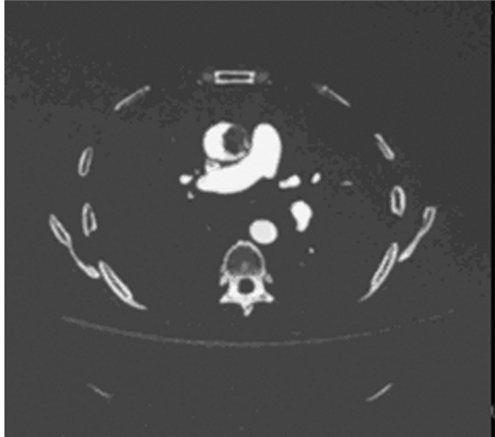
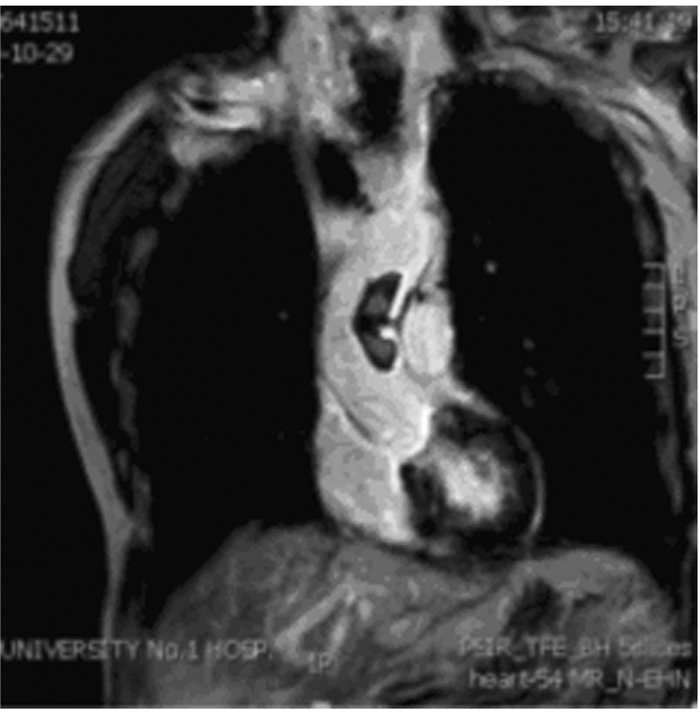
1 临床资料患者,男性,56岁,身高168 cm,体质量61 kg,2017年2月来本院常规体检。生命指征:心率100 min-1,血压140/100 mmHg,室温下动脉血氧饱和度95%,体温36.5℃。既往无高血压病史,未口服降压药物。无胸背部疼痛病史,无心房颤动病史。查体未闻及心脏杂音,四肢运动感觉未见异常。化验回报人脑利钠肽(brain natriuretic peptide,BNP)131 ng·L-1,凝血酶原时间10.3 s,D二聚体及肌钙蛋白正常。超声心动图及主动脉CTA检查(图 1)提示升主动脉腔内存在一处占位性病变,病变大小为2.2 cm×2.2 cm×4.5 cm,边界清楚。头部CT提示双侧多发腔隙性脑梗死。双下肢动脉彩超无异常。进一步行MRI检查(图 2),结果回报病变的性质可能为血栓。
|
| 图 1 升主动脉腔内巨大血栓患者术前主动脉CTA结果 Figure 1 Result ofaortic CTA before operation of patient with huge thrombus in ascending aorta |
|
|
|
| 图 2 升主动脉腔内巨大血栓患者术前主动脉MRI结果 Figure 2 Result of aortic MRI before operation of patient with huge thrombus in ascending aorta |
|
|
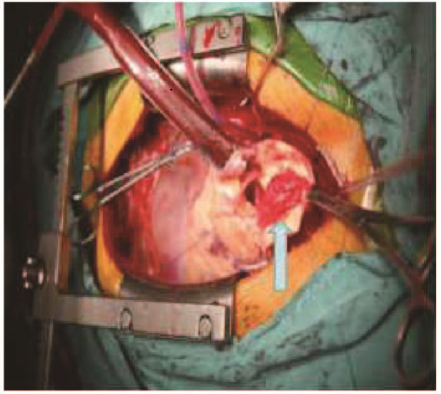
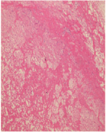
治疗方法包括病变切除和人工血管置换。应用右股动脉和右心房插管,建立体外循环。升主动脉与周围组织无黏连。阻断升主动脉,切开后可见一光滑的、红色团块样物质(图 3和4,见插页六),通过蒂部附着于升主动脉内膜,附着处位于主动脉瓣环上方3.0 cm处,局部无夹层改变,无心内膜炎改变或其他病变。主动脉瓣呈三叶,无关闭不全。术中行肿物Enbloc切除,并置换升主动脉,范围自主动脉瓣环上方1.0 cm至头臂干动脉近端3.0 cm处。术后病理检查结果显示:血栓机化(图 5,见插页六)。
|
| 图 3 术中病变和主动脉内壁的图片 Figure 3 Photograph of mass lesion and aortic wall in operation (blue arrow) |
|
|

|
| 图 4 术中切除后标本的图片 Figure 4 Photograph of mass specimen after excision in operation |
|
|
|
| 图 5 术后切除标本的病理检查结果(HE, ×40) Figure 5 Pathological result of specimen after operation(HE, ×40) |
|
|
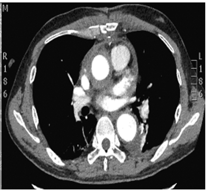
患者术后10 d顺利出院,无脏器或肢体梗死。复查超声心动图显示:左室射血分数(left ventricular ejection fractions,LVEF)54%,主动脉瓣功能良好。主动脉CTA显示无血栓复发(图 6)。
|
| 图 6 升主动脉腔内巨大血栓患者术后主动脉CTA结果 Figure 6 Result of aortic CTA after operation of patient with huge thrombus in ascending aorta |
|
|
主动脉内血栓并非是缺血性脑卒中栓子的主要来源。在手术前多诊断为主动脉内占位性病变,即使借助磁共振检查,也无法完全确认其病变性质。如果术前考虑病变为血栓,并且病变位于胸降主动脉、腹主动脉及其分支内,可考虑给予抗凝药物[4-5]或植入覆膜支架[6-7],进行腔内隔绝。经常使用的抗凝药物包括低分子肝素和华法林。同时需要监测国际标准化比值(international ratio,INR),复查经胸超声心动图(transthoracic echocardiography,TTE)或主动脉CTA。通常血栓会逐渐减小并消失。如果病变位于升主动脉或主动脉弓内,病变性质考虑为血栓,尤其是经主动脉CTA及TTE检查,证实病变呈“漂动”状态[1-2, 8-10],需在体外循环下行急诊开胸手术。术后治疗主要集中在栓塞引起的并发症。在一项关于主动脉内占位性病变的研究中,患者因急性下肢缺血行取栓治疗后,病理检查证实病变性质不是血栓,而是黏液瘤[10]。提示外科医生应通过主动脉CTA扩大检查范围,寻找原发病灶,尽早再次手术,避免肿瘤继续脱落或发生恶变。本病例的不同之处在于,患者术前并无栓塞症状。由于不确定病变的性质,也无法确定其为血栓,故未进行抗凝治疗[1];考虑此病变体积巨大,位置在升主动脉腔内,一旦脱落将导致严重的脏器栓塞,故采取积极的手术治疗[5]。术后无栓塞并发症发生。
由于主动脉内占位性病变危害较大,故早期发现和早期治疗尤为重要[10]。这一病例充分说明主动脉CTA检查的重要性。主动脉CTA检查因其便捷性和高敏感性,被推荐作为首选检查方法。术前主动脉CTA检查可为医生提供病变的准确位置和形状[2]。同时还可用于评估肺部影像,尤其是患者术前并发有呼吸系统疾病的情况。当病变位于升主动脉内时,患者往往需要在体外循环辅助下行急诊手术,又无法进行详细的肺功能检查。借助主动脉CTA检查提供的肺部影像,大致评估其对体外循环的耐受程度。
多数情况下,主动脉内膜滋养动脉破裂后会造成局部出血;血肿逐渐扩大,形成主动脉夹层;或经积极药物治疗后逐渐吸收。应用计算流体力学技术的研究[11-12]结果显示:射血时管腔内血流速度分布并不均匀,从升主动脉至左锁骨下动脉根部区域,以小弯侧后壁流速较快,且由近心端向远心端逐渐增加;从壁面剪切力分析,升主动脉剪切力从大弯向小弯逐渐升高,分支血管根部剪切力较高,近侧壁以低剪切力为主。该例患者血栓发生部位的血流速度不快,剪切力又低于分支血管的根部,故未形成典型的主动脉夹层,而是血肿被局部组织限制住;反复出血后血肿体积逐渐增大,突入升主动脉腔内,形成占位性病变而少量血栓脱落,造成腔隙性脑梗死。
综上所述,对于主动脉腔内血栓的患者,主动脉CTA是首选的检查手段。血栓切除和人工血管植入可以有效降低缺血性卒中的风险。
| [1] | Madershahian N, Kuhn-Regnier F, Mime L, et al. A loose cannon:free-floating thrombus in ascending aorta[J]. J Cardiac Surg, 2009, 24: 198–199. DOI:10.1111/jcs.2009.24.issue-2 |
| [2] | de Maat GE, Vigano G, Mariani MA, et al. Catching a floating thrombus:a case report on the treatment of a large thrombus in the ascending aorta[J]. J Cardiothorac Surg, 2017, 12: 34. DOI:10.1186/s13019-017-0600-x |
| [3] | Barry N, Tsui J, Dick J, et al. Aortic tumor presenting as acute lower limb ischemia[J]. Circulation, 2011, 123: 1785–1787. DOI:10.1161/CIRCULATIONAHA.110.985689 |
| [4] | Dhillon P, Murdoch D, Jayasinghe R, et al. A case of mobile aortic arch thrombus with systemic embolisation-a management dilemma[J]. Heart Lung Circu, 2014, 23: e88–e91. DOI:10.1016/j.hlc.2013.09.009 |
| [5] | Yang SY, Yu J, Zeng WJ, et al. Aortic floating thrombus detected by computed tomography angiography incidentally:Five cases and a literature review[J]. J Thoracic Cardiovasc Surg, 2017, 153(4): 791–803. DOI:10.1016/j.jtcvs.2016.12.015 |
| [6] | Daniel J, Joseph M, Zachary M. Endovascular management of a mobile thoracic aortic thrombus following recurrent distal thromboembolism:a case report and literature review[J]. Vasc Endovasc Surg, 2014, 48(3): 246–250. DOI:10.1177/1538574413513845 |
| [7] | Luebke T, Aleksic M, Brunkwall J. Endovascular therapy of a symptomatic mobile thrombus of the thoracic aorta[J]. Eur J Vasuc Endovasc Surg, 2008, 36: 550–552. DOI:10.1016/j.ejvs.2008.07.004 |
| [8] | Maden J, Goran Panic, Vesna Lackovic, et al. Butterfly-shaped thrombus in the ascend aorta[J]. Euro J Cardio-thorac Surg, 2006(30): 936. |
| [9] | TeranishiH, TsubataM, AraiY, et al. Floating thrombus in ascending aorta[J]. J Cardiac Surg, 2016, 31: 549–550. DOI:10.1111/jocs.v31.8 |
| [10] | Jacvorski L, Fihjal Kowski M, Rogowski J. Giant thrombus in ascending aorta[J]. J Thorac Cardiovasc Surg, 2013, 145(6): 1668–1669. DOI:10.1016/j.jtcvs.2013.01.009 |
| [11] | Zhang T, Xiong J, Hu XZ, et al. Application of computational fluid dynamics in hemodynamic research of aortic arch[J]. Zhonghua YiXue ZaZhi, 2013, 93(5): 380–384. |
| [12] | Li W, Shen C, Zhang X, et al. Role of computational fluid dynamics in thoracic aortic diseases research:technical superiority and application prospect[J]. Zhonghua Wai Ke Za Zhi, 2015, 53(8): 637–640. |
 2018, Vol. 44


